
BioMed Central
Page 1 of 8
(page number not for citation purposes)
Theoretical Biology and Medical
Modelling
Open Access
Research
Identification and isolation of embryonic stem cells in reproductive
endocrinology: theoretical protocols for conservation of human
embryos derived from in vitro fertilization
Eric Scott Sills1, Takumi Takeuchi2, Noriko Tanaka2, Queenie V Neri2 and
Gianpiero D Palermo*2,3
Address: 1Georgia Reproductive Specialists LLC, Division of Reproductive Endocrinology and Infertility, Department of Obstetrics and
Gynecology, Atlanta Medical Center; Atlanta, Georgia 30342 USA, 2Cornell Center for Reproductive Medicine and Infertility, Weill Medical
College of Cornell University, New York, New York 10021 USA and 3HT-336, 505 East 70th Street, New York, New York 10021 USA
Email: Eric Scott Sills - dr.sills@ivf.com; Takumi Takeuchi - ttakeuchi@med.cornell.edu; Noriko Tanaka - not2003@med.cornell.edu;
Queenie V Neri - qneri@med.cornell.edu; Gianpiero D Palermo* - qneri@med.cornell.edu
* Corresponding author
Abstract
Background: Embryonic stem cells (ESC) are pluripotent cells obtained from the inner cell mass
(ICM) of blastocysts derived from in vitro culture associated with reproductive endocrinology
therapy. Human ESCs are regarded as highly significant since they retain the capacity to differentiate
into any of approximately 200 unique cell types. Human ESC research is controversial because to
acquire such cells, the ICM of human blastocysts must be manipulated in a way that renders
embryos nonviable and unsuitable for transfer in utero. Techniques to yield competent ESCs with
conservation of source blastocysts would satisfy many objections against ESC research, but at
present such approaches remain largely untested.
Results and discussion: We contrast experimental culture of single blastomeres obtained by 1)
non-destructive biopsy of embryos destined for transfer, and 2) isolation of karyotypically normal
blastomeres from disaggregated ("dead") embryos considered unsuitable for transfer, and evaluate
these approaches with regard to production of ESCs. Pluripotency was confirmed by morphological
criteria and by quantification of divergent homeodomain proteins specific to undifferentiated cell
development. Following ESC isolation and identification, assessment was conducted according to a
novel ESC grading system, also proposed here.
Conclusion: The role of reproductive endocrinology in ESC research remains paramount. In this
report, we hypothesize new and expand on existing strategies having the potential to enhance
human ESC isolation, identification and in vitro maintenance.
Background
While the definitive characterization of murine embry-
onic stem cells was first reported in 1981, embryonic stem
cells (ESC) were not isolated and fully described in
humans until much later [1]. Without question, the scarce
supply of human ESCs combined with the technical
Published: 18 July 2005
Theoretical Biology and Medical Modelling 2005, 2:25 doi:10.1186/1742-4682-2-
25
Received: 01 April 2005
Accepted: 18 July 2005
This article is available from: http://www.tbiomed.com/content/2/1/25
© 2005 Sills et al; licensee BioMed Central Ltd.
This is an Open Access article distributed under the terms of the Creative Commons Attribution License (http://creativecommons.org/licenses/by/2.0),
which permits unrestricted use, distribution, and reproduction in any medium, provided the original work is properly cited.

Theoretical Biology and Medical Modelling 2005, 2:25 http://www.tbiomed.com/content/2/1/25
Page 2 of 8
(page number not for citation purposes)
challenges associated with interspecies translation of stem
cell derivation contributed to the long interval between
these reports. To obtain human ESCs, embryos produced
during in vitro fertilization (IVF) are maintained in
extended culture to the blastocyst stage (4–5d post fertili-
zation) when the polarized inner cell mass (ICM) devel-
ops. The outer trophoectoderm is removed via
immunosurgery, thus exposing the ICM for disaggrega-
tion and plating on a feeder cell layer for further culture.
Importantly, this disruptive process renders the embryo
non-viable and unsuitable for in utero transfer [2].
Homogenous human ESC colonies may be derived from
subsequent isolation and re-plating of the ICM cells,
which are then screened for stemness by a variety of recog-
nized markers.
Once in stable culture, ESCs are capable of either symmet-
ric (clonogenic) or asymmetric fission. Symmetric ESC
division yields a self-renewing supply of pluripotent ESCs,
while asymmetric division produces one cell identical to
the parent ESC plus one differentiated cell. The mecha-
nism(s) responsible for modulating these specific ESC fis-
sion patterns remain poorly understood. In any case, since
it is not yet possible to de-differentiate committed somatic
cells to reacquire pluripotency, embryos associated with
IVF have thus far been the only source for human ESCs.
The fact that live human embryos must be destroyed to
produce human ESCs presents a substantial ethical obsta-
cle for the advancement of human ESC research. With the
vast therapeutic promise of human ESC seen against the
destruction of human embryos required to realize such
aspirations, compelling arguments have been articulated
both in support of and in opposition to human ESC
research [3,4].
It must be admitted that thus far human ESCs have pro-
vided no reproducible, safe, unique and previously unat-
tainable treatment for any human disease. Nevertheless,
interest in exploration of the full therapeutic possibility of
human ESCs continues to grow and is no longer confined
to medical scientists and reproductive endocrinologists –
indeed, it now includes public opinion leaders and medi-
cal consumers as well [5]. Yet the concerns of human ESC
research opponents are not without ethical justification;
these objections could be substantially assuaged if safe
and effective laboratory protocols could be developed that
offered human ESCs whilst preserving (or at least not
destroying) the blastocysts from which they originated. In
this paper we present results from pilot studies based on
some theoretical approaches, in a manner to facilitate
human ESC research and to promote respect for human
embryos obtained from clinical reproductive endocrinol-
ogy practice.
Human embryonic stem cells: theoretical approaches
Blastomere biopsy and culture
Prior to the blastocyst stage, a human embryo at 2–3d
post fertilization consists of just 4–8 cells, all of which are
totipotent. In contrast to the pluripotent cells obtained
from the blastocyst ICM, one or two of these blastomeres
may be biopsied without compromising the integrity of
the sampled embryo [6]. Blastomeres obtained for PGD
are generally fixed and processed with fluorochromes to
detect aneuploidy by partial karyotype analysis, although
the process has more recently advanced to testing for sin-
gle gene disorders via polymerase chain reaction [7] and
single cell whole genome amplification by multiple dis-
placement amplification [8]. Such processing irrevocably
alters the blastomere destined for PGD – the viability of
this cell is sacrificed in return for the vital genomic infor-
mation provided by PGD. However, assuming two dis-
tinct blastomeres were extracted at a well-timed embryo
biopsy for PGD, and since in the absence of mosaicism
each blastomere retains the potential to develop into a
complete organism [9], the possibility exists that at least
one sampled blastomere obtained for PGD could be
maintained in culture specifically for ESC production. As
with traditional PGD protocols, genetic data needed from
PGD could still be obtained and inform embryo transfer
decisions, while the second blastomere could provide a
potential source of human ESCs with no measurable
adverse affect on the developmental integrity of the biop-
sied embryo.
Utilizing a murine embryo model, we evaluated this con-
cept where two blastomeres were isolated from a single 8-
cell embryo via standard microsurgical biopsy techniques
[10]. Zona-free murine blastomeres were then washed,
individually plated and cultured as previously described
[11]; embryos from which the biopsies were taken were
maintained in standard culture (control group). All cells
were monitored × 12 h to assess cleavage, differentiation,
and attachment to the feeder cell monolayer, as applicable
(Figure 1 and 2). With proper culture conditions we
observed advancement to morphologically normal blast-
ocyst stage in both groups. Next, cells resembling an ICM
that originated from the intact/source embryo group and
the single blastomere culture group were disaggregated
from their respective blastocysts and re-plated on to fresh
feeder cells for confirmation and further analysis; no cells
were frozen. This work carries forward a theoretical
approach suggested more than a decade ago [12], and
demonstrates that a blastomere biopsy and culture
approach can supply a single totipotent cell for subse-
quent ESC culture without harming the source embryo.
Blastomere donation from non-viable ("dead") embryos
Human embryo assessment plays a central role in IVF to
identify embryos with the best prognosis for transfer, but

Theoretical Biology and Medical Modelling 2005, 2:25 http://www.tbiomed.com/content/2/1/25
Page 3 of 8
(page number not for citation purposes)
what is less clear is the fate of embryos judged not suitable
for transfer or cryopreservation due to arrested growth or
gross developmental abnormality. Despite the absence of
formal guidelines governing human embryology practice,
many IVF centers carefully monitor embryos over several
days before making the determination that they should be
neither transferred nor cryopreserved based on non-via-
bility. Indeed, even with cryopreservation as late as post-
fertilization day 7, human livebirths have been achieved
[13]. However, as previous investigators have noted
[2,14], a consensus definition of embryo non-viability or
death remains elusive and it is reasonable to expect that
the concept of embryo death will formalize gradually in a
process similar to that which led to the 1981 Uniform
Determination of Death Act [15]. In the meantime, most
major IVF clinics already obtain written informed consent
from patients to discard any human embryos deemed
non-viable or dead.
Interestingly, IVF laboratories have confronted this chal-
lenge and produced an informal if not exactly uniform
process to declare a human embryo "dead". Since the life
of any developing organism is more than the sum of its
cellular parts, it has been suggested that the defining vital
characteristics of a 4- or 8-cell human embryo must
include continued and integrated cellular division,
growth, and differentiation [16]. And by extension,
embryos that have irreversibly lost this basic capacity
(even if individual constituent cells may remain alive)
should be properly regarded as organismically dead.
Therefore our investigations were based on assessment of
fresh (non-cryopreserved) 4–8 cell embryos demonstrat-
ing developmental arrest observed over an 8-day in vitro
culture interval. Among such non-viable embryos des-
tined for discard, a high rate of chromosomal error has
been found in some, but not all, blastomeres [17]. It is the
salvage of any normal blastomeres within a "dead"
embryo that holds particular promise for human ESC
research. Specifically, if embryos classified as non-viable
and unsuitable for transfer or cryopreservation were disag-
gregated (rather than discarded) and plated as single
totipotent blastomeres as described above, then the possi-
bility exists that at least some karyotypically normal cell
colonies could develop and serve as a reliable human ESC
source. While the attempt to produce blastocysts from iso-
lated blastomeres in vitro is not new [18], we feel this
approach has received limited attention and merits fur-
ther exploration to advance human ESC research, particu-
larly since this source of ESCs would not derive from
human embryos otherwise destined for transfer or
cryopreservation.
We investigated the efficacy of a novel methodology with
murine embryos that failed to meet viability standards,
and were therefore unsuitable for transfer or cryopreserva-
tion. Embryos used in this pilot study displayed arrested
growth and were classified as nonviable no later than the
8-cell stage. Embryos were disaggregated into single blast-
omeres by brief exposure to trypsin under micromanipu-
lation control. Next, blastomeres were individually plated
on a feeder cell layer and cultured in an experimental
medium supplemented with β-mercaptoethanol, amino
acids, nucleosides, antibiotics, L-glutamine with 2000 IU/
ml mouse recombinant leukemia inhibiting factor in 6%
CO2 at 37°C. Fully-expanded or hatching mouse blasto-
cysts were plated as controls. The salvaged blastomeres
and normal blastocysts were monitored daily to evaluate
differentiation, cleavage and attachment to the feeder cell
layer. Although some blastomeres obtained from the dead
embryos failed to progress, a few ICM-like clusters devel-
oped from single blastomeres. These were isolated (as
were ICMs derived from the intact blastocysts) and disag-
gregated into single cells by trypsinization and replated on
to fresh feeder cell layers. These ESC lines were assessed
for pluripotency by morphological criteria as well as alka-
line phosphatase activity, Oct-4, and TROMA-1 [19],
which validated stemness in this experiment.
Impact of mosaicism on ESC derivation
Soon after the first clinical experience with preimplanta-
tion genetic diagnosis was reported [20], it was suggested
that blastomere mosaicism might contribute to the clini-
cal error rate observed in PGD [21]. The precept that not
all blastomeres are necessarily equivalent has subse-
quently emerged as a recognized tenet in human embry-
ology; it figures prominently in the informed consent for
patients contemplating PGD [22]. Currently, a technique
to determine the extent of embryo mosaicism without dis-
assembling the embryo (and thus rendering it nonviable)
does not exist. Accordingly, mosaicism presents poten-
tially serious weaknesses for the two proposed ESC tech-
niques described here, since the effectiveness of each
approach is affected by the extent of blastomere mosai-
cism, which cannot be known a priori.
Nevertheless, for the two theoretical ESC protocols we
present, the impact of embryo mosaicism is not the same
and each instance deserves separate consideration. For
example, if the PGD+blastomere biopsy and culture
method were applied to embryos with extensive blast-
omere heterogeneity, this approach would be unlikely to
produce chromosomally normal cells for subsequent in
vitro ESC culture. If, however, embryos with very limited
or no mosaicism are used for the proposed PGD+blast-
omere culture process, human ESC production could pro-
ceed with much greater likelihood given the uniformity of
all sampled cells. Given the unknown extent of embryo
mosaicism, limitations of single blastomere biopsy have
been recognized [22] and some researchers have recom-
mended confirmatory PGD by sampling a second
Theoretical Biology and Medical Modelling 2005, 2:25 http://www.tbiomed.com/content/2/1/25
Page 4 of 8
(page number not for citation purposes)
blastomere [23]. In contrast, among blastomeres obtained
from the disaggregation of nonviable embryos, it would
be reasonable to expect a higher frequency of mosaicism.
In such a setting, even limited mosaicism would yield the
desirable result based on the presence of at least one
genetically normal constituent blastomere within an
organismically dead embryo.
Objective assessment of ESC colonies
Although considerable resources are required to harvest
and propagate ESCs, effective methods to verify stemness
and monitor quality in such cells are also needed to bring
the full range of therapeutic possibility into focus. In an
effort to develop an assessment system for ESCs, our
center cultured murine blastocysts on mouse fibroblasts
in experimental media supplemented with 2000 IU/ml
mouse recombinant leukemia inhibitory factor. At 4–5d,
the ICMs were mechanically isolated and disaggregated by
trypsin. Cell passages were repeated × 2–3d as needed,
according to colony confluency.
Our ESC colonies were then graded on the basis of three
factors: 1) colony number, 2) colony density, and 3) col-
ony quality. We determined colony character by morpho-
logical assessment using an inverted microscope with
phase-contrast optics. Typically, ESCs are large and dem-
onstrate a high nuclear:cytoplasm ratio (Figure 3). Each
colony was classified according to the proportion of stem
cells present within the colony, where >70% (good), 40–
70% (average), or <40% (poor) were the three divisions.
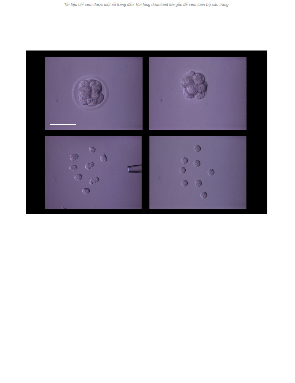
Blastomere isolation sequenceFigure 1
Blastomere isolation sequence. An intact 8-cell mouse embryo (a) was subjected to pronase digestion to remove the zona pel-
lucida (b). Single blastomeres were disaggregated by microdissection (c) and after stabilization in culture were monitored for
further treatment (d). Scale bar = 100 microns.
ab
cd
Theoretical Biology and Medical Modelling 2005, 2:25 http://www.tbiomed.com/content/2/1/25
Page 5 of 8
(page number not for citation purposes)
For all ESC colonies, alkaline phosphatase activity and
Oct-4 were used as markers of totipotency. TROMA-1 anti-
body (monoclonal) directed against cytokeratin-like fila-
ments of trophectoderm and endodermal cells served as a
negative marker. As an additional control these markers
were tested on expanded blastocysts. Specimens were
fixed with 4% paraformaldehyde and permeabilized with
0.2% Triton X-100. Alkaline phosphatase activity in fixed
cells was detected via azo-dye with Texas-red filter under
fluorescence microscopy. ESCs were exposed to Oct-4 pol-
yclonal antibody (1:100 dilution) and monoclonal
TROMA-1 antibody (1:6 dilution), followed by rinse with
PBS/BSA to remove unbound antibody. From these exper-
iments, we obtained 16 murine ESC lines from 164 source
blastocysts. Assessments via alkaline phosphatase and
Oct-4 to verify pluripotency of the ESC lines were in agree-
ment (χ2 = 0.105), while TROMA-1 identified endoderm
and trophoblast. Pluripotency was successfully confirmed
in at least some cells from each colony studied, and we
were able to establish concordance between morphologi-
cal criteria and marker activity. Further studies will be
helpful to show if additional parameters can refine this
ESC scoring system.
Stem cell research: social and political factors
Public sharing of information about the basic science of
ESCs has proven to be important, since those who are
aware of the stem cell debate tend to be more supportive
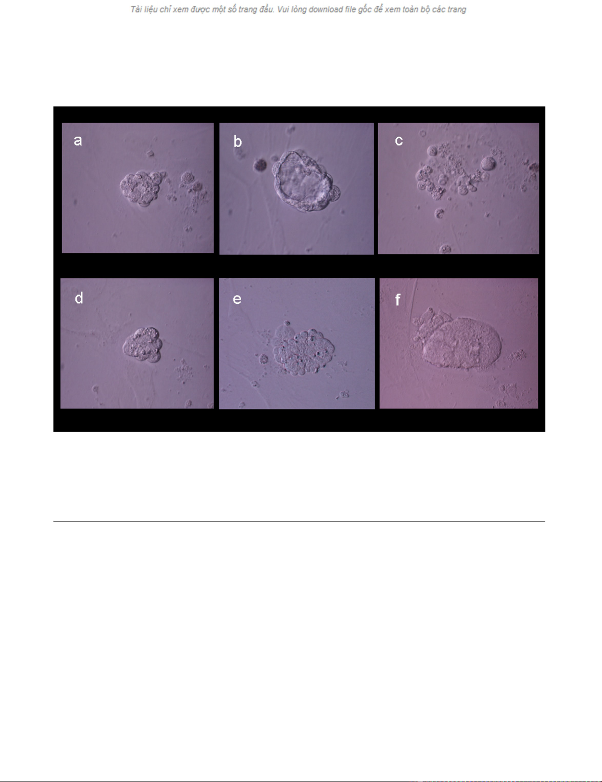
Evolution of experimental blastomere growth observed on feeder cell layer on culture days 2, 3, and 4Figure 2
Evolution of experimental blastomere growth observed on feeder cell layer on culture days 2, 3, and 4. Top row shows a single
blastomere undergoing cleavage (a) and forming a "unilaminar vesicle" on day 3 (b). Cellular arrest and degeneration were evi-
dent by day 4 (c). Bottom row shows another cleaving blastomere (d), which formed a cellular aggregate on day 3 (e) and later
developed an inner cell mass-like structure (f).


