
Open Access
Available online http://arthritis-research.com/content/7/2/R324
R324
Vol 7 No 2
Research article
Endothelin-1 in osteoarthritic chondrocytes triggers nitric oxide
production and upregulates collagenase production
Christina Alexandra Manacu1, Johanne Martel-Pelletier2, Marjolaine Roy-Beaudry1, Jean-
Pierre Pelletier2, Julio C Fernandes3, Fazool S Shipkolye1, Dragoslav R Mitrovic4 and
Florina Moldovan1,5
1Research Center, Sainte-Justine Hospital, Montreal, Quebec, Canada
2Osteoarthritis Research Unit, Centre Hospitalier de l'Université de Montréal, Hopital Notre-Dame, Montreal, Quebec, Canada
3Orthopaedics Research Laboratory, Department of Orthopaedics, Centre hospitalier Sacre-Coeur, Montreal, Quebec, Canada
4INSERM U-606, Hôpital Lariboisière, Paris, France
5Faculty of Dentistry, Université de Montréal, Quebec, Canada
Corresponding author: Florina Moldovan, florina.moldovan@umontreal.ca
Received: 20 Apr 2004 Revisions requested: 19 May 2004 Revisions received: 10 Nov 2004 Accepted: 1 Dec 2004 Published: 17 Jan 2005
Arthritis Res Ther 2005, 7:R324-R332 (DOI 10.1186/ar1489)http://art hritis-research .com/content /7/2/R324
© 2005 Manacu et al., licensee BioMed Central Ltd.
This is an Open Access article distributed under the terms of the Creative Commons Attribution License (http://creativecommons.org/licenses/by/
2.0), which permits unrestricted use, distribution, and reproduction in any medium, provided the original work is properly cited.
Abstract
The mechanism of endothelin-1 (ET-1)-induced nitric oxide (NO)
production, MMP-1 production and MMP-13 production was
investigated in human osteoarthritis chondrocytes. The cells
were isolated from human articular cartilage obtained at surgery
and were cultured in the absence or presence of ET-1 with or
without inhibitors of protein kinase or LY83583 (an inhibitor of
soluble guanylate cyclase and of cGMP). MMP-1, MMP-13 and
NO levels were then measured by ELISA and Griess reaction,
respectively. Additionally, inducible nitric oxide synthase (iNOS)
and phosphorylated forms of p38 mitogen-activated protein
kinase, p44/42, stress-activated protein kinase/Jun-N-terminal
kinase and serine-threonine Akt kinase were determined by
western blot. Results show that ET-1 greatly increased MMP-1
and MMP-13 production, iNOS expression and NO release.
LY83583 decreased the production of both metalloproteases
below basal levels, whereas the inhibitor of p38 kinase,
SB202190, suppressed ET-1-stimulated production only.
Similarly, the ET-1-induced NO production was partially
suppressed by the p38 kinase inhibitor and was completely
suppressed by the protein kinase A kinase inhibitor KT5720 and
by LY83583, suggesting the involvement of these enzymes in
relevant ET-1 signalling pathways. In human osteoarthritis
chondrocytes, ET-1 controls the production of MMP-1 and
MMP-13. ET-1 also induces NO release via iNOS induction. ET-
1 and NO should thus become important target molecules for
future therapies aimed at stopping cartilage destruction.
Keywords: endothelin-1, metalloproteases, nitric oxide, osteoarthritis, signalling pathways
Introduction
Cartilage degradation in osteoarthritis (OA) and rheuma-
toid arthritis constitutes a major structural change in the
joint, which may severely impair its function and cause pain
and disability. This degradation is accompanied by the
release in the synovial fluid of degraded matrix constituents
that primarily result from an increased matrix catabolism [1].
Various factors are directly involved in this process.
Endothelin-1 (ET-1), a potent vasoconstrictor and promi-
togen peptide for many cell types, including chondrocytes,
was recently identified as one such factor [2,3].
ET-1 binds to the specific endothelin A or endothelin B
receptors expressed on chondrocytes [4] and triggers a
cascade of intracellular events, including phospholipase C
activation [5], an increase in intracellular calcium [6,7],
prostaglandin production [8] and nitric oxide (NO) release
[9]. The effect of ET-1 on DNA and protein synthesis in
chondrocytes is biphasic. The potent initial stimulatory
DMEM = Dulbecco's modified Eagle's medium; ELISA = enzyme-linked immunosorbent assay; ET-1 = endothelin-1; FCS = foetal calf serum; IL =
interleukin; iNOS = inducible nitric oxide synthase; L-NIL = L-N6(1-iminoethyl)lysine; MAP = mitogen-activated protein; MEK1/2 = mitogen-activated
protein kinase kinase 1/2; MMP = metalloprotease; NO = nitric oxide; OA = osteoarthritis; PKA = protein kinase A; SAP/JNK = stress-activated pro-
tein kinase/Jun-N-terminal kinase; TUNEL = terminal deoxynucleotidyl transferase-medulated dUTP nick end labelling.

Arthritis Research & Therapy Vol 7 No 2 Manacu et al.
R325
effect of ET-1 decreases progressively with time and is fol-
lowed by an inhibition [3,8]. The inhibitory effect seems to
be mediated by NO and cGMP, both produced in response
to ET-1 stimulation [8,9]. Additionally, we have recently
demonstrated that ET-1 is significantly increased locally in
OA cartilage and synovial membrane when compared with
normal tissues. In OA cartilage, ET-1 is involved in cartilage
catabolism through metalloprotease (MMP) regulation and
the induction of type II collagen breakdown [2].
MMPs are a family of structurally related zinc-dependent
neutral endopeptidases classified into subgroups of colla-
genases, gelatinases, stromelysins, membrane-type MMPs
and other MMPs [10]. When activated, MMPs degrade a
broad spectrum of substrates, including collagens and
other matrix macromolecules. As a whole, MMPs play an
important role in the extracellular matrix remodelling that
occurs under physiological and pathological conditions.
Among all the MMPs, we have recently demonstrated an
induction in the synthesis, secretion and activation of two
collagenases (MMP-1 and MMP-13) by ET-1 [2]. These
MMPs play an active role in the progression of OA pathol-
ogy as they are the most effective at initiating collagen
destruction during the inflammatory process and the
remodelling phase of the disease [11,12].
Another deleterious agent in joint cartilage is the NO radi-
cal [13,14], which downregulates DNA [8] and matrix syn-
thesis [14] and upregulates matrix degradation via
increased MMP synthesis [15]. Indeed, inhibition of NO
production was shown to slow down the progression of
OA. It has been demonstrated that, in vitro, NO could also
upregulate MMP synthesis and activity in joint chondro-
cytes and cartilage [15]. In vivo in an OA animal model,
selective inhibition of the inducible nitric oxide synthase
(iNOS) provides a protective effect on OA joint tissues
more specifically in regard to the degradation of the extra-
cellular matrix and the downregulation of MMP [16].
The aim of the present study was to further investigate the
role of ET-1 in human OA chondrocytes, focusing on NO,
MMP-1 and MMP-13 production as well as the relevant sig-
nalling pathways activated by ET-1 in human OA chondro-
cytes in regard to these factors.
Materials and methods
Specimens
Human cartilage was obtained with the consent of 12 OA
patients (mean ± standard error of the mean age, 58 ± 6
years) undergoing total knee replacement. The Institutional
Ethics Committee Board of Notre Dame Hospital in Mon-
treal, Canada approved the study protocol. Tissue speci-
mens were embedded in paraffin, were sectioned and
stained with Safranin O and fast green, and were evaluated
using the Mankin histological/histochemical scale [17].
Only tissues corresponding to a moderate degree of OA
severity (Mankin 3–7) were included in this study. Cartilage
was sectioned from the tibial plateaus, rinsed and finely
chopped, and the cells released by enzymatic digestion
performed as previously described [2,11]. The cells were
seeded in culture flasks at the density of 104 cells/cm2 and
were grown to confluence in DMEM (Gibco BRL, Burling-
ton, ON, Canada) containing 10% heat-inactivated FCS
(Hyclone, Logan, UT, USA) and 1% penicillin/streptomycin
(Gibco BRL). Only first-passage-cultured cells were used.
MMP-1 and MMP-13 quantification
MMP-1 and MMP-13 protein levels were determined in the
culture media using specific ELISA assays. The ELISA
assay (Amersham Biosciences Corp., Baie d'Urfé, QC,
Canada) for MMP-1 specifically detected the total human
MMP-1 (i.e. active MMP-1, the latent enzyme and the
enzyme complexed with inhibitors such as tissue inhibitor
of matrix metalloproteinases 1). The sensitivity of this assay
is 1.7 ng/ml, and there is no significant cross-reactivity or
interference with MMP-3, MMP-2 and MMP-9. The MMP-
13 ELISA assay (R&D Systems Inc., Minneapolis, MN,
USA) is a monoclonal polyclonal-based assay specific for
both the active and latent MMP-13. Its sensitivity is 0.032
ng/ml, and there is no cross-reactivity with MMP-1, MMP-2,
MMP-3, MMP-7, MMP-8, MMP-9 and MT1-MMP. Results
are expressed as nanograms per 5 × 10⁵ cells.
The effect of ET-1, protein kinase inhibitors and a
guanylate cyclase inhibitor (LY83583) on MMP-1, MMP-
13 and NO production
MMP-1 production, MMP-13 production and NO produc-
tion were studied in the absence of and in the presence of
ET-1, using various inhibitors: 1 µM SB 202190 (inhibitor
of p38 mitogen-activated protein [MAP] kinase), 10 µM PD
98059 (a selective mitogen-activated protein kinase kinase
1/2 [MEK1/2] inhibitor), 100 nM Wortmannin (a phosphati-
dyl inositol 3 kinase inhibitor), 4 µM KT5720 (a protein
kinase A [PKA] inhibitor), or 2 µM LY83583 (an inhibitor of
NO-dependent soluble guanylate cyclase inhibitor). All
inhibitors were purchased from Calbiochem EDM Bio-
sciences Inc. (San Diego, CA, USA), and the active con-
centrations chosen are based on the literature or were
assayed in preliminary experiments [18,19]. ET-1 was pur-
chased from (Sigma-Aldrich, Oakville, ON, Canada). Con-
fluent OA chondrocytes were preincubated for 30 min with
these inhibitors and then 10 nM ET-1 was added for 24
hours. Following incubation, the MMP-13 and MMP-1 pro-
tein levels and NO levels were determined in the media of
six independent cultures as described in the following.
NO determination
Nitrite (NO2-), a stable end product of NO, was measured
in the media of cultured cells using a spectrophotometric
method based on the Griess reaction [20]. To examine the

Available online http://arthritis-research.com/content/7/2/R324
R326
effects of ET-1 on NO production, a dose–response curve
was performed by incubating OA chondrocytes for 24
hours with increased concentrations (0–100 nM) of ET-1,
or by pretreating with protein kinase inhibitors or a guan-
ylate cyclase inhibitor and ET-1 as already described. NO
production was also evaluated in the presence of the iNOS
inhibitor L-NIL (L-N6 (1-iminoethyl)lysine) (Calbiochem
EDM Biosciences Inc.). Chondrocytes were preincubated
for 30 min with 0–50 µM L-NIL and were then incubated for
24 hours with 10 nM ET-1. The media were collected and
the released NO levels were determined. Results are
expressed as nanomoles per 5 × 10⁵ cells ± standard error
of the mean or as a percentage of the control cultures.
Western blot
Confluent OA chondrocytes were incubated in the pres-
ence of or in the absence (control) of 10 nM ET-1, and the
cells were lysed in 0.2 ml lysis buffer (25 mM HEPES, 5
mM MgCl2, 1 mM EDTA, 1 mM PMSF, 10 µg/ml pepstatin,
10 µg/ml leupeptin, pH 7.5). The protein concentration of
the lysate was determined with the Bradford dye assay
(Bio-Rad Laboratories, Hercules, CA, USA). For western
blot, 10 µg lysate protein was separated by electrophoresis
on a 10% SDS discontinuous gradient polyacrylamide gel.
Separated proteins were then transferred electrophoreti-
cally onto a nitrocellulose membrane (Hybond C extra;
Amersham, Pharmacia Biotech, Chalfont St Giles, UK). The
membranes were immersed overnight in the Super Block
Blocking buffer (Pierce, Rockford, IL, USA), rinsed and
incubated for 24 hours at 4°C with one of the mouse mon-
oclonal primary antibodies (New England Biolabs, Missis-
sauga, ON, Canada) specifically recognizing
phosphorylated p38 or total p38 (dilution, 1/1000), phos-
phorylated p44/42 (dilution, 1/5000), phosphorylated Akt
(dilution, 1/2000), phosphorylated stress-activated protein
kinase/Jun-N-terminal kinase (SAP/JNK) (dilution, 1/1000),
or actin C-terminal fragment (dilution, 1/5000). iNOS was
detected with a rabbit polyclonal antibody (dilution, 1/
1000; Santa Cruz Biotechnology Inc., Santa Cruz, CA,
USA).
Following incubation with primary antibody, membranes
were carefully washed and reincubated for 1 hour at 4°C
with a second antibody (anti-rabbit IgG). Anti-mouse horse-
radish peroxidase-conjugated IgG (dilution, 1/25,000) was
used for the detection of the monoclonal antibody, and
sheep anti-rabbit horseradish peroxidase-conjugated IgG
(dilution, 1/40,000) was used for the polyclonal antibody.
Detection was performed using the Super Signal Ultra
Western blot chemiluminescence system (Pierce) [11].
Apoptosis
Apoptosis was investigated in OA chondrocytes cultured
on Lab-Tec chamber slides (Nalge Nunc International,
Naperville, IL, USA). At confluence, the cells were rinsed
and incubated at 37°C for 72 hours in DMEM containing
2.5% heat-inactivated FCS in the absence of or in the pres-
ence of 10 nM human recombinant ET-1. Apoptotic cells
were detected by in situ staining using the TUNEL method
(Trevigen Inc., Gaithersburg, MD, USA). Both pro-apop-
totic Bad and anti-apoptotic Bcl2 proteins were deter-
mined by immunocytochemical detection using specific
anti-Bad and anti-Bcl2 antibodies (Upstate Biotechnology,
Lake Placid, NY, USA). The results are expressed as the
mean percentage of positively stained cells according to a
previously published method [21,22].
Statistical analysis
Data are expressed as the mean ± standard error of the
mean of five or six independent cultures. Statistical signifi-
cance was assessed by the Mann–Whitney test, and P <
0.05 was considered significant.
Results
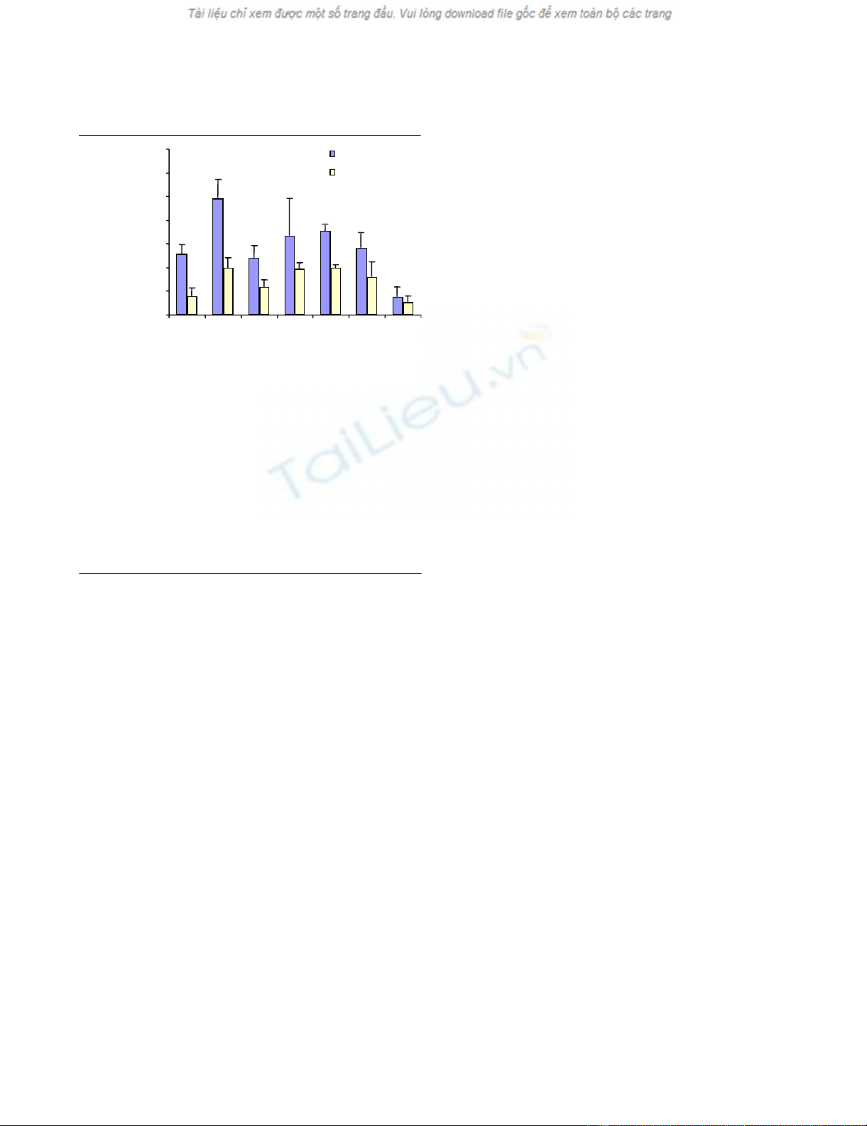
ET-1 induces MMP-1 and MMP-13 production
The effects of ET-1 and those of various inhibitors on MMP-
1 production and MMP-13 production are shown in Fig. 1.
At 10 nM ET-1 the production of both enzymes was signif-
icantly increased (P < 0.005). SB202190, a p38 inhibitor,
completely suppressed the ET-1-stimulated production of
both enzymes, whereas the phosphatidyl inositol 3 kinase
inhibitor Wortmannin and the PKA inhibitor KT5720 par-
tially but significantly (P < 0.01) decreased the level of
MMP-13 only. Interestingly, the most potent inhibitor of
MMP-1 and MMP-13 production was LY83583, an inhibi-
tor of NO-dependent soluble guanylate cyclase and of
cGMP. This agent not only suppressed the ET-1-induced
stimulation, but also decreased the level of both enzymes
below the basal level: a significant difference was found for
both MMP-13 and MMP-1 when compared with the ET-1
stimulation (P < 0.005) and for MMP-13 when compared
with the control (P < 0.05). Although a decrease in MMP-
13 was noted with the MEK1/2 kinase inhibitor PD98059
at the concentration tested, it did not reach statistical sig-
nificance. With this inhibitor, no effect was found on MMP-
1 production.
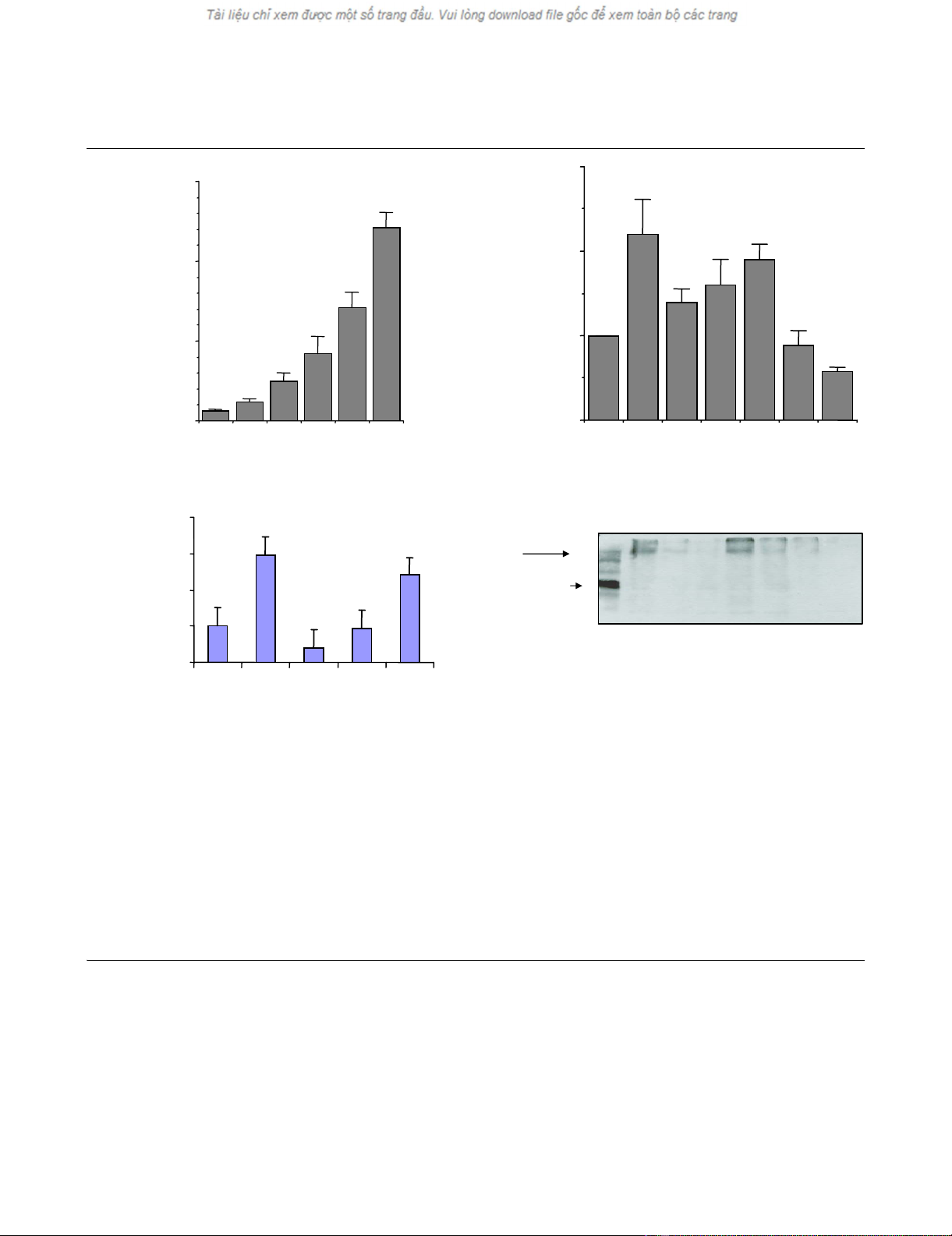
ET-1 induces NO production
The effects of ET-1 on NO release and on iNOS expression
are shown in Fig. 2. Figure 2a shows that ET-1 greatly stim-
ulated NO production and was released in a concentration-
dependent manner. Incubation with increasing concentra-
tions of ET-1, from 0.1 to 100 nM, augmented almost 12-
fold the linear accumulation of NO. To determine the mech-
anism involved in the ET-1-induced NO production, the
effects of the major intracellular signalling pathways were
investigated. Figure 2b shows that the ET-1-induced NO
release was significantly inhibited by p38 inhibition and
prevented by KT5720, a PKA inhibitor. No significant effect
was noted for MEK1/2 inhibition by PD98059 and by
Arthritis Research & Therapy Vol 7 No 2 Manacu et al.
R327
Wortmannin. Moreover, the guanylate cyclase inhibitor
LY83583 reduced the NO production as significant differ-
ences were found when compared with either the ET-1
stimulation (P < 0.05) or with the control (P < 0.05), and
this inhibitor also decreased both the endogenous and ET-
1-induced iNOS level (Fig. 2d). The ET-1-induced NO
release occurs via iNOS as shown in Figure 2c: complete
inhibition of iNOS by 50 µM allosteric iNOS inhibitor L-NIL,
as expected, almost completely inhibited NO release. Fig-
ure 2d shows the effects of various inhibitors on iNOS
expression, as determined by western blot analysis of cell
extracts. The 24-hour incubation of cells with ET-1 results
in an increase of iNOS protein (Fig. 2d, lane 2). The ET-1-
induced iNOS protein expression was completely sup-
pressed by SB202190 and LY83583, and was partially
suppressed by Wortmannin and KT5720. PD98059 had
no effect.
Intracellular protein kinase phosphorylation in the
presence of ET-1
Figure 3a–d show the effects of ET-1 on the phosphoryla-
tion of p38, Akt, p44/42 and SAP/JNK kinases as detected
by western blot of cell extracts. ET-1 at 10 nM induced
p38, Akt, p44/42, and SAP/JNK phosphorylation in a time-
ordered manner. For p38, the maximal effect following cell
exposure to ET-1 was obtained at 10 min. For Akt, the max-
imal effect was observed at 2 min of cell exposure and this
effect persisted during 30 min, followed by a decline at 45
min. At this time (45 min), both p38 kinase and Akt phos-
phorylated forms were diminished. The maximal effect was
obtained at 15 min for p44/42 kinase and at 45 min for
SAP/JNK. The SAP/JNK phosphorylated forms were not
detected at 60 min, whereas that of p44/42 decreased but
was still present even at 60 min.
ET-1 did not affect apoptosis
As ET-1 induces NO release and because the accumula-
tion of NO causes apoptosis, we explored this potential
effect. OA chondrocytes incubated in the absence of (con-
trol) or in the presence of ET-1 (10 nM) for 72 hours
showed that ET-1 did not affect apoptosis (TUNEL reac-
tion; data not shown) or the production of either anti-apop-
totic Bcl2 or pro-apoptotic Bad proteins. A similar
percentage of positively stained cells was found for Bcl2
(42.8 ± 5.1% and 43.2 ± 4.3% for the control and for ET-
1, respectively) and for Bad (10.1 ± 3.8% and 9.5 ± 2.1%,
respectively).
Discussion
This study shows an overproduction of NO, MMP-1 and
MMP-13 in human OA chondrocytes stimulated by ET-1.
This result goes beyond previous results [2], which showed
that human OA synovial tissue and joint cartilage express
the ET-1 gene and overproduce ET-1, resulting in an exces-
sive synthesis of MMP-1 and MMP-13 in the same tissues.
In addition, the result goes beyond these findings and
enlightens on the mechanism by which ET-1 accomplishes
this action. Strong evidence was obtained for the key role
played by NO, whose production and release were also
upregulated by ET-1.
NO induces smooth muscle cell relaxation by activating sol-
uble guanylate cyclase and by increasing the intracellular
concentration of cGMP. LY83583 suppresses the effect of
NO by inhibiting this NO-dependent production of cGMP
[23]. In the present study, LY83583 was also shown to
strongly inhibit MMP-1 and MMP-13 production by unstim-
ulated and ET-1-stimulated OA chondrocytes, showing the
key role of cGMP for the synthesis of these enzymes. This
finding confirms a previous observation that cGMP is nec-
essary for protein synthesis [9], and brings further evidence
that an excess of NO is harmful to cells.
It is generally accepted that progressive tissue destruction
in rheumatoid arthritis and in OA results from an excessive
breakdown mediated by various proteolytic enzymes and
other catabolic agents such as free radicals and NO
[1,13,24,25]. Our results suggest that ET-1 should also be
added to the list of potential deleterious agents that may
account for articular cartilage destruction in rheumatic dis-
eases. The action of ET-1 seems to be dual via an increase
in MMP and NO production. ET-1-induced stimulation of
Figure 1
Effect of protein kinase inhibitors and LY83583 on endothelin-1 (ET-1)-induced MMP-13 and MMP-1 production by human osteoarthritis chondrocytesEffect of protein kinase inhibitors and LY83583 on endothelin-1 (ET-1)-
induced MMP-13 and MMP-1 production by human osteoarthritis
chondrocytes. Confluent monolayer chondrocytes were preincubated
30 min at 37°C with SB 202190 (1 µM), PD98059 (10 µM), Wortman-
nin (100 nM), KT5720 (4 µM) or LY83583 (2 µM) for 30 min at 37°C,
and were then challenged with ET-1 for 24 hours. MMP-13 and MMP-1
proteins were measured in the culture media using specific ELISA
assays. P values indicate significant differences comparing experimen-
tal conditions with ET-1 treatment alone (*) and versus the control cul-
tures (#). Values are expressed as the mean ± standard error of the
mean of five independent experiments performed in duplicate. Signifi-
cant differences: #, * P < 0.05; ##, ** P < 0.01; ###, *** P < 0.005.
0
5
10
15
20
25
30
35
MMP-13
MMP-1
ET-1 (10 nM) – + + + + + +
Wortmannin
SB202190
PD98059
KT5720
LY 83583
pg/5×10
5
cells
CONTROL
***
***
**
**
###
#
***
***
###
##
Available online http://arthritis-research.com/content/7/2/R324
R328
MMP-1 and MMP-13, as well as the induction of iNOS
gene expression with subsequent NO overproduction by
OA chondrocytes, may interfere with the proinflammatory
cytokine pathways. Indeed, we and other workers have
shown that IL-1β upregulates the synthesis of ET-1 [3],
which in turn can induce IL-1β gene transcription and con-
sequently the production of the protein [26]. We previously
demonstrated [2] that MMP-13 expression was induced
similarly by ET-1 and IL-1β; however, although they both
enhanced MMP-1 expression, the effect of IL-1β was more
potent on this enzyme. Interestingly, using a specific immu-
noassay measuring the C telopeptide of type II collagen
fragments on OA cartilage explants, we also found that the
level of the cleaved collagen fragments were significantly
increased in the presence of both IL-1β and ET-1 with a
more potent effect observed for ET-1. This could be
Figure 2
Effect of endothelin-1 (ET-1) on nitric oxide (NO) release and inducible nitric oxide synthase (iNOS) expression by human osteoarthritis (OA) chondrocytesEffect of endothelin-1 (ET-1) on nitric oxide (NO) release and inducible nitric oxide synthase (iNOS) expression by human osteoarthritis (OA)
chondrocytes. NO was measured in the culture media, and iNOS protein was detected in cell extracts and revealed by western blot using specific
antiserum, as described in Materials and methods. (a) Concentration-dependent ET-1-induced NO accumulation in the culture media from confluent
human OA chondrocytes treated with ET-1 (0–100 nM) at 37°C for 24 hours. (b) Effect of protein kinase inhibitors and of guanylate cyclase inhibitor
on ET-1-induced NO release in OA chondrocytes. Confluent monolayer chondrocytes were preincubated with SB 202190 (1 µM), PD98059 (10
µM), Wortmannin (100 nM), KT5720 (4 µM) or LY83583 (2 µM) for 30 min at 37°C and then challenged with ET-1 for 24 hours, and NO was deter-
mined in the culture media. (c) Effect of iNOS inhibition on NO release induced by ET-1 in human OA chondrocytes. The chondrocytes were pre-
treated with the allosteric inhibitor of iNOS, L-N6 (1-iminoethyl)lysine (L-NIL) (0–50 µM), for 30 min and were then incubated with ET-1 (10 nM) for
an additional 24 hours. The NO level was measured in the culture media. (d) Effect of protein kinase inhibitors and LY83583 on ET-1-induced iNOS
in human OA chondrocytes. Chondrocytes were preincubated with SB 202190 (1 µM), PD98059 (10 µM), Wortmannin (100 nM), KT5720 (4 µM)
or LY83583 (2 µM) for 30 min at 37°C and then challenged with ET-1 for 24 hours, and iNOS was then quantified. M.W., molecular weight. (a)–
(c) Values are the mean ± standard error of the mean of six independent experiments performed in duplicate. (d) Representative blot of three inde-
pendent experiments. P values indicate the significant difference between ET-1 treated cells and cells treated with indicated inhibitors + ET-1 (*) and
versus control (#). Significant differences: #, * P < 0.05, ###, *** P < 0.005.
ET-1 (nM) 0 0.1 1 10 50 100
Wortmannin
PD98059
SB202190
KT5720
LY83583
0
100
200
300
ET-1 (10 nM)
++++++
NO (nmol/5×10
5
cells)
0.0
0.5
1.0
1.5
(a) (b)
0
100
200
300
400
++++–
–50
–10 1
L-NIL (µM)
ET-1 (10 nM)
CONTROL
iNOS
(130 kDa)
ET-1 (10 nM) M.W.
++++++
Wortmannin
PD98059
SB202190
KT5720
LY83583
63.2
CONTROL
***
*
*
#
###
###
#
*
*
#
(c) (d)
NO (% of control)
NO (% of control)
–
–


