CHAPTER 3

Figure Anatomy
✎
33
Understanding human anatomy will help you achieve greater
expressive ability in figure drawing. By understanding the many dif-
ferent aspects of the human form, you can better grasp how the fig-
ure works as a whole. For example, if you feel along the bone on the lower
part of your jaw, you will notice that there is a small indentation about
halfway between the chin and the back of the jaw. This indentation is to allow
a blood vessel to pass under the jaw. The indentation helps to protect the ves-
sel. The significance of this little indentation is that it affects the curvature of
the jaw. The jawbone is actually concave here, rather than convex. A slender
person who has little fat around the jaw will show this distinct feature of the
jaw more clearly than a heavy person will. Knowing this little aspect of the
figure can help the artist who wishes to express a thin person.
In a way, the study of anatomy increases your figure drawing arsenal. By
studying the underlying structure, you can develop a greater feel for the sur-
face, expanding creative possibilities. In essence, the human form is made up
of soft and hard tissue held together by tendons and ligaments. The bones
form the underlying structure of the body and in some cases act as protection
for delicate internal organs. Around and over the bones are muscles that are
used to drive movement. The whole system is controlled by an extensive
nervous system.
The human form is capable of extreme movement and flexibility. The muscles
that power body movement expand and contract, causing surface changes to
appear. Take a look at your arm. Hold it out in front of you with the palm
down. Now twist your arm so the palm is facing up. Notice how the muscle
beneath the skin move as the arm rotates. As the arm rotates, some muscles in
the forearm will expand and twist, while others will contract to cause the

34
Figure Drawing with Virtual Models
movement. Now bend your arm
up at the elbow. The muscles of
the upper arm will bunch to pull
the arm up. These muscles are
the biceps, so named because
there are two muscles.
Drawing from life, an artist is
often confronted with a number
of organic surfaces. It is helpful if
the artist understands not only
why the surface changes in
movement, but also what the
underlying structure is doing
during those changes. This will
help the artist to recognize the
subtle aspects of the figure that
might go unnoticed if the artist
didn’t have the proper instruc-
tion in anatomy.
The Skeleton
In Chapter 2 you created a simpli-
fied skeletal structure to use as a
base for drawing the figure. We
called it drawing from the inside
out. Now you will have the oppor-
tunity to better understand the
actual skeletal structure of the
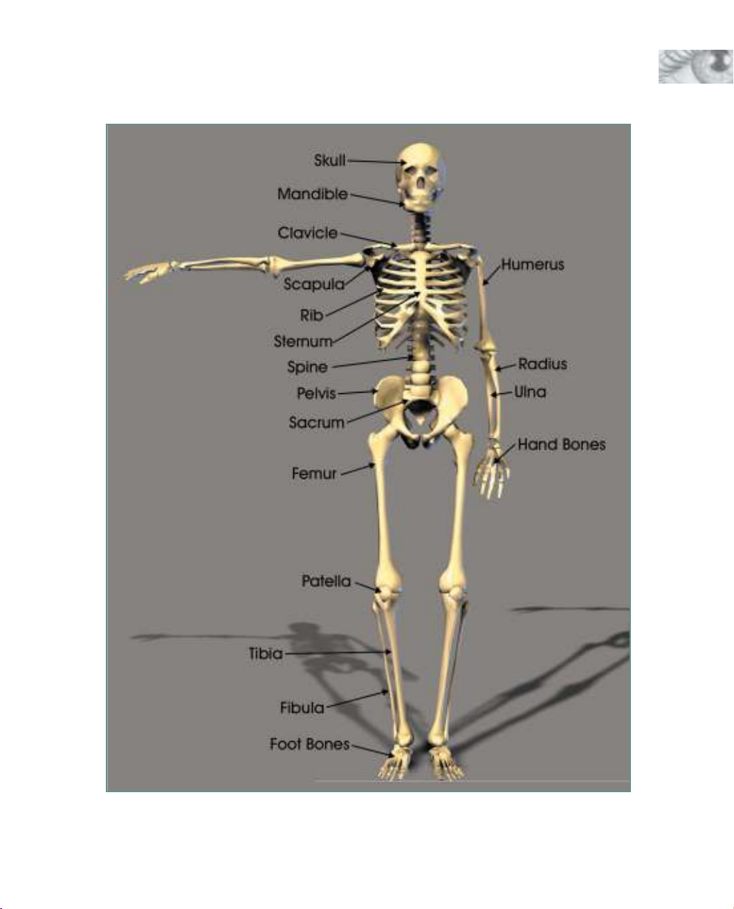
human body. Figure 3.1 shows the
male skeleton. This skeleton comes
with Figure Artist’s bigger cousin,
Poser, and is available as additional
content for Figure Artist.
The human skeleton contains more
than 200 individual bones.
Although it might not be essential
for you to learn the names of all
200 bones, you should become
familiar with some of the major
bones listed here.
✎Skull. The skull is the
bony framework upon
which the head is built.
It is composed of eight
cranial and 14 facial
bones. The cranial
bones are the dome-
shaped bones that
cover the top of your
head. They are very
near the surface. If you
press on the top of
your head, you can feel
the hard surface of the
bone just below the
skin. The shape of the
cranium pretty much
determines the shape
of the head. The facial
bones make up the
bones of the front of
the head and con-
tribute greatly to how a
person looks.
✎Mandible. The
mandible, sometimes
referred to as the jaw-
bone, is actually one of
the facial bones of the
skull. It is the moveable
bone on the lower part
of the head. It is hinged
to the rest of the skull
up near the ears. It is
important to note
where the bone is
hinged because that
controls the bone
movement.
✎Clavicle. The clavicle is
located on the upper
front of the chest near
the neck. It is a very
pronounced bone near
the surface, connecting
the arms to the chest.
There are two clavicle
bones—one on each
side of the body. The
clavicle’s flexible move-
ment allows for the
variety of movement in
the shoulder.
✎Scapula. The scapula is
a plate-like triangular
bone located on the
right and left side of
the upper back. It is
sometimes called the
shoulder blade. It has
quite a range of move-
ment under the skin
and is more pro-
nounced in a slender
person. It will also pro-
trude more in a person
with poor posture.
35
Figure Anatomy
Figure 3.1 The skeleton forms the structure upon which the body is built.

36
Figure Drawing with Virtual Models
✎Ribcage. The ribs are
actually a group of
bones that surround
the chest cavity and
serve as protection for
the delicate organs
housed in that area. All
together, the ribs form
a somewhat egg-
shaped structure that is
open at the bottom and
more closed at the top.
The ribcage also acts
as an anchor for many
of the muscles of the
upper back and chest.
✎Sternum. The sternum
is located in the center
of the chest and con-
nects the ribs of the left
and right sides by way
of cartilage, which
gives the chest the flex-
ibility to expand and
shrink with breathing.
The sternum has a dis-
tinctive dagger shape
and is sometimes
referred to as the
breastbone.
✎Spine. The spine is a
column of bones that
extends from the skull
to the pelvis. The spine
is a very flexible com-
bination of bones and
cartilage that encloses
and protects the spinal
cord. The spine is also
the structure that holds
the upper body erect.
There are 33 separate
irregularly shaped
bones called vertebrae
in the spinal column.
The top bone of the
spinal column is called
the Atlas, and the next
is called the Axis. The
shape of the Atlas
allows the head to nod
yes, and the shape of
the Axis allows the
head to shake no. The
vertebrae at the top of
the spinal column are
smaller than those near
the bottom. They con-
nect the ribcage in the
back and support most
of the major muscles of
the back. They can be
seen as a row of ridges
when a person bends
forward.
✎Pelvis. The pelvis is
located in the lower
body and forms your
hips. There are actually
two pelvic bones—one
on either side of the
body. They are joined
together in the back by
the sacrum and in the
front by a muscle
called the pubic sym-
physis. The pelvis
serves to support the
body by anchoring the
spinal column, and it
also protects many of
the delicate organs of
the lower body. The
pelvis on a female is
wider, and the central
opening is larger than
on a male. This differ-
ence helps the female
to support a baby dur-
ing pregnancy. The
wider opening allows
for the baby to be born
because the baby must
pass through the
mother’s pelvis.
✎Sacrum. The sacrum is
a V-shaped bone that is
actually several verte-
brae fused together as
a person reaches adult-
hood. This bone
attaches the spinal col-
umn to the pelvis
bones.
✎Femur. The femur is
the large bone that
runs from the hip to the
knee. It would be the
largest bone in the
body except that there
are two of them, and
since they are usually
the same size, they
both share that honor.
The femurs are the
largest, longest, and
strongest bones in the
body. They support the
massive thigh muscles


![Tác phẩm sứ nghệ thuật đẹp đến ngỡ ngàng: Tuyển chọn [Năm hiện tại]](https://cdn.tailieu.vn/images/document/thumbnail/2013/20130626/chipinz/135x160/3361372260442.jpg)


