
BioMed Central
Page 1 of 15
(page number not for citation purposes)
Respiratory Research
Open Access
Research
Inhibition of c-Jun NH2-terminal kinase or extracellular
signal-regulated kinase improves lung injury
Hui Su Lee1, Hee Jae Kim1, Chang Sook Moon1, Young Hae Chong2 and
Jihee Lee Kang*1
Address: 1Department of Physiology, Division of Cell Biology, Ewha Medical Research Institute, Ewha Womans University College of Medicine,
911-1 Mok-6-dong, Yangcheon-ku, Seoul 158-056, Korea and 2Department of Microbiology, Division of Cell Biology, Ewha Medical Research
Institute, Ewha Womans University College of Medicine, 911-1 Mok-6-dong, Yangcheon-ku, Seoul 158-056, Korea
Email: Hui Su Lee - huisulee@hanmail.com; Hee Jae Kim - kitty7808@hanmail.net; Chang Sook Moon - 94cmoon@hanmail.net;
Young Hae Chong - younghae@ewha.ac.kr; Jihee Lee Kang* - jihee@ewha.ac.kr
* Corresponding author
JNKERKLPSacute lung injuryNF-κB
Abstract
Background: Although in vitro studies have determined that the activation of mitogen-activated
protein (MAP) kinases is crucial to the activation of transcription factors and regulation of the
production of proinflammatory mediators, the roles of c-Jun NH2-terminal kinase (JNK) and
extracellular signal-regulated kinase (ERK) in acute lung injury have not been elucidated.
Methods: Saline or lipopolysaccharide (LPS, 6 mg/kg of body weight) was administered
intratracheally with a 1-hour pretreatment with SP600125 (a JNK inhibitor; 30 mg/kg, IO), or
PD98059 (an MEK/ERK inhibitor; 30 mg/kg, IO). Rats were sacrificed 4 hours after LPS treatment.
Results: SP600125 or PD98059 inhibited LPS-induced phosphorylation of JNK and ERK, total
protein and LDH activity in BAL fluid, and neutrophil influx into the lungs. In addition, these MAP
kinase inhibitors substantially reduced LPS-induced production of inflammatory mediators, such as
CINC, MMP-9, and nitric oxide. Inhibition of JNK correlated with suppression of NF-κB activation
through downregulation of phosphorylation and degradation of IκB-α, while ERK inhibition only
slightly influenced the NF-κB pathway.
Conclusion: JNK and ERK play pivotal roles in LPS-induced acute lung injury. Therefore, inhibition
of JNK or ERK activity has potential as an effective therapeutic strategy in interventions of
inflammatory cascade-associated lung injury.
Background
Lipopolysaccharide (LPS) causes acute lung injury associ-
ated with the activation of macrophages, an increase in
alveolar-capillary permeability, neutrophil influx into the
lungs, and parenchymal injury [1]. This pulmonary
response contributes to the pathogenesis of various acute
inflammatory respiratory diseases. Mitogen-activated pro-
tein (MAP) kinases are crucial in intracellular signal trans-
duction, mediating cell responses to a variety of
inflammatory stimuli, such as LPS, tumor necrosis factor
Published: 27 November 2004
Respiratory Research 2004, 5:23 doi:10.1186/1465-9921-5-23
Received: 27 August 2004
Accepted: 27 November 2004
This article is available from: http://respiratory-research.com/content/5/1/23
© 2004 Lee et al; licensee BioMed Central Ltd.
This is an Open Access article distributed under the terms of the Creative Commons Attribution License (http://creativecommons.org/licenses/by/2.0),
which permits unrestricted use, distribution, and reproduction in any medium, provided the original work is properly cited.

Respiratory Research 2004, 5:23 http://respiratory-research.com/content/5/1/23
Page 2 of 15
(page number not for citation purposes)
(TNF) and interleukin (IL)-1. Recently, various in vitro
studies have shown that pharmacological inhibitors of
MAP kinases strongly affect the production of inflamma-
tory mediators [2,3]. Through the use of specific inhibi-
tors, the potential role of these kinases in inflammatory
lung diseases is beginning to be studied. Treatment with
p38 MAP Kinase inhibitors has been proposed as a selec-
tive intervention to reduce LPS-induced lung inflamma-
tion due to decreases in neutrophil recruitment to the air
spaces [4,5]. However, the functions of c-Jun NH2-termi-
nal kinase (JNK) and extracellular signal-regulated kinase
(ERK) in LPS-induced lung injury remain unclear.
Cytokine-induced neutrophil chemoattractant (CINC)
has been shown, in rodent models of lung injury, to play
an important role in neutrophil migration into the lung
[6]. Matrix metalloproteinases (MMPs), including MMP-
9, allow activated neutrophils to permeate subsequent
extracellular matrix (ECM) barriers after adhesion, and
also for transendothelial cell migration, since these prote-
olytic enzymes digest most of the ECM components in the
basement membranes and tissue stroma [7]. Another
inflammatory mediator, nitric oxide (NO), has been
linked to a number of physiologic processes, including
leukocyte-dependent inflammatory processes and oxi-
dant-mediated tissue injury [8,9]. Like CINC and MMP-9,
overproduction of NO, which is dependent on the activity
of inducible NO synthase, has been reported to contribute
to endothelial or parenchymal injury, as well as to induce
an increase in microvascular permeability, resulting in
lung injury [10,11]. These inflammatory mediators are
produced in response to LPS, TNF and IL-1 [6,11] and are
regulated at the transcription level by nuclear factor-kappa
B (NF-κB) [6,12].
NF-κB activation is regulated by phosphorylation of the
inhibitor protein, IκB-α, which dissociates from NF-κB in
the cytoplasm. The active NF-κB can then translocate to
the nucleus, where it binds to the NF-κB motif of a gene
promoter and functions as a transcriptional regulator. In
vivo activation of NF-κB, but not other transcription fac-
tors, has also been demonstrated in alveolar macrophages
from patients with acute respiratory distress syndrome
(ARDS) [13]. Our previous study indicated that NF-κB
activation is an important mechanism underlying both
LPS-induced NO production, and also MMP-9 activity
and resulting neutrophil recruitment [14]. Therefore, the
activation of NF-κB binding to various gene promoter
regions appears to be a key molecular event in the initia-
tion of LPS-induced pulmonary disease.
Once activated, MAP kinases appear to be capable of fur-
ther signal transduction through kinase phosphorylation,
as well as modulating phosphorylation of transcription
factors [15-17]. Activator protein (AP)-1, another tran-
scription factor mediating acute inflammation, is acti-
vated through MAP kinase signaling cascades in response
to various factors, such as LPS, cytokines, and various
stresses and in turn regulates genes encoding inflamma-
tory cytokines, such as TNF-α, IL-1, IL-6, and IL-8 [18].
Davis [19] reported that activated JNK is capable of bind-
ing the NH2-terminal activation domain of c-Jun, activat-
ing AP-1 by phosphorylating its component c-Jun. AP-1
can then translocate into the nucleus to promote tran-
scription of downstream genes. However, action of MAP
kinases on the upstream of NF-κB activation remains con-
troversial [20-22]. Here, using a selective JNK inhibitor,
SP600125, and the downstream MEK inhibitor of ERK,
PD98059, we focused on the roles of JNK and ERK in LPS-
induced acute lung injury and production of CINC, MMP-
9, and NO. In addition, we investigated the regulatory
effects of these MAP kinases on the NF-κB activation path-
way during acute lung injury.
Methods
Experimental Animals
Specific pathogen-free male Sprague-Dawley rats (280–
300 g) were purchased from Daehan Biolink Co. (Eum-
sung-Gun, Chungbuk, Korea). The Animal Care Commit-
tee of the Ewha Medical Research Institute approved the
experimental protocol. The rats were cared for and han-
dled according to the National Institute of Health (NIH)
Guide for the Care and Use of Laboratory Animals.
Experimental Protocols
Six groups of specific pathogen-free male Sprague-Dawley
rats (280–300 g) were used: (1) controls received an
intratracheal (IT) instillation of 0.5 ml of LPS-free saline
(0.9 % NaCl); (2) an LPS-treated group received an IT
instillation of 6 mg/kg body weight of LPS (Escherichia coli
lipopolysaccharide, 055:B5, Sigma Chemical Co., St.
Louis, MO) in 0.5 ml LPS-free saline; (3) an LPS-
SP600125 group was injected with SP600125 (Calbio-
chem, La Jolla, CA) 1 hour before the IT instillation of 6
mg/kg body weight of LPS in 0.5 ml of LPS-free saline. (4)
a saline-SP600125 group was injected with SP600125 1
hour before IT instillation of 0.5 ml of LPS-free saline (0.9
% NaCl); (5) an LPS-PD98059 group was injected with
PD98059 (BIOMOL Research Laboratories, Plymouth,
PA) 1 hour before IT instillation of 6 mg/kg body weight
of LPS in 0.5 ml of LPS-free saline. (6) a saline-PD98059
group was injected with PD98059 1 hour before IT instil-
lation of 0.5 ml of LPS-free saline (0.9 % NaCl).
SP600125 or PD98059 was injected intraorally via a size
8 French feeding tube at a dose of 30 mg/kg body weight
[5,23]. For IT instillation, rats were treated with enflurane
anesthesia. The trachea was then exposed after a 1 cm
midline cervical incision, and LPS or saline was injected
intratracheally through a 24-gauge catheter. LPS or saline
administration was immediately followed by 3

Respiratory Research 2004, 5:23 http://respiratory-research.com/content/5/1/23
Page 3 of 15
(page number not for citation purposes)
insufflations of 1 ml of air through the catheter and by
rotating the animals to attempt to homogeneously distrib-
ute LPS or saline in the lungs. After a few minutes, the rats
recovered from the anesthesia and were immediately
placed in a chamber. Animals were sacrificed 4 hours after
LPS treatment, and the following parameters were moni-
tored: (1) phosphorylation of JNK, ERK, and p38 MAP
kinase in lung tissue; (2) cell differential count, and meas-
urement of protein content and lactate dehydrogenase
(LDH) activity in bronchoalveolar lavage (BAL) fluid; (3)
cytokine-induced neutrophil chemoattractant (CINC)
expression, matrix metalloproteinase (MMP)-9 activity or
expression and nitrite production in lung tissue, BAL fluid
or the supernatants of alveolar macrophage cultures; (4)
DNA binding activity of nuclear factor-kappa B (NF-κB) in
lung tissue and alveolar macrophages; (5) serine phos-
phorylation and degradation of IκB-α in lung tissue. In
addition, phosphorylation of JNK and ERK was also deter-
mined at 2, 4, 14 or 24 hours after LPS treatment to deter-
mine the kinetics of the kinase activation in lung tissue.
Isolation of BAL cells, Lung Tissue, and Cell Counts
Four hours after LPS treatment, the rats were sacrificed,
and BAL was then performed through a tracheal cannula
with aliquots of 8 ml each using ice-cold Ca2+/Mg2+-free
phosphate-buffered medium (145 mM NaCl, 5 mM KCl,
1.9 mM NaH2PO4, 9.35 mM Na2HPO4, and 5.5 mM dex-
trose; pH 7.4) for a total of 80 ml for each rat. The bron-
choalveolar lavagate was centrifuged at 500 × g for 5 min
at 4°C and cell pellets washed and resuspended in phos-
phate-buffered medium. Cell counts and differentials
were determined using an electronic coulter counter with
a cell sizing analyzer (Coulter Model ZBI with a chan-
nelizer 256; Coulter Electronics, Bedfordshire, UK), as
described by Lane and Mehta [24]. Red blood cells, lym-
phocytes, neutrophils, and alveolar macrophages were
distinguished by their characteristic cell volumes [25]. The
recovered cells were 98% viable, as determined by trypan
blue dye exclusion. Following lavage, lung tissue was
removed, immediately frozen in liquid nitrogen, and
stored at -70°C.
Measurement of Total Protein and lactate dehydrogenase
(LDH) Activity
To assess the permeability of the bronchoalveolar-capil-
lary barrier, total protein was measured according to the
method of Hartree [26], using bovine serum albumin as
the standard. Total protein and LDH activity were meas-
ured in the first aliquot of the acellular BAL fluid. LDH
activity, a cytosolic enzyme used as a marker for cytotox-
icity, was measured at 490 nm using an LDH determina-
tion kit according to the manufacturer's instructions
(Roche Molecular Biochemicals, Mannheim, Germany).
LDH activity was expressed as U/L, using an LDH
standard.
Western Blot Analysis
Lung tissue homogenate samples (55 µg or 100 µg pro-
tein/lane for JNK, ERK, p38 MAP kinase, IκB-α and CINC)
or aliquots of acellular BAL fluid (70 µl/lane for CINC and
MMP-9) were separated on a 10% or 20% SDS-polyacry-
lamide gel. Separated proteins were electrophoretically
transferred onto nitrocellulose paper and blocked for 1
hour at room temperature with Tris-buffered SAL contain-
ing 3% BSA. The membranes were then incubated with an
anti-rabbit phospho-JNK/JNK antibody, anti-rabbit phos-
pho-ERK/ERK, anti-rabbit phospho-p38 MAP kinase/p38
MAP kinase, antiserum against rat CINC, anti-human
MMP-9 monoclonal antibody or anti-rabbit phospho-
IκBα (Ser32)/IκBα at room temperature for 1 hour. Anti-
body labeling of protein bands was detected with
enhanced chemiluminescence (ECL) reagents according
to the supplier's protocol.
Zymographic Analysis of MMP-9
The gelatinolytic activities in BAL fluid, or the superna-
tants of alveolar macrophage cultures, were determined
using zymography with gelatin copolymerized with acry-
lamide in the gel according to previously published meth-
ods [14]. To obtain the supernatants of alveolar
macrophage cultures, lavage cells were resuspended in
RPMI-1640 medium (Mediatech, Washington, DC), con-
taining 2 mM glutamine, 100 units/ml mycostatin with-
out fetal bovine serum (FBS). Aliquots of 1 ml, containing
106 alveolar macrophages, were added to 24-well plates
(Costar, Cambridge, MA) and incubated at 37°C in a
humidified atmosphere of 5% CO2 for 2 hours. The non-
adherent cells were then removed, and adherent cells were
counted and further incubated in 1 ml RPMI medium.
After a 24 hour incubation, the supernatant was collected
and filtered.
Aliquots of BAL fluid and the culture supernatants, nor-
malized for equal volume (8 µl) or amount of protein (8
µg), were electrophoresed on a 10% SDS-PAGE gel with
0.1% gelatin as a substrate without boiling under non-
reducing conditions. After removing SDS with 2.5% Tri-
ton X-100 for 2 hours, gels were incubated for 20 hours at
37°C in 50 mM Tris-Cl (pH 7.4) containing 10 mM CaCl2
and 0.02% NaN3. The gels were then stained for 1 hour in
7.5% acetic acid/10% propanol-2 containing 0.5%
Coomassie Brilliant Blue G250 and destained in same
solution without dye. Positions of gelatinolytic activity are
unstained on a darkly stained background. The clear
bands on the zymograms were photographed on the neg-
ative (Polaroid's 665 film) and the signals were quantified
by densitometric scanning using an UltroScan XL laser
densitometer (LKB, Model 2222-020) to determine the
intensity of MMP-9 activity as arbitrary densitometric
units. To confirm MMP-9 activity, aliquots of BAL fluid
were analyzed by Western blotting with anti-human

Respiratory Research 2004, 5:23 http://respiratory-research.com/content/5/1/23
Page 4 of 15
(page number not for citation purposes)
MMP-9 monoclonal antibody, which was raised against
MMP-9 secreted by human HT1080 fibrosarcoma cells
[27] and cross-reacts with rat MMP-9 [28].
Nitrite Assay in BAL fluid and Alveolar Macrophage
Culture
NO levels in the first aliquot of the acellular BAL fluid,
and the supernatants of alveolar macrophage cultures,
were measured using a nitrite assay. Direct measurement
of NO is difficult due to the very short half-life [29]. How-
ever, the stable oxidation end product of NO production,
nitrite, can be readily measured in biological fluids and
has been used in vitro and in vivo as an indicator of NO
production [30]. Briefly, lavage cells were resuspended in
RPMI-1640 medium (Mediatech, Washington, DC), con-
taining 2 mM glutamine, 100 units/ml mycostatin, and
10% FBS. Aliquots of 1 ml, containing 106 alveolar mac-
rophages were added to 24-well plates (Costar, Cam-
bridge, MA) and incubated at 37°C in a humidified
atmosphere of 5% CO2 for 2 hours. The non-adherent
cells were then removed by vigorous washing with two 1
ml of RPMI medium. After incubating the cells for 24
hours, the supernatant was collected and filtered.
Nitrite was assayed after adding 100 µl Greiss reagent (1%
sulfanilamide and 0.1% naphthylethylenediamide in 5%
phosphoric acid) to 50 µl samples of BAL fluid and cell
culture. Optical density at 550 nm (OD550) was measured
using a microplate reader. Nitrite concentrations were cal-
culated by comparison with OD550 of standard solutions
of sodium nitrite prepared in cell culture medium. Data
were presented as µM of nitrite.
Nuclear Extracts
Nuclear extracts were prepared by a modified method of
Sun et al. [31]. Lavage cells were resuspended in Dul-
becco's modified Eagle's medium (DMEM; Mediatech,
Washington, DC), supplemented with 5% FBS (HyClone,
Logan, UT), 2 mM glutamine, and 1,000 units/ml penicil-
lin-streptomycin. DMEM medium (5 ml), containing 5 ×
106 alveolar macrophages, was added to 6-well plates and
incubated at 37°C, in a humidified atmosphere of 5%
CO2 for 2 hours. The nonadherent cells were then
removed with two 1 ml aliquots of DMEM. At the end of
the incubation, adherent cells (> 95% alveolar macro-
phages) were harvested and then resuspended in hypot-
onic buffer A (100 mM HEPES, pH 7.9, 10 mM KCl, 0.1
M ethylenediaminetetraacetic acid [EDTA], 0.5 mM dithi-
othreitol [DTT], 1% Nonidet P-40, and 0.5 mM phenyl-
methylsulfonyl fluoride [PMSF]) for 10 min on ice, then
vortexed for 10 s. Nuclei were pelleted by centrifugation at
12,000 rpm for 30 s. Nuclear extracts were also prepared
from lung tissue by the modified method of Deryckere
and Gannon [32]. Aliquots of frozen tissue were mixed
with liquid nitrogen and ground to powder using a mortar
and pestle. The ground tissue was placed in a Dounce tis-
sue homogenizer (Kontes Co., Vineland, NJ) in the pres-
ence of 4 ml of buffer A to lyse the cells. The supernatant
containing intact nuclei was incubated on ice for 5 min,
and centrifuged for 10 min at 5,000 rpm. Nuclear pellets
obtained from alveolar macrophages or lung tissue were
resuspended in buffer C (20 mM HEPES, pH 7.9, 20%
glycerol, 0.42 M NaCl, 1 mM EDTA, and 0.5 mM PMSF)
for 30 min on ice. The supernatants containing nuclear
proteins were collected by centrifugation at 10,000 rpm
for 2 min, and stored at -70°C.
Electrophoretic Mobility Shift Assay (EMSA)
Binding reaction mixtures (10 µl), containing 5 µg (4 µl)
nuclear extract protein, 2 µg poly (dI-dC)•poly (dI-dC)
(Sigma Co., St. Louis. MO), and 40,000 cpm 32P-labeled
probe in binding buffer (4 mM HEPES, pH 7.9, 1 mM
MgCl2, 0.5 mM DTT, 2% glycerol, and 20 mM NaCl), were
incubated for 30 min at room temperature. The protein-
DNA complexes were separated on 5% non-denaturing
polyacrylamide gels in 1 × TBE buffer, and autoradio-
graphed. Autoradiographic signals for activated NF-κB
were quantitated by densitometric scanning using an
UltroScan XL laser densitometer (LKB, Model 2222-020,
Bromma, Sweden) to determine the intensity of each
band.
The oligonucleotide used as a probe for EMSA was a dou-
ble-stranded DNA fragment, containing the NF-κB con-
sensus sequence (5'-
CCTGTGCTCCGGGAATTTCCCTGGCC-3'), labeled with
[α-32P]-dATP (Amersham, Buckinghamshire, UK), using
DNA polymerase Klenow fragment (Life Technologies,
Gaithersburg, MD). Cold competition was performed by
adding 100 ng unlabeled double-stranded probe to the
reaction mixture.
Statistical Analysis
Values were expressed as means ± standard errors. Data
were compared among the groups by one-way ANOVA
followed by a Tukey's post hoc test. A P value of < 0.05 was
considered to be statistically significant.
Results
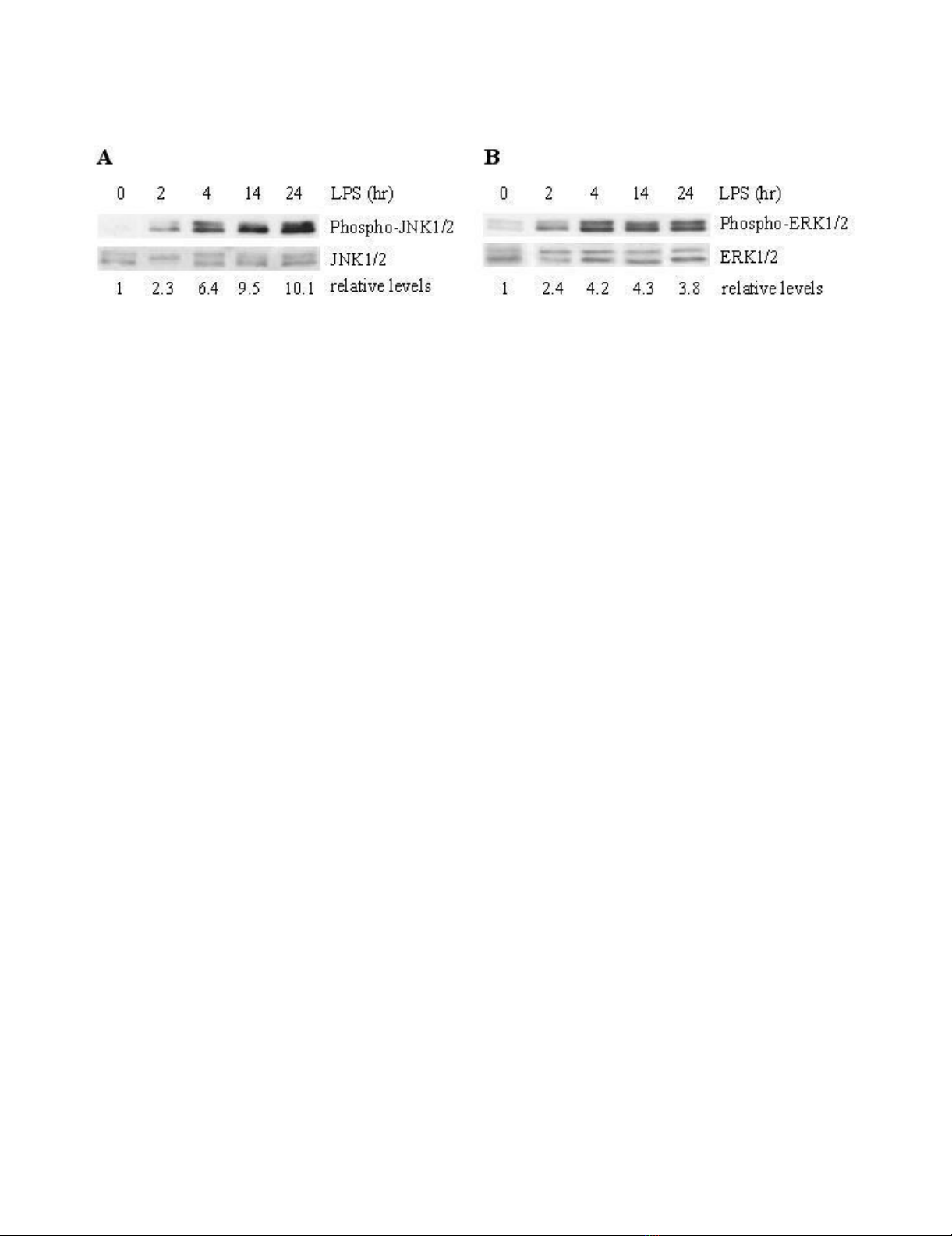
Phosphorylation of JNK and ERK in Lung Tissue
To determine JNK and ERK activation in the lung tissue
from LPS treated animals, Western blot analysis with a
phospho-specific JNK antibody or ERK antibody was
employed. Figures 1A and 1B showed time courses of LPS-
induced phosphorylation, or activation, of JNK1/2 and
ERK1/2. Phosphorylation of these MAP kinases substan-
tially increased beginning 4 hours after LPS treatment,
and progressively further increased (JNK activation) or
were maintained (ERK activation) for up to 24 hours after
LPS treatment. SP600125 pretreatment partially inhibited
Respiratory Research 2004, 5:23 http://respiratory-research.com/content/5/1/23
Page 5 of 15
(page number not for citation purposes)
LPS-induced phosphorylation of JNK1/2 in lung tissue at
4 hours after LPS treatment (Figure 2A), but this inhibitor
had little effect on the activation of ERK1/2 (2C) and p38
MAP kinse (2E). PD98059 pretreatment specifically
inhibited the activation of ERK1/2 (Figure 2D), but nei-
ther the activation of JNK1/2 (2B) nor p38 MAP kinase
(2F). Both JNK and ERK activation were barely detectable
in the animals treated with saline or saline-kinase
inhibitors.
Total Protein and LDH Activity in BAL Fluid and
Neutrophil Influx into Lungs
BAL protein contents (Figure 3A) and LDH activity (Figure
3B) in LPS-treated animals were significantly increased (p
< 0.05). BAL protein increased 2.9-fold, and LDH activity
increased 4.7-fold. This indicates that IT LPS treatment of
rats induced acute lung injury. However, SP600125 or
PD98059 pretreatment significantly inhibited LPS-
induced changes in protein contents, by 63 and 74%,
respectively, and BAL LDH activity by 71 and 86%, respec-
tively (P < 0.05). There were no significant differences in
these parameters between saline-SP600125, saline-
PD98059, and saline control animals (p < 0.05).
BAL cells were differentially analyzed, in order to evaluate
the effects of these kinase inhibitors on LPS-induced neu-
trophil influx. As shown in Figure 3C, neutrophil counts
of the total lung lavage cells in LPS-treated animals signif-
icantly increased by a factor of 26, compared to values in
saline-treated animals, indicating a significant increase in
neutrophil influx into the alveolar spaces (p < 0.05).
SP600125 or PD98059 significantly suppressed BAL neu-
trophil counts by 53 or 46 %, respectively (vs LPS animals,
p < 0.05). The BAL neutrophil counts in saline-kinase
inhibitor animals were not significantly different from
those of the saline control animals (p < 0.05).
CINC, MMP-9 and NO Production in Lungs or Alveolar
Macrophages
CINC, MMP-9 and NO were chosen in our experiments as
representative inflammatory mediators, because of their
important roles in neutrophil influx and lung damage,
and also because their gene regulation is dependent on
NF-κB. Figure 4 illustrates representative Western blots of
lung tissue and BAL fluid for CINC. CINC protein expres-
sion was undetectable in the samples of saline control ani-
mals, but was markedly increased by LPS treatment for 4
hours. By densitometric analysis, CINC protein in lung
tissue (Figure 4A and 4Clane 2) and BAL fluid (Figure 4B
and 4Dlane 2) from LPS animals was approximately 7-
and 2.5-fold higher than in saline control animals, respec-
tively. SP600125 or PD98059 significantly decreased the
level of LPS-induced CINC expression, by 50 and 62%,
respectively, in lung tissue (Figure 4A and 4Clane 3, p <
0.05) and, by 76 and 97%, respectively, in BAL fluid (Fig-
ure 4B and 4Dlane 3, p < 0.05). These kinase inhibitors
alone had little effect on CINC levels in the lung tissue
and lavage fluid.
BAL fluid (Figure 5A and 5D), and the supernatants from
alveolar macrophage cultures (Figure 5B and 5E), were
analyzed for evidence of MMP-9 activity, using gelatin
zymography. The BAL fluid from the saline control ani-
mals showed undetectable gelatinolytic bands. LPS treat-
ment induced a distinct increase in the amount of
gelatinolytic activity and the most prominent band was
found to be a 92 kD species in the BAL fluid, correspond-
ing to a molecular weight identical to MMP-9 [25,26].
This was confirmed to be MMP-9 by Western blot analysis
with the antiMMP-9 monoclonal antibody (Figure 5C and
5Flane 2). In the supernatants from alveolar macrophage
cultures of saline control animals, MMP-9 activity was
also barely detectable, but was also markedly increased in
Time course of phosphorylation of JNK (A) and ERK (B), in lung tissue from rats treated with saline (0 time) or LPS (2–24 h)Figure 1
Time course of phosphorylation of JNK (A) and ERK (B), in lung tissue from rats treated with saline (0 time) or LPS (2–24 h).
Western blots with anti-phospho-JNK/JNK antibody or phospho-ERK/ERK antibody were employed in order to monitor JNK
or ERK phosphorylation. Relative values for levels of phosphorylated JNK1/2 or ERK1/2 normalized to JNK1/2 or ERK1/2 are
indicated below the gel. Results are representative results from 5 rats in each group.


