
BioMed Central
Page 1 of 12
(page number not for citation purposes)
Respiratory Research
Open Access
Research
A role for MCP-1/CCR2 in interstitial lung disease in children
Dominik Hartl1, Matthias Griese1, Thomas Nicolai1, Gernot Zissel2,
Christine Prell1, Dietrich Reinhardt1, Dolores J Schendel3 and
Susanne Krauss-Etschmann*1
Address: 1Childrens' Hospital of the Ludwig-Maximilians-University, Munich, Germany, 2Department of Pneumology, Medical Center, Albert-
Ludwigs-University, Freiburg, Germany and 3Institute of Molecular Immunology and Immune Monitoring Platform, GSF National Research
Center for Environment and Health, Munich, Germany
Email: Dominik Hartl - dominic.hartl@med.uni-muenchen.de; Matthias Griese - mathias.griese@med.uni-muenchen.de;
Thomas Nicolai - thomas.nicolai@med.uni-muenchen.de; Gernot Zissel - zissel@med1.ukl.uni-freiburg.de;
Christine Prell - christine.prell@med.uni-muenchen.de; Dietrich Reinhardt - dietrich.reinhardt@med.uni-muenchen.de;
Dolores J Schendel - schendel@gsf.de; Susanne Krauss-Etschmann* - susanne.krauss-etschmann@med.uni-muenchen.de
* Corresponding author
ChemokinesMCP-1CCR2Bronchoalveolar LavageChildrenInterstitial Lung Diseases
Abstract
Background: Interstitial lung diseases (ILD) are chronic inflammatory disorders leading to
pulmonary fibrosis. Monocyte chemotactic protein 1 (MCP-1) promotes collagen synthesis and
deletion of the MCP-1 receptor CCR2 protects from pulmonary fibrosis in ILD mouse models. We
hypothesized that pulmonary MCP-1 and CCR2+ T cells accumulate in pediatric ILD and are related
to disease severity.
Methods: Bronchoalveolar lavage fluid was obtained from 25 children with ILD and 10 healthy
children. Levels of pulmonary MCP-1 and Th1/Th2-associated cytokines were quantified at the
protein and the mRNA levels. Pulmonary CCR2+, CCR4+, CCR3+, CCR5+ and CXCR3+ T cells
were quantified by flow-cytometry.
Results: CCR2+ T cells and MCP-1 levels were significantly elevated in children with ILD and
correlated with forced vital capacity, total lung capacity and ILD disease severity scores. Children
with lung fibrosis had significantly higher MCP-1 levels and CCR2+ T cells in bronchoalveolar lavage
fluid compared to non-fibrotic children.
Conclusion: The results indicate that pulmonary CCR2+ T cells and MCP-1 contribute to the
pathogenesis of pediatric ILD and might provide a novel target for therapeutic strategies.
Background
Interstitial lung diseases (ILD) are chronic inflammatory
disorders characterized by restrictive lung disease and dif-
fuse pulmonary infiltrates. Although the precise incidence
is not known, ILD are less frequent in children than adults
[1-3]. Lungs of ILD patients show inflammation with alve-
olar wall thickening by leukocytes and pulmonary fibro-
sis. Despite immunosuppressive treatment and
Published: 11 August 2005
Respiratory Research 2005, 6:93 doi:10.1186/1465-9921-6-93
Received: 19 April 2005
Accepted: 11 August 2005
This article is available from: http://respiratory-research.com/content/6/1/93
© 2005 Hartl et al; licensee BioMed Central Ltd.
This is an Open Access article distributed under the terms of the Creative Commons Attribution License (http://creativecommons.org/licenses/by/2.0),
which permits unrestricted use, distribution, and reproduction in any medium, provided the original work is properly cited.

Respiratory Research 2005, 6:93 http://respiratory-research.com/content/6/1/93
Page 2 of 12
(page number not for citation purposes)
supportive measures, the progressive course leading to
irreversible lung fibrosis sometimes can not be prevented.
Therefore, the development of additional therapeutic
strategies is of high importance.
Monocyte chemotactic protein 1 (MCP-1, CCL2) is pro-
duced in response to inflammatory stimuli by a variety of
cells, including monocytes/macrophages, lymphocytes
and airway epithelial cells [4-6]. MCP-1 stimulates colla-
gen synthesis and production of the pro-fibrotic factor
transforming growth factor β (TGF-β) in fibroblasts, while
MCP-1 antisense oligonucleotides reduce TGF-β produc-
tion[7,8]. Application of MCP-1 into murine lungs
induces an inflammatory cytokine response and pulmo-
nary leukocyte accumulation. In adult patients with ILD,
increased levels of MCP-1 were observed in serum[9,10]
and bronchoalveolar lavage fluid (BALF) [11-14].
Although MCP-1 was originally described for its chemo-
tactic activity on monocytes, in vitro studies revealed an
even higher activity on T cells[15]. This occurs through
MCP-1 binding to its sole receptor CCR2[16]. Deletion of
the CCR2-gene or receptor blockade with anti-CCR2 anti-
bodies leads to a dramatic inhibition of leukocyte accu-
mulation in murine lungs[17]. Furthermore, CCR2-/-
mice are protected from fluorescein (FITC) or bleomycin
induced lung fibrosis[18]. Thus far, CCR2+ T cells in BALF
of patients with fibrotic lung diseases have not been
determined.
In addition to the MCP-1/CCR2 axis, Th2 cytokines seem
to mediate pulmonary fibrosis [19-22]. IL-4 stimulates
fibroblast proliferation and collagen synthesis[23,24],
while IFN-γ inhibits this process [25-28]. In a Th2 mouse
model fibroblasts expressed more CCR2 protein and
higher levels of MCP-1 and TGF-β as compared to fibrob-
lasts from a Th1-mouse model[8]. Furthermore, increased
levels of IL-4 were observed in animal models of pulmo-
nary fibrosis[29] and lungs of patients with idiopathic
pulmonary fibrosis (IPF)[30] or cryptogenic fibrosing
alveolitis[31].
The contribution of MCP-1 to ILD has been investigated
exclusively in adults. However, the spectrum of ILD differs
considerably between adults and children and some
forms are unique to children while others, such as idio-
pathic pulmonary fibrosis (IPF), are extremely rare in
childhood[32].
Therefore, we asked whether levels of MCP-1 and frequen-
cies of CCR2+ T cells are increased in BALF of children
with ILD and, if so, how levels of MCP-1 and CCR2+ T
cells relate to disease severity in pediatric ILD.
To address these questions levels of MCP-1 and frequen-
cies of CCR2+ T cells in BALF were compared between chil-
dren with ILD and children without lung disease.
To evaluate the contribution of the pulmonary Th1/Th2
micromilieu to the pathogenesis of pediatric ILD, CCR4+
and CCR3+ (Th2) and CCR5+ and CXCR3+ (Th1) cells
were determined in BALF together with an array of pulmo-
nary Th1- and Th2-associated cytokines.
Our results indicate that pulmonary CCR2+ T cells and lev-
els of MCP-1 are characteristic components in BALF of
children with ILD. A pathophysiological role in pediatric
ILD seems likely as their levels relate to restrictive lung
function and ILD disease severity.
Methods
Characterization of the patients
Children attending the Department of Pulmonology and
Allergology of the University Children's Hospital of
Munich during 1999–2004 were considered for inclusion
in this study. Children suspective of ILD underwent a
comprehensive clinical evaluation, including patient his-
tory, physical examination, routine laboratory tests, lung
function testing, chest radiography, high resolution com-
puted tomography (HRCT) and bronchoalveolar lavage
(BAL). Children were assigned to the ILD group according
to the criteria of Fan[33]: (i) ≥3 months of respiratory
symptoms characteristic for ILD, i.e. non-productive
cough, dyspnoea, tachypnea, crackles and/or rales, exer-
cise intolerance and/or hypoxemia, (ii) diffuse infiltrates
on chest radiographs and HRCT and (iii) restrictive lung
function (decreased forced vital capacity (FVC) and total
lung capacity (TLC)) according to the ATS criteria[34].
The diagnosis of the specific form of ILD was established
by patient history, physical examination, HRCT, BAL and/
or lung biopsy according to consensus criteria[33,35].
Two thoracic radiologists independently evaluated all
lobes on HRCT for ground glass opacity and pulmonary
fibrosis as described previously[36,37]. A pathologist spe-
cialized on pediatric ILD[38] evaluated the lung sections
systematically[39,40]. Furthermore, the disease severity of
each ILD patient was characterized using the clinical ILD
score of Fan[41]: 1 = asymptomatic, no desaturation; 2 =
symptomatic but normoxic (>90%) under all conditions;
3 = symptomatic with desaturation during sleep or with
exercise; 4 = symptomatic with desaturation at rest. None
of the included children had familial idiopathic pulmo-
nary fibrosis. Patients with congenital heart disease or sus-
pected or proven bacterial pulmonary infection were
excluded from the study.
Twenty-five children with ILD (median age: 7 ± 3.6 years;
male/female = 16/9) were included (Table 1).

Respiratory Research 2005, 6:93 http://respiratory-research.com/content/6/1/93
Page 3 of 12
(page number not for citation purposes)
Ten age-matched children were selected as the control
group (median age: 7.5 ± 2.9 years, m/f: 6/4). These chil-
dren were considered as healthy, i.e. had no systemic dis-
ease, had no suspected or proven pulmonary disease and
were free of respiratory tract infections. These children
underwent elective tonsillectomy under general anaesthe-
sia. BAL was performed prior to the surgical procedure.
Ten age-matched children with chronic severe asthma
(median age: 8.7 ± 1.6 years, m/f: 5/5), from a previous
study[42], who were comparable to the ILD group in
terms of gender and age were included as disease control
group. All parents and/or patients gave their informed
consent prior to bronchoscopy and the institutional
review board approved the study protocol.
Bronchoalveolar lavage
Bronchoscopy with BAL was performed as described pre-
viously[43]. Residual BALF cells were used for flow cytom-
etry. The BALF recovery and the viability of cells did not
differ significantly between the patient groups. Cellular
profiles are shown in Table 2.
Flow cytometry
BALF cells were analyzed by four-colour flow cytometry
(FACSCalibur, Becton-Dickinson, Heidelberg, Germany)
as described previously[42]. The following antibodies
were used: CD4-allophycocyanine (APC) mouse IgG1,
CD8-phycocyanine 5 (PC5) mouse IgG1 (Immunotech,
Marseille, France), CD69-PE mouse IgG1, CCR5-PE
mouse IgG2a, CCR4-PE mouse IgG2a (BD Pharmingen,
Heidelberg, Germany), CCR2-PE mouse IgG2b, CXCR3-
Table 1: Patients' characteristics
No Sex Age
[years]
Interstitial
lung disease
Diagnosis
finding
Radiographic findings Fibrotic
changes
(CT)
ILD
Score*
Dyspnoe Cough Cyanosis Exercise
Intolerance
Failure to
thrive
Medication FVC
[% of pred.]
TLC %
[of pred.]
1 F 7 LIP CT, LB • diffuse interstitial involvement + 4 ++ + + + + CS, AZT 34 56
• reticular-nodular pattern
• follicular bronchiolitis
2 M 14 U-ILD, IPH CT, BAL patchy interstitial involvement - 2 + - - - - CS 77 89
3 M 8 U-ILD CT, LB • ground-glass opacity + 3 ++ - - + - 46 74
4 M 4 IPH CT, BAL, LB interstitial involvement - 3 + - - - - 77 168
5 M 16 U-ILD CT, BAL interstitial involvement + 2 + - - + + 76 95
6 F 7 U-ILD CT, BAL interstitial involvement - 2 + + - - + AZT 50 68
7 M 4 CPI CT, LB • diffuse infiltrates + 3 + - - + + AZT 58 64
• ground-glass opacity
8 F 3 NSIP CT, LB • interstitial involvement + 3 ++ - + + + CS n.d. n.d.
• alveolar infiltrates
9 M 8 Sarcoidosis CT, BAL, LB • interstitial involvement + 2 ++ + - + + CS 56 63
• perivascular nodules
10 F 8 U-ILD CT, BAL, LB ground-glass opacity - 1 - + - + - 76 87
11 F 8 CPI CT, LB • interstitial involvement + 2 + + - + + CS 37 74
• ground-glass opacity
12 M 9 U-ILD CT interstitial involvement - 2 + - - - - 70 98
13 M 5 NSIP CT, LB • interstitial involvement + 3 ++ - - + - CS 61 76
• ground-glass opacity
14 F 6 U-ILD CT reticular-nodular pattern + 3 ++ + - - - AZT 60 68
15 F 4 U-ILD CT interstitial involvement + 2 + + - - + n.d. n.d.
16 M 12 U-ILD CT interstitial involvement - 2 + - - - - 68 75
17 M 3 PAP† CT, BAL, LB • ground glass opacity - 4 +++ + + + + CS n.d. n.d.
18 M 6 NSIP CT, BAL, LB • alveolar infiltrates + 4 +++ + + + + CS, AZT 63 72
PAP • ground glass opacification
19 F 3 PAP† CT, BAL, LB • ground glass opacity + 4 ++ - + + + CS n.d. n.d.
• alveolar infiltrates
20 F 9 NSIP CT, LB • interstitial involvement + 3 ++ + + + + CS, AZT 55 74
• honeycombing
21 M 7 U-ILD CT reticular-interstitial pattern + 3 + + - + + AZT, MT 38 59
22 M 7 Cholesterol CT, BAL, LB • interstitial involvement + 4 +++ + + + + CS 16 24
pneumonitis† • reticular-interstitial pattern
23 M 4 U-ILD CT, LB • interstitial involvement - 2 + - - + + CS 102 99
• honeycombing
24 M 8 U-ILD CT interstitial involvement - 2 + + - + - CS 63 78
25 M 7 NSIP CT, LB • interstitial involvement + 3 + + - + - CS 60 76
ILD-NC: children with interstitial lung disease without systemic corticosteroid treatment; ILD-C: children with interstitial lung disease with systemic
corticosteroid treatment;
U-ILD: undefined/idiopathic interstitial lung disease: no specific diagnosis could be made; PAP: pulmonary alveolar proteinosis; CGD: chronic
granulomatous disease; IPH: idiopathic pulmonary hemosiderosis; LIP: lymphocytic interstitial pneumonia; CPI: Chronic pneumonitis of infancy
CS: corticosteroids, AZT: azathioprine, MT: methotrexat
n.d.: lung function testing not done (children < 5 years); † symbolizes patients who died due to respiratory failure.
CT: Computed tomography; BAL: Bronchoalveolar lavage; LB: Lung biopsy
* ILD score according to Fan[41]

Respiratory Research 2005, 6:93 http://respiratory-research.com/content/6/1/93
Page 4 of 12
(page number not for citation purposes)
fluorescein isothiocyanate (FITC) mouse IgG1 and CCR3-
FITC rat IgG2a (R&D Systems, Wiesbaden, Germany).
Mouse IgG1-FITC, mouse IgG1-PE, mouse IgG2a-PE,
mouse IgG2b-PE (Immunotech, Marseille, France) and rat
IgG2a-FITC (kindly provided by Dr. E. Kremmer, GSF-
Institute of Molecular Immunology, Munich, Germany)
were used as isotype controls.
Detection of MCP-1 and cytokines
Levels of MCP-1 and Th1 (IL-2, IFN-γ), Th2 (IL-4, IL-5, IL-
10) and pro-inflammatory cytokines (TNF-α, IL-6) were
quantified by a multiplex, particle-based assay (Bio-Rad
Laboratories, Minneapolis, USA) as described previ-
ously[42]. The detection limits for all cytokines were 1.5–
2.5 pg/ml (min.) and 1000 pg/ml (max.).
Quantitative RT-PCR
BALF cells were lysed in Trizol LS Reagent (Invitrogen, Life
Technologies, Karlsruhe, Germany) and were stored at -
20°C until mRNA extraction. Total mRNA was isolated
according to the manufacturer's instructions and reverse
transcribed into cDNA. Contamination with genomic
DNA was excluded by mRNA controls without reverse
transcriptase in the cDNA synthesis reaction. The follow-
ing oligonucleotide primers were used: MCP-1 (5-
TGAAGCTCGCACTCTCGCCT-3; 5- GTGGAGTGAGTGT-
TCAAGTC-3); and GAPDH (5-GAGGTGAAGGTCG-
GAGTC-3; 5-AAGATGGTGATGGGATTTC-3). Expression
levels were determined in duplicates by Real time RT-PCR
using SYBR green and the iCycler iQ detection system
(Biorad, Hercules, CA, USA) according to the
manufacturer's instructions. Threshold cycle (CT) values
for genes of interest were normalized to GAPDH and used
to calculate the relative mRNA expression.
Statistical analysis
The non-parametric Mann-Whitney U test was applied.
Correlations were tested with Spearman's rank correlation
test. A probability of p < 0.05 was regarded as signifi-
cant[44] (SPSS statistical program, version 11.5, SPSS Inc.
Chicago, USA).
Results
MCP-1 levels and CCR2+ T cells in BALF
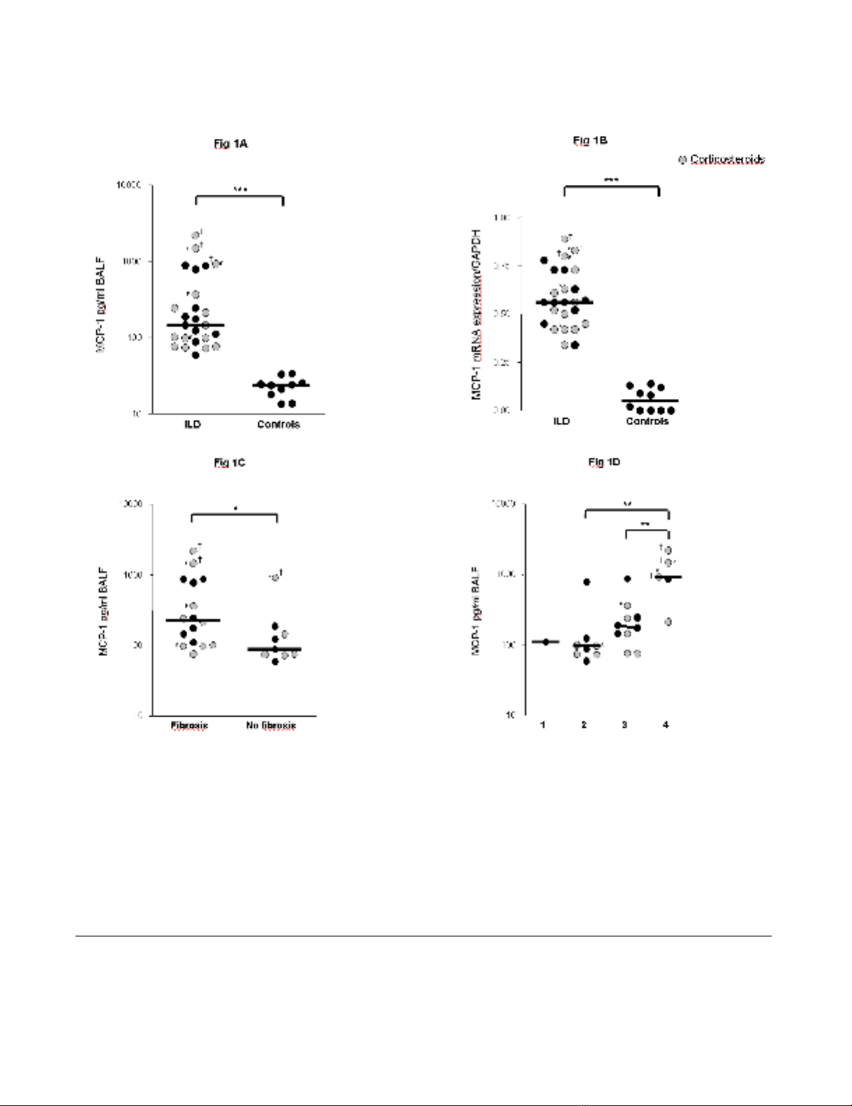
Levels of MCP-1 were significantly higher in children with
ILD (n = 25) as compared to the control group at protein
and mRNA level (Figure 1A, B). MCP-1 protein and
mRNA expression levels correlated positively with each
other (r = 0.72, p < 0.01). ILD children with pulmonary
fibrosis had significantly higher MCP-1 levels in BALF as
compared to children with non-fibrotic ILD (Figure 1C).
MCP-1 levels related positively to the stage of disease (Fig-
ure 1D). The highest levels of MCP-1 were observed in the
three patients who died after respiratory failure (Table 1;
P17, P19, P22). Furthermore, MCP-1 levels correlated
negatively with restrictive lung function parameters (TLC,
FVC) (Figures 2A, B).
To test whether increased MCP-1 levels are associated with
increased frequencies of CCR2+ T cells, BALF lymphocytes
were quantified by flow cytometry. CCR2 was expressed
on a higher percentage of CD4+ than CD8+ T cells. The
majority of CCR2+ T cells showed an activated phenotype
(75% CCR2+CD69+). Children with ILD had significantly
higher percentages of CCR2+CD4+and CCR2+CD8+ T cells
Table 2: Bronchoalveolar lavage cells
ILD-NC ILD-C Control
Total cells × 103/ml 230 (2.1–1124)** 144 (11–268)* 89 (83–97)
Recovery (%) 55 (25–86) 49 (34–75) 54 (35–70)
Neutrophils (%) 10.5 (1–44)* 8.5 (3–30)* 2 (0–3)
Eosinophils (%) 1 (0–6) 1.5 (0–3) 0 (0–1)
Mast cells (%) 2 (0–43) 2 (1–4) 0 (0-0)
Plasma cells (%) 0 (0–4) 0 (0–4) 0 (0-0)
Macrophages (%) 60 (7–97)* 49 (26–77)* 94 (81–92)
Lymphocytes (%) 24 (2–54)** 22 (5–34)** 4 (2–13)
CD4+ T cells (%)†23 (9–45) 29 (9–82) 23 (15–28)
CD8+ T cells (%)†29 (6–62) 27 (2–83) 25 (15–31)
CD4/8 ratio 0.7 (0.3–6) 1.1 (0.1–55) 0.7 (0.4–0.9)
results are expressed as medians with ranges shown in parenthesis.
ILD-NC: children with interstitial lung disease without systemic corticosteroid treatment;
ILD-C: children with interstitial lung disease with systemic corticosteroid treatment;
*p < 0.05, **p < 0.01 as compared to the control group, Mann-Whitney-U Test.
Total cells and differential cell count were obtained from cytospin slides, CD4+, CD8+ and CD4/CD8 T cells using flow cytometry.
†CD4+ T cells and CD8+ T cells are shown as the percentage of total lymphocytes in BALF, i.e. cells gated in the lymphocyte population.
Neutrophils, eosinophils, mast cells, plasma cells, macrophages and lymphocytes are shown as percentage of total cells in BALF.
Respiratory Research 2005, 6:93 http://respiratory-research.com/content/6/1/93
Page 5 of 12
(page number not for citation purposes)
MCP-1 levels in children with ILDFigure 1
MCP-1 levels in children with ILD. MCP-1 levels in bronchoalveolar lavage fluid (BALF) of children with interstitial lung dis-
eases (ILD) and healthy controls are shown at the (A) protein and at the (B) mRNA level. (C) MCP-1 levels in BALF of ILD chil-
dren with and without pulmonary fibrosis. Pulmonary fibrosis was assessed by computed tomography according to [36,37]. (D)
MCP-1 levels in ILD children related to ILD disease severity according to the criteria of Fan [33]. 1 = asymptomatic, no desat-
uration; 2 = symptomatic but normoxic (> 90%) under all conditions; 3 = symptomatic with desaturation during sleep or exer-
cise; 4 = symptomatic with desaturation at rest; MCP-1 protein levels were quantified in BALF by a multiplex, particle-based
assay (Bio-Rad Laboratories, Minneapolis, USA) as described previously [42]. MCP-1 mRNA levels were quantified in BALF
cells by Real time RT-PCR using SYBR green and the iCycler iQ detection system (Biorad, Hercules, CA, USA) and were nor-
malized to GAPDH. Median values are shown by horizontal bars. Differences between the patient groups were tested with the
Mann-Whitney U test; * p < 0.05, *** p < 0.001; Children with systemic corticosteroid therapy are shown as grey circles. P:
Pulmonary alveolar proteinosis; S: Sarcoidosis; † symbolize children who died due to respiratory failure.


