
BioMed Central
Page 1 of 13
(page number not for citation purposes)
Virology Journal
Open Access
Research
Phosphorylation of HIV Tat by PKR increases interaction with TAR
RNA and enhances transcription
Liliana Endo-Munoz1, Tammra Warby1, David Harrich2 and
Nigel AJ McMillan*1
Address: 1Centre for Immunology and Cancer Research, University of Queensland, Princess Alexandra Hospital, Brisbane, Australia and
2Queensland Institute of Medical Research, Royal Brisbane Hospital, Brisbane, Australia
Email: Liliana Endo-Munoz - lmunoz@cicr.uq.edu.au; Tammra Warby - t.warby@ugrad.unimelb.edu.au; David Harrich - davidH@qimr.edu.au;
Nigel AJ McMillan* - n.mcmillan@uq.edu.au
* Corresponding author
Abstract
Background: The interferon (IFN)-induced, dsRNA-dependent serine/threonine protein kinase,
PKR, plays a key regulatory role in the IFN-mediated anti-viral response by blocking translation in
the infected cell by phosphorylating the alpha subunit of elongation factor 2 (eIF2). The human
immunodeficiency virus type 1 (HIV-1) evades the anti-viral IFN response through the binding of
one of its major transcriptional regulatory proteins, Tat, to PKR. HIV-1 Tat acts as a substrate
homologue for the enzyme, competing with eIF2α, and inhibiting the translational block. It has been
shown that during the interaction with PKR, Tat becomes phosphorylated at three residues: serine
62, threonine 64 and serine 68. We have investigated the effect of this phosphorylation on the
function of Tat in viral transcription. HIV-1 Tat activates transcription elongation by first binding to
TAR RNA, a stem-loop structure found at the 5' end of all viral transcripts. Our results showed
faster, greater and stronger binding of Tat to TAR RNA after phosphorylation by PKR.
Results: We have investigated the effect of phosphorylation on Tat-mediated transactivation. Our
results showed faster, greater and stronger binding of Tat to TAR RNA after phosphorylation by
PKR. In vitro phosphorylation experiments with a series of bacterial expression constructs carrying
the wild-type tat gene or mutants of the gene with alanine substitutions at one, two, or all three of
the serine/threonine PKR phosphorylation sites, showed that these were subject to different levels
of phosphorylation by PKR and displayed distinct kinetic behaviour. These results also suggested a
cooperative role for the phosphorylation of S68 in conjunction with S62 and T64. We examined
the effect of phosphorylation on Tat-mediated transactivation of the HIV-1 LTR in vivo with a series
of analogous mammalian expression constructs. Co-transfection experiments showed a gradual
reduction in transactivation as the number of mutated phosphorylation sites increased, and a 4-fold
decrease in LTR transactivation with the Tat triple mutant that could not be phosphorylated by
PKR. Furthermore, the transfection data also suggested that the presence of S68 is necessary for
optimal Tat-mediated transactivation.
Conclusion: These results support the hypothesis that phosphorylation of Tat may be important
for its function in HIV-1 LTR transactivation.
Published: 28 February 2005
Virology Journal 2005, 2:17 doi:10.1186/1743-422X-2-17
Received: 30 November 2004
Accepted: 28 February 2005
This article is available from: http://www.virologyj.com/content/2/1/17
© 2005 Endo-Munoz et al; licensee BioMed Central Ltd.
This is an Open Access article distributed under the terms of the Creative Commons Attribution License (http://creativecommons.org/licenses/by/2.0),
which permits unrestricted use, distribution, and reproduction in any medium, provided the original work is properly cited.

Virology Journal 2005, 2:17 http://www.virologyj.com/content/2/1/17
Page 2 of 13
(page number not for citation purposes)
Background
Since its isolation in 1983 [1,2], human immunodefi-
ciency virus type 1 (HIV-1) continues to cause 5 million
new infections each year, and since the beginning of the
epidemic, 31 million people have died as a result of HIV/
AIDS [3]. One of the major mechanisms employed by the
immune system to counteract the effects of viral infections
is through an antiviral cytokine – type 1 interferon (IFN).
However, while IFN is able to inhibit HIV-1 infection in
vitro [4], it has not been effective in the treatment of HIV-
1 infections in vivo. Furthermore, the presence of increas-
ing levels of IFN in the serum of AIDS patients while viral
replication continues and the disease progresses [5-7]
indicates that HIV-1 must employ a mechanism to evade
the antiviral effects of IFN.
In response to viral infection, IFN induces a number of
genes including the dsRNA-dependent protein kinase R
(PKR). PKR exerts its anti-viral activity by phosphorylating
the alpha subunit of translation initiation factor 2
(eIF2α), which results in the shut-down of protein synthe-
sis in the cell [8]. The importance of PKR in the host anti-
viral response is suggested by the fact that most viruses
including vaccinia [9], adenovirus [10], reovirus [11],
Epstein-Barr virus [12], poliovirus [13], influenza [14],
hepatitis C [15,16], human herpes virus [17-19], and
SV40 [20], employ various mechanisms to inhibit its
activity. HIV-1 is no exception and we and others have
shown that PKR activity is inhibited by HIV via the major
regulatory protein, Tat [21-23]. Productive infection by
HIV-1 results in a significant decrease in the amounts of
PKR [23] and HIV-1 Tat protein has been shown to act as
a substrate homologue of eIF2α, preventing the phospho-
rylation of this factor and allowing protein synthesis and
viral replication to proceed in the cell [21,22]. During the
interaction between Tat and PKR the activity of the
enzyme is blocked by Tat and Tat itself is phosphorylated
by PKR [21] at serine 62, threonine 64 and serine 68 [22].
HIV-1 Tat is a 14 kDa viral protein involved in the regula-
tion of HIV-1 transcriptional elongation [24-26] and in its
presence, viral replication increases by greater than 100-
fold [27,28]. It functions to trigger efficient RNA chain
elongation by binding to TAR RNA, which forms the ini-
tial portion of the HIV-1 transcript [29]. The interaction
between Tat and TAR is critical for virus replication and
mutations in Tat that alter the RNA-binding site result in
defective viruses. Furthermore, virus replication can be
strongly inhibited by the overexpression of TAR RNA
sequences that act as competitive inhibitors of regulatory
protein binding [30].
While a number of reports have shown that PKR and Tat
protein interact, and furthermore, that Tat is phosphor-
ylated by PKR, none have yet addressed the issue of the
functional consequences for the phosphorylation of the
Tat protein. Here we examine the phosphorylation of Tat
by PKR and its effect on TAR RNA binding and HIV-1 tran-
scription, and show that the phosphorylation of Tat
results in Tat protein binding more strongly to TAR RNA.
Removal of the residues reported to be phosphorylated by
PKR resulted in decreased Tat phosphorylation and a sig-
nificant loss of Tat-mediated transcriptional activity.
Results
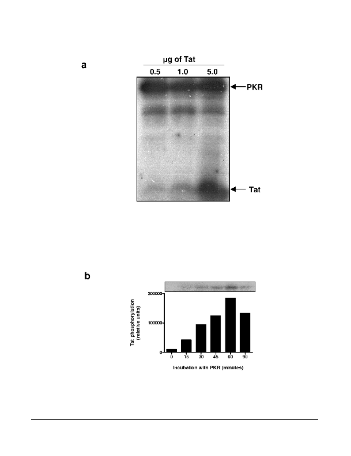
The phosphorylation of HIV-1 Tat by PKR increases its
interaction with TAR RNA
We first confirmed the capability of our PKR preparation
immunoprecipitated from HeLa cells to phosphorylate
synthetic Tat protein (aa 1–86) (Figure 1a), and we deter-
mined the optimal phosphorylation time of Tat by PKR as
60 minutes (Figure 1b). We also confirmed that Tat was
not phosphorylated by PKR in the absence of ATP, or by
ATP alone (data not shown).
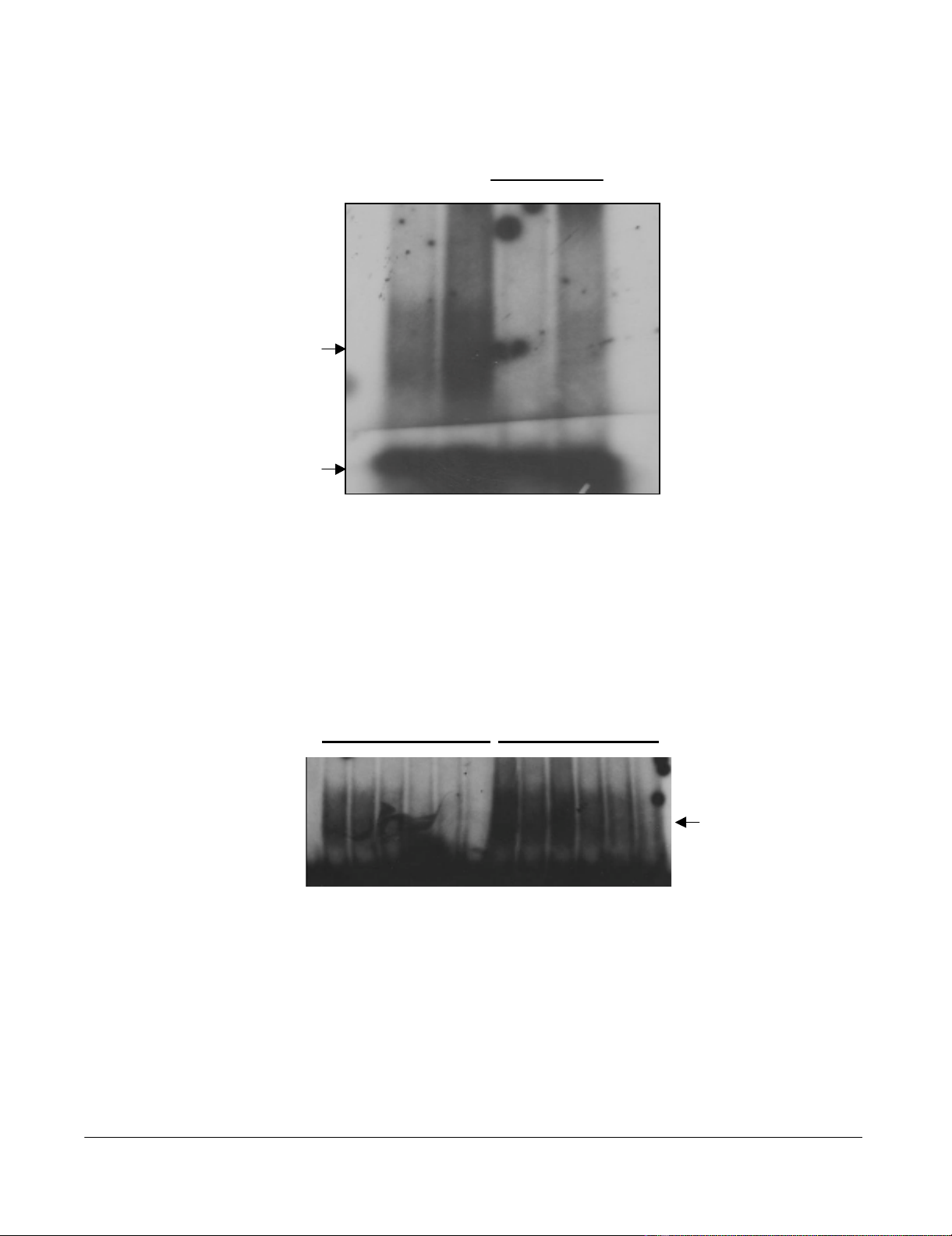
To address the issue of the consequences of PKR phospho-
rylation on Tat function we investigated the ability of
phosphorylated Tat (herein called Tat-P) and normal Tat
(Tat-N) to bind to HIV-1 TAR RNA. Synthetic Tat protein
(aa 1–86) was phosphorylated in vitro using PKR previ-
ously immunoprecipitated from HeLa cells. An electro-
phoretic mobility shift assay (EMSA) was performed to
observe any difference in the binding of Tat-N and Tat-P
to TAR RNA (Figure 2a). It can be seen that Tat-N was able
to form a specific Tat-TAR complex that could be effec-
tively competed off using a 7.5-fold excess of cold TAR
RNA. Tat-P was also able to form a specific Tat-TAR com-
plex that clearly contained more TAR RNA than non-phos-
phorylated Tat. This complex could also be competed off
using cold TAR but some residual complex was left sug-
gesting that the Tat-P-TAR complex was more resistant to
competition with cold TAR than the Tat-N-TAR complex.
As Tat-P appeared to bind more readily to TAR, we next
investigated the differences in the binding efficiency of
Tat-N and Tat-P with TAR RNA. EMSA were performed in
the presence of increasing concentrations of NaCl (from
25–1000 mM). The progressive dissociation of the Tat-N-
TAR RNA complex with increasing concentrations of salt
in the buffer was observed (Figure 2b, lanes 2–7) while
Tat-P-TAR complexes under the same conditions were
clearly more stable (lanes 8–13). For example, at 500 mM
NaCl the Tat-N-TAR complex was almost completely dis-
sociated (lane 6) while the Tat-P-TAR complex was still
clearly observed (lane 12). Even at the maximum salt con-
centration (1000 mM), the Tat-P-TAR complex can still be
seen (lane 13), while the Tat-N-TAR complex was com-
pletely dissociated. These results suggest that Tat86 phos-
phorylated by PKR binds TAR RNA more efficiently and
more strongly than normal Tat.
Virology Journal 2005, 2:17 http://www.virologyj.com/content/2/1/17
Page 3 of 13
(page number not for citation purposes)
Phosphorylation of HIV-1 Tat86 by PKRFigure 1
Phosphorylation of HIV-1 Tat86 by PKR. (a) PKR was immunoprecipitated from HeLa cell extracts and activated with
synthetic dsRNA in the presence of γ-32P-ATP. This activated 32P-PKR was used to phosphorylate 0.5, 1 and 5 µg of synthetic
Tat86 in the presence of γ-32P-ATP, at 30°C for 15 minutes. Proteins were separated by 15% SDS-PAGE. (b) PKR immunopre-
cipitated from HeLa cell extracts, and activated with dsRNA and ATP, was used to phosphorylate 2 µg of synthetic Tat86 at
30°C for the times indicated.
Virology Journal 2005, 2:17 http://www.virologyj.com/content/2/1/17
Page 4 of 13
(page number not for citation purposes)
EMSA of Tat-N, Tat-P and TAR RNA showing dissociation of the Tat-TAR complex with increasing salt concentrationFigure 2
EMSA of Tat-N, Tat-P and TAR RNA showing dissociation of the Tat-TAR complex with increasing salt con-
centration. (a) PKR immunoprecipitated from HeLa cell extracts, and activated with dsRNA and ATP, was used to phospho-
rylate 2 µg of synthetic Tat86 at 30°C for 1 h, in the presence (Tat-P) or absence (Tat-N) of γ-32P-ATP. TAR RNA was
synthesized in vitro from pTZ18TAR80 using a commercial kit, and either γ-32P-dCTP or unlabelled dCTP. The Tat-TAR RNA
binding reaction was allowed to proceed in binding buffer at 30°C for 10 minutes. Each reaction contained 200 ng of either
Tat-N or Tat-P, and approximately 70 000 cpm of 32P-TAR RNA (lanes 1 and 2), or approximately 70 000 cpm of 32P-TAR
RNA and 7.5 × the volume of unlabelled TAR RNA (lanes 3 and 4). The Tat-TAR complexes formed were resolved on a 5%
acrylamide/0.25X TBE gel. (b) The Tat-TAR binding reactions were performed at 30°C for 10 minutes in binding buffer con-
taining various concentrations of NaCl: 25 mM (lanes 2 and 8), 50 mM (lanes 3 and 9), 100 mM (lanes 4 and 10), 200 mM (lanes
5 and 11), 500 mM (lanes 6 and 12), and 1000 mM (lanes 7 and 13). Lanes 2–7 show the dissociation of the Tat-N-TAR com-
plex, and lanes 8–13 show the dissociation of the Tat-P-TAR complex. Lane 1 is TAR RNA only.
Tat-N Tat-P
Tat-TAR
12345678 910111213
Tat-N Tat-P Tat-N Tat-P
+ cold TAR
Tat-TAR
Free TAR
a
b

Virology Journal 2005, 2:17 http://www.virologyj.com/content/2/1/17
Page 5 of 13
(page number not for citation purposes)
Efficient phosphorylation of Tat requires particular
residues
Brand et al. [22] reported that PKR was able to phosphor-
ylate Tat at amino acids serine-62, threonine-64 and ser-
ine-68. We therefore wished to know if any of these
residues were critically important in the ability of Tat to
bind TAR RNA. To this end, we created a series of Tat pro-
teins containing mutations of all possible combinations
of S62, T64 and T68 and investigated the phosphorylation
of the resulting mutant Tat protein. A series of seven Tat
mutants were made using alanine scanning (Figure 3a)
and cloned into the bacterial expression vector pET-
DEST42, which contains a C-terminal 6 × His tag to allow
purification using metal affinity chromatography. The
resulting constructs were validated by sequencing before
the mutant Tat proteins were expressed and purified (Fig-
ure 3b). Protein yields varied between 40–170 g/mL and
all mutants were full length, as confirmed by western blot-
ting using an anti-His antibody (data not shown).
Activated PKR was used to phosphorylate each of the Tat
mutants as above and the reaction was allowed to proceed
for 2, 5, 10, 15, 30, 45 and 60 minutes. The phosphor-
ylated proteins were analyzed by SDS-PAGE and visual-
ized by autoradiography (Figure 4). As can be seen from
the figure, the phosphorylation of each protein by PKR
varied and was the most efficient for wild-type Tat and the
least efficient for the triple mutant, Tat S62A.T64A.T68A,
where no sites for PKR phosphorylation were available.
Scanning densitometry and non-linear regression analysis
was performed and the extent of phosphorylation after 15
minutes was measured for each protein and expressed as
a percentage of the wild-type protein (which is set to
100%) (Figure 5a). This time was chosen from non-linear
regression analysis of the wild-type protein that indicated
enzymatic phosphorylation of the wild-type protein was
active at this time point. Non-linear regression analysis
was performed to calculate the maximal phosphorylation
for each protein (Pmax), and the time required to reach
half-maximal phosphorylation (K0.5) (Figure 5b).
Phosphorylation of the single mutants was rapid and spe-
cific with maximal phosphorylation values (Pmax) for S62,
T64 and T68 of 98.6%, 87.5% and 81.6% respectively
compared to the wild type (Pmax = 82.8%) and K0.5 values
of 10.9 min, 5.2 min and 0.8 min (wild-type = 5.5 min).
This observation was also applicable to the Tat S62A.T64A
mutant, which exhibited 87% phosphorylation (Figure
5a) (Pmax = 82.1%, K0.5 = 5.5 min). However, the percent-
age of phosphorylation at 15 minutes for the other double
mutants and for the triple mutant decreased to 68% for
Tat T64A.S68A, 48% for Tat S62A.S68A, and 56% for Tat
S62A.T64A.S68A. These values also correlated well with
the higher Pmax values (172.8%, 256.8% and 189.7%
respectively) and K0.5 values (54.9 min, 109.7 min and
62.2 min respectively) for each mutant, indicating slower,
less efficient and non-specific phosphorylation.
The phosphorylation of HIV-1 Tat by PKR enhances viral
transcription
To examine the effect of Tat phosphorylation on its trans-
activation ability mammalian expression constructs con-
taining the Tat mutants were prepared and transfected
into HeLa cells. To measure Tat-specific transcription, we
co-transfected with pHIV-LTR-CAT as well as with β-actin-
luciferase to normalize for transfection efficiency. The
transfection reaction was optimized for DNA concentra-
tion, transfection reagent concentration, and time. The
results for three separate transfections are shown in Figure
6 and expressed as percentage of wild-type Tat. As
expected, no transactivation of the HIV-1 LTR was
observed in the untransfected control or in the absence of
pHIV-LTR-CAT, and basal transcription was present at low
levels (0.08-fold) in the absence of Tat. We observed sig-
nificant decreases in transactivation with mutant Tat, even
when a single phosphorylation site was mutated. There
was a general trend to low activity as more mutations were
introduced. Thus, the average transactivation by the single
mutants, Tat 62A, T64A and S68A, was 58%, transactiva-
tion by the double mutants, Tat S62A.T64A, T64A.S68A
and S62A.S68A, was 41%, while the triple mutant, Tat
S62A.T64A.S68A, exhibited only 24% transactivation.
The differences in LTR activation observed for the individ-
ual single mutants were not large, indicating that the
absence of any one of these phosphorylation residues
reduced the ability of Tat to activate the HIV-1 LTR but
that no single residue was more important than the other.
As in the phosphorylation data, Tat S62A.T64A behaved
similarly to the single mutants. The mutations that had
the greatest effect were the T64A.S68A, S62A.S68A, and
the triple mutant. Of the three residue combinations, the
absence of T64 and S68 together had the greatest negative
effect on transactivation, inducing a 3-fold decrease,
which was comparable to that observed for the triple
mutant (4-fold).
The absence of S62 in combination with S68 also had a
marked effect on transactivation, reducing it 2.5-fold. On
the other hand, the absence of S62 in combination with
T64 reduced transactivation 1.8-fold. This suggests that
the absence of S62 and T64 either singly or in combina-
tion is not as important for Tat-mediated transactivation
as when these residues are absent in combination with
S68, and may indicate a more important role for S68 in
Tat transactivation. These data correlate with observations
previously obtained in PKR phosphorylation experiments
with these Tat mutants.


%20--%3e%3cdefs%3e%3cstyle%3e%20.st0%20{%20fill:%20%23fff;%20}%20.st1%20{%20fill:%20%237800fa;%20}%20%3c/style%3e%3c/defs%3e%3cpath%20class='st1'%20d='M117.78,12.18H43.11c2.9,3.47,4.65,7.94,4.65,12.82,0,5.6-2.3,10.66-6.01,14.29h76.02l7.22-13.56-7.22-13.56Z'/%3e%3cg%3e%3cpath%20class='st0'%20d='M53.58,26.17h-.59v-1.46h.59v-4.96h2.83c1.78,0,2.67.94,2.67,2.82v5.76c0,1.87-.89,2.81-2.67,2.81h-2.83v-4.96ZM55.36,21.37v3.34h1.1v1.46h-1.1v3.34h1.01c.61,0,.91-.37.91-1.1v-5.93c0-.74-.3-1.1-.91-1.1h-1.01Z'/%3e%3cpath%20class='st0'%20d='M65.99,31.14h-1.8l-.31-2.07h-2.19l-.31,2.07h-1.64l1.82-11.39h2.62l1.82,11.39ZM65.28,18.04c-.25.46-.51.77-.75.94-.21.15-.47.22-.79.22-.26,0-.57-.07-.92-.22l-.38-.15c-.14-.05-.26-.07-.37-.07-.3,0-.53.18-.71.54l-.91-.68c.25-.46.51-.77.75-.94.21-.14.48-.21.79-.21.26,0,.57.07.92.21l.38.15c.14.05.26.07.37.07.3,0,.53-.18.71-.54l.91.68ZM61.91,27.52h1.73l-.87-5.76-.87,5.76Z'/%3e%3cpath%20class='st0'%20d='M74.53,26.89v1.52c0,1.91-.89,2.86-2.67,2.86s-2.67-.95-2.67-2.86v-5.93c0-1.91.89-2.86,2.67-2.86s2.67.95,2.67,2.86v1.11h-1.69v-1.22c0-.75-.31-1.12-.93-1.12s-.93.37-.93,1.12v6.15c0,.74.31,1.11.93,1.11s.93-.37.93-1.11v-1.63h1.69Z'/%3e%3cpath%20class='st0'%20d='M81.4,31.14h-1.8l-.31-2.07h-2.19l-.31,2.07h-1.64l1.82-11.39h2.62l1.82,11.39ZM75.9,19.2l1.52-1.91h1.71l1.51,1.91h-1.61l-.76-.95-.75.95h-1.61ZM77.32,27.52h1.73l-.87-5.76-.87,5.76ZM83.1,15.99l-1.76,1.91h-1.26l1.17-1.91h1.86Z'/%3e%3cpath%20class='st0'%20d='M84.86,19.75c1.78,0,2.67.94,2.67,2.82v1.48c0,1.87-.89,2.81-2.67,2.81h-.85v4.28h-1.79v-11.39h2.64ZM84.01,21.37v3.86h.85c.58,0,.87-.36.87-1.08v-1.71c0-.71-.29-1.07-.87-1.07h-.85Z'/%3e%3cpath%20class='st0'%20d='M93.51,19.75c1.78,0,2.67.94,2.67,2.82v1.48c0,1.87-.89,2.81-2.67,2.81h-.85v4.28h-1.79v-11.39h2.64ZM92.66,21.37v3.86h.85c.58,0,.87-.36.87-1.08v-1.71c0-.71-.29-1.07-.87-1.07h-.85Z'/%3e%3cpath%20class='st0'%20d='M98.8,31.14h-1.79v-11.39h1.79v4.88h2.03v-4.88h1.83v11.39h-1.83v-4.88h-2.03v4.88Z'/%3e%3cpath%20class='st0'%20d='M105.36,24.55h2.46v1.62h-2.46v3.34h3.09v1.63h-4.88v-11.39h4.88v1.63h-3.09v3.18ZM108.17,17.29l-1.76,1.91h-1.26l1.17-1.91h1.86Z'/%3e%3cpath%20class='st0'%20d='M112.2,19.75c1.78,0,2.67.94,2.67,2.82v1.48c0,1.87-.89,2.81-2.67,2.81h-.85v4.28h-1.79v-11.39h2.64ZM111.35,21.37v3.86h.85c.58,0,.87-.36.87-1.08v-1.71c0-.71-.29-1.07-.87-1.07h-.85Z'/%3e%3c/g%3e%3ccircle%20class='st1'%20cx='25'%20cy='25'%20r='20'/%3e%3cpath%20class='st0'%20d='M32.78,19.27c2.92,0,4.43,2.55,5.28,5.33l.71,2.17c.14.38-.33.75-.71.75h-5.61c.19-.33.24-.71.09-1.08l-.75-2.45c-.43-1.32-.99-2.64-1.79-3.77.75-.57,1.65-.94,2.78-.94h0ZM25,18.38c3.25,0,4.9,2.78,5.89,5.89l.76,2.45c.14.42-.33.8-.8.8h-11.69c-.42,0-.94-.38-.8-.8l.75-2.45c.99-3.11,2.64-5.89,5.89-5.89h0ZM25,11.35c1.74,0,3.11,1.37,3.11,3.11s-1.37,3.11-3.11,3.11-3.11-1.41-3.11-3.11,1.41-3.11,3.11-3.11h0ZM17.27,19.27c1.08,0,1.98.38,2.73.94-.8,1.13-1.37,2.45-1.74,3.77l-.8,2.45c-.14.38-.05.75.09,1.08h-5.56c-.42,0-.9-.38-.75-.75l.71-2.17c.9-2.78,2.41-5.33,5.33-5.33h0ZM17.27,12.91c1.51,0,2.78,1.27,2.78,2.83s-1.27,2.83-2.78,2.83-2.83-1.27-2.83-2.83,1.27-2.83,2.83-2.83h0ZM32.78,12.91c1.56,0,2.78,1.27,2.78,2.83s-1.23,2.83-2.78,2.83-2.83-1.27-2.83-2.83,1.27-2.83,2.83-2.83h0ZM27.07,28.56v.09c0,.57-.24,1.08-.61,1.46h0v.05c-.38.33-.9.57-1.46.57s-1.08-.24-1.46-.61h0c-.38-.38-.61-.9-.61-1.46v-.09h1.41v.09c0,.19.05.38.19.47v.05c.09.09.28.19.47.19s.38-.09.47-.19v-.05c.14-.09.24-.28.24-.47t-.05-.09h1.41ZM30.99,28.56v.09c0,1.65-.66,3.16-1.74,4.24-1.08,1.08-2.59,1.79-4.24,1.79s-3.16-.71-4.24-1.79l-.05-.05c-1.04-1.08-1.7-2.55-1.7-4.2v-.09h1.41v.09c0,1.27.47,2.4,1.27,3.25h.05c.85.85,1.98,1.37,3.25,1.37s2.4-.52,3.25-1.37c.85-.8,1.37-1.98,1.37-3.25v-.09h1.37ZM34.99,28.56v.09c0,2.78-1.13,5.28-2.92,7.07-1.79,1.79-4.29,2.92-7.07,2.92s-5.23-1.13-7.07-2.92c-1.79-1.79-2.92-4.29-2.92-7.07v-.09h1.41v.09c0,2.4.94,4.53,2.5,6.08,1.56,1.56,3.72,2.5,6.08,2.5s4.52-.94,6.08-2.5c1.56-1.56,2.5-3.68,2.5-6.08v-.09h1.41Z'/%3e%3c/svg%3e)