
Open Access
Available online http://arthritis-research.com/content/10/2/R42
Page 1 of 8
(page number not for citation purposes)
Vol 10 No 2
Research article
Essential role of platelet activation via protease activated receptor
4 in tissue factor-initiated inflammation
Nathalie Busso1, Veronique Chobaz-Péclat1, Justin Hamilton2,3, Pieter Spee4, Nicolai Wagtmann4
and Alexander So1
1Laboratoire de Rhumatologie, Centre Hospitalier Universitaire Vaudois, 1011 Lausanne, Switzerland
2Cardiovascular Research Institute, University of California at San Francisco, Parnassus Avenue, 94143, San Francisco, California, USA
3Monash University, Australian Centre for Blood Diseases, 89 Commercial Rd, Melbourne, Victoria 3004. Australia
4Biopharmaceuticals Biology, Novo Nordisk R&D, 2760 Bagsvaerd, Denmark
Corresponding author: Alexander So, alexanderkai-lik.so@chuv.ch
Received: 19 Dec 2007 Revisions requested: 7 Feb 2008 Revisions received: 26 Feb 2008 Accepted: 15 Apr 2008 Published: 15 Apr 2008
Arthritis Research & Therapy 2008, 10:R42 (doi:10.1186/ar2400)
This article is online at: http://arthritis-research.com/content/10/2/R42
© 2008 Busso et al.; licensee BioMed Central Ltd.
This is an open access article distributed under the terms of the Creative Commons Attribution License (http://creativecommons.org/licenses/by/2.0),
which permits unrestricted use, distribution, and reproduction in any medium, provided the original work is properly cited.
Abstract
Introduction Tissue factor (TF) activation of the coagulation
proteases enhances inflammation in animal models of arthritis
and endotoxemia, but the mechanism of this effect is not yet fully
understood – in particular, whether this is primarily due to fibrin
formation or through activation of protease activated receptors
(PARs).
Methods We induced extravascular inflammation by injection of
recombinant soluble murine TF (sTF1–219) in the hind paw. The
effects of thrombin inhibition, fibrinogen and platelet depletion
were evaluated, as well as the effects of PAR deficiency using
knockout mice deficient for each of the PARs.
Results Injection of soluble TF provoked a rapid onset of paw
swelling. Inflammation was confirmed histologically and by
increased serum IL-6 levels. Inflammation was significantly
reduced by depletion of fibrinogen (P < 0.05) or platelets (P =
0.015), and by treatment with hirudin (P = 0.04) or an inhibitor
of activated factor VII (P < 0.001) compared with controls. PAR-
4-deficient mice exhibited significantly reduced paw swelling (P
= 0.003). In contrast, a deficiency in either PAR-1, PAR-2 or
PAR-3 did not affect the inflammatory response to soluble TF
injection.
Conclusion Our results show that soluble TF induces acute
inflammation through a thrombin-dependent pathway and both
fibrin deposition and platelet activation are essential steps in this
process. The activation of PAR-4 on platelets is crucial and the
other PARs do not play a major role in soluble TF-induced
inflammation.
Introduction
The links between inflammation and coagulation have been the
subject of intense research. On the one hand, inflammation
activates the coagulation cascade and is prothrombotic; on
the other hand, coagulation can also initiate and perpetuate
inflammation. The molecules that are implicated in this cross-
talk include tissue factor (TF), fibrin, the TF-generated coagu-
lation proteases activated factor X and thrombin, and the pro-
tease activated receptors (PARs). In rheumatoid arthritis, we
and other workers have shown that joint inflammation is
accompanied by massive activation of coagulation proteases
[1,2], and fibrin deposition can perpetuate inflammation in a
murine model of RA [3]. Inhibition of thrombin activation and
factor VII can also reduce synovial inflammation in these mod-
els [4,5].
TF is a glycoprotein that binds the serine protease activated
factor VII (FVIIa) to initiate coagulation. Two major forms of TF
are recognized; one cell bound, and the other in plasma or sol-
uble form. Most of the known biological functions are attrib-
uted to the cell-bound form, but there are reports that soluble
forms of TF may play a role in coagulation or hemostasis [6]
and may be a link between tissue inflammation and thrombosis
[7]. Soluble tissue factor (sTF) by itself can induce inflamma-
tory arthritis when injected into mouse joints [8,9].
ELISA = enzyme-linked immunosorbent assay; FVIIa = activated factor VII; NF = nuclear factor; PAR = protease activated receptor; PEG = polyeth-
ylene glycol; sTF = soluble tissue factor; TAT = thrombin–antithrombin III; TF = tissue factor.

Arthritis Research & Therapy Vol 10 No 2 Busso et al.
Page 2 of 8
(page number not for citation purposes)
The precise mechanisms linking TF-dependent coagulation
activation to extravascular inflammation are not fully under-
stood. Thrombin activation of PAR-1 and PAR-4 can lead to G-
protein-mediated cellular activation, as well as to NF-κB-medi-
ated expression of P-selectin, E-selectin, vascular cell adhe-
sion molecule 1 and intracellular adhesion molecule 1
adhesion molecules that favor leukocyte migration and activa-
tion in the vascular lining [10]. Fibrin, the final product of the
coagulation cascade, can also be proinflammatory. Fibrin
induces endothelial expression of adhesion molecules [11],
and the fibrin degradation products are neutrophil chemotax-
ins [12]. Fibrin deposition in human glomerulonephritis and
arthritis is associated with more severe disease [13,14]; in ani-
mal models of glomerulonephritis, arthritis and nerve injury
fibrin exacerbates inflammation and tissue damage [3,15-17].
To assess the inflammatory effects of TF and the mechanisms
involved, we studied the effects of TF when injected extravas-
cularly. We showed that sTF injected into the mouse footpad
is a potent proinflammatory stimulus and is critically depend-
ent on both platelet PAR-4 and fibrin. These findings provide
a better understanding of the role of TF activation in inflamma-
tion, and suggest potential targets for interrupting this path-
way in disease states.
Materials and methods
Production of soluble tissue factor
sTF (residues 1 to 219 of murine TF) was expressed as inclu-
sion bodies in Escherichia coli, harvested, and refolded essen-
tially as previously described by Stone and colleagues [18]
and Freskgard and colleagues [19]. Briefly, following expres-
sion, cells were harvested by centrifugation and resuspended
in 100 ml of 50 mM Tris, 2 mM ethylenediamine tetraacetic
acid, 0.1% Triton X-100, pH 8.0, and were lysed by sonication
– after which, cell debris and inclusion bodies were recovered
by centrifugation. The pellet was washed twice with 10 mM
Tris, 1 mM ethylenediamine tetraacetic acid, 3% Tween 20,
pH 7.5 and twice with H2O before it was dissolved in 6 M gua-
nidine HCl, 50 mM Tris, 250 mM NaCl, pH 8.0. Refolding of
the material was accomplished by dilution in 50 mM Tris, 250
mM NaCl, pH 8.5. The resulting solution was then concen-
trated and buffer exchanged into 20 mM Tris, 10 mM NaCl, pH
8.0 by diafiltration and then applied on a Q-sepharose ion-
exchange column (Amersham Biosciences, Otelfingen Swit-
zerland). The column was washed with 20 mM Tris, 20 mM
NaCl, pH 8.0 and eluted using a 12-column volume gradient
from 20 to 300 mM NaCl in 20 mM Tris, pH 8.0.
The resulting material was essentially pure at this point, as
judged by SDS-PAGE and Coomassie staining. Endotoxin
assay showed that the preparation was endotoxin free.
Animals
PAR-1-deficient mice [20], PAR-2-deficient mice [21], PAR-3-
deficient mice [22] and PAR-4-deficient mice [23] were bred
from heterozygous mice, in a mixed Ola/C57Bl/6 background
(PAR-1, PAR-3 and PAR-4 knockouts) or on a C57Bl/6 back-
ground (PAR-2, backcrossed >8 generations), and were used
between 8 and 10 weeks old. Age-matched +/+ or +/- litter-
mates were used as controls.
Footpad inflammation
Ten microliters of sTF (0.2 to 5 μg/footpad) was administered
into the intraplantar region of the right mouse hindfootpad. The
contralateral footpad was injected with vehicle control (phos-
phate-buffered saline). Footpad swelling was evaluated using
a caliper. Institutional approval was obtained for all animal
experiments.
Histological analysis
At least five mice per group were sacrificed, and the footpads
were dissected and fixed in 10% buffered formalin for 7 days.
Fixed tissues were decalcified for 3 weeks in 15% ethylenedi-
amine tetraacetic acid, dehydrated and embedded in paraffin.
Sagittal sections (8 μm) of the hind footpad were stained with
Safranin-O and were counterstained with fast green/iron
hematoxylin.
Immunohistochemistry
Immunostaining was performed essentially as described else-
where [3]. Fibrin immunostaining in the footpad was graded
independently by two observers unaware of animal treatment
on a scale of zero (no fibrin at all) to six (maximum of fibrin
staining). Lymphocyte and macrophage infiltrations and
endothelial cells in mouse synovium were detected using anti-
CD3, anti-MAC-2, or anti-intracellular adhesion molecule anti-
bodies, respectively, on paraffin-embedded sections as
described previously [24].
Thrombin–antithrombin III determination
The levels of thrombin–antithrombin III (TAT) complex in
mouse plasma were measured by an ELISA kit designed for
human TAT (Enzygnost TAT; Dade-Behring, Marburg, Ger-
many), which cross-reacts with murine TAT. The content of
murine TAT in plasma was calculated according to the human
TAT standard curve.
IL-6 measurements
Determination of IL-6 in serum was performed by ELISA
(Amersham Biosciences, Otelfingen, Switzerland).
Platelet depletion and platelet counts
Sixteen hours before injection with sTF, an intraperitoneal
injection of 100 μl of 1/100 rabbit anti-mouse platelet serum
(Accurate Chemicals, Westbury, NY, USA) was performed.
Control mice received an injection of diluted normal rabbit
serum. Platelet counts were performed using an automatic
blood cell machine (Coulter Electronics, Miami, FL, USA). This
dose of rabbit antimouse platelet serum resulted in >98%
Available online http://arthritis-research.com/content/10/2/R42
Page 3 of 8
(page number not for citation purposes)
reduction in average number of circulating platelets after 24
hours and 40 hours.
Anticoagulation treatments and systemic
defibrinogenation
PEG-hirudin (Polyethylene glycol) (Knoll AG-BASF Pharma,
Ludwigshafen, Germany) at 1 mg/kg was administered subcu-
taneously 1 hour before sTF injection. Active-site inhibited
FVIIa (ASIS; Novo Nordisk, Bagsvaerd, Denmark) at 200 μg/
kg was injected into the footpad just before sTF. Ancrod
(Sigma Chemical Company, Buchs, Switzerland) at 100 U/kg
was administered intraperitoneally
1 hour and 24 hours before injection with sTF, and resulted in
>95% reduction in murine plasma fibrinogen as quantified by
western blot. Equivalent amounts of phosphate-buffered
saline were injected as control.
Statistical analysis
Data are reported as mean values ± standard error of the
mean. The Wilcoxon/Kruskal–Wallis (rank sum) test for
unpaired variables was used to compare differences between
groups with a non-Gaussian distribution. The unpaired Stu-
dent's t test was used to compare groups with normally distrib-
uted values. All statistical calculations were performed using
the JMP package (JMP version 4.02; SAS Institute, Cary NC
27513).
Results
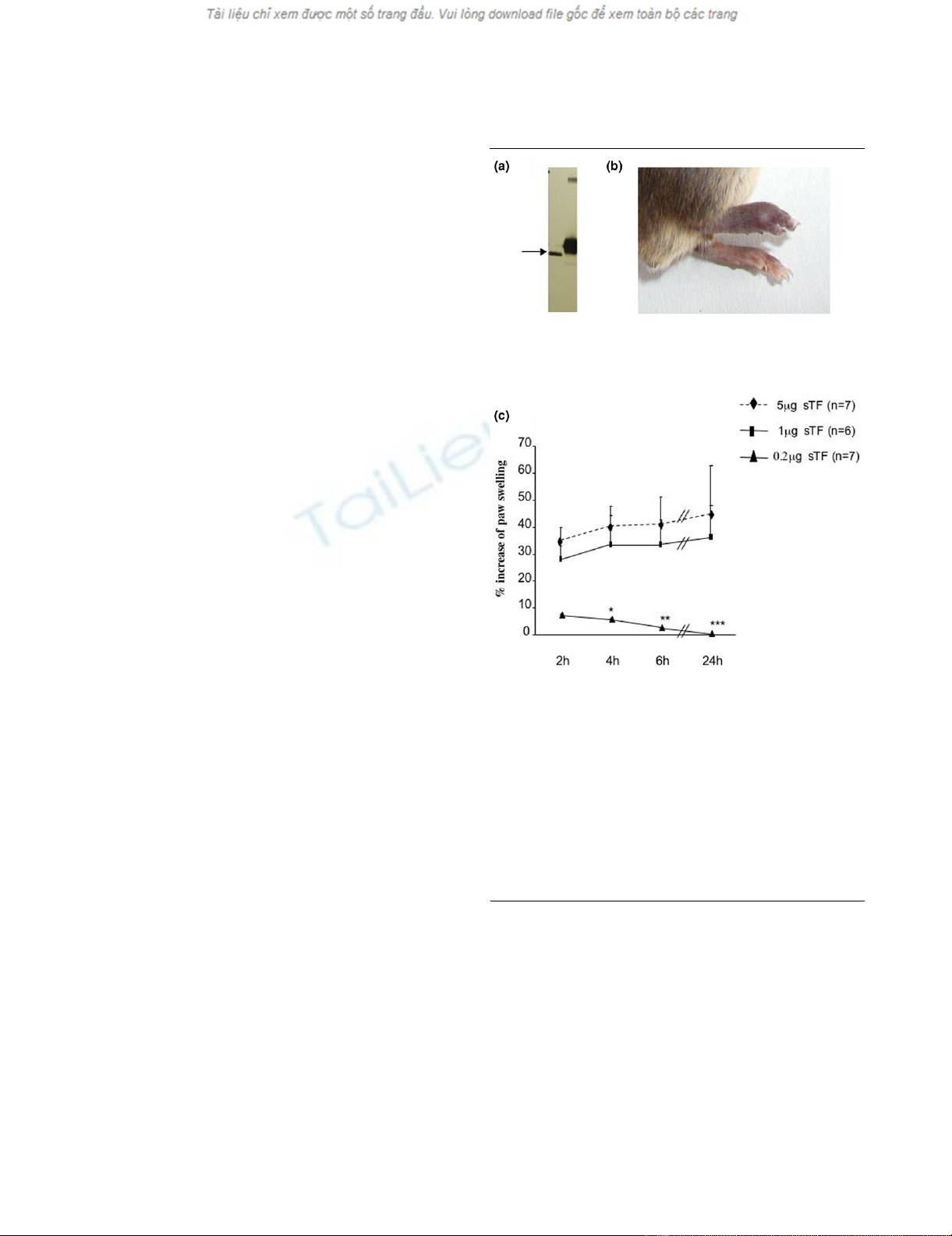
Soluble tissue factor-induced footpad inflammation
We produced a recombinant form of TF, corresponding to the
extracellular domain of murine TF (amino acids 1 to 219). The
soluble recombinant protein migrated as a single band of ≈30
kDa (Figure 1a) and was purified as a single peak on mass
spectrometry (data not shown).
Injection of sTF into the footpad of C57Bl/6 mice provoked an
acute inflammatory response. Edema and erythema developed
rapidly following injection (Figure 1b). The inflammatory
response was quantified by measuring the footpad thickness.
Paw swelling was maximal around 2 to 4 hours after injection
(Figure 1c) and was sustained over 24 hours. Footpad swell-
ing was dose dependent and the maximal effect was observed
at 5 μg/injection. sTF blocked by prior incubation with inacti-
vated FVIIa (ASIS; Novo Nordisk) did not induce footpad
inflammation, thus confirming that it was sTF induced (data not
shown). Serum IL-6 levels confirmed that inflammation was
increased by sTF injection, and this was abrogated in ASIS-
treated animals (Table 1).
Histological analysis showed pronounced edema and cellular
infiltration (Figure 2a). Infiltrating inflammatory cells were pre-
dominantly macrophages (Figure 2c), with some CD3-positive
T cells (Figure 2d). Fibrin staining was prominent (Figure 2f).
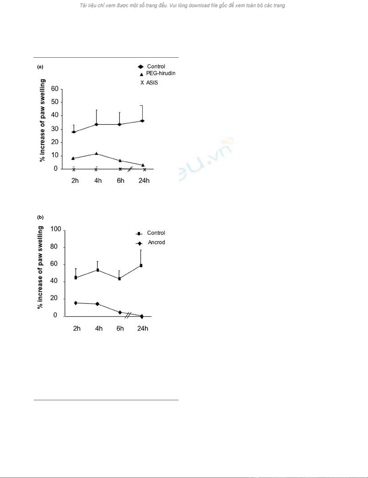
Role of thrombin, factor VII and fibrin
We tested the effects of a specific thrombin inhibitor (PEG-
hirudin), a FVIIa inhibitor (ASIS; Figure 3a), and a defibrinogen-
ating agent (ancrod; Figure 3b) administrated prior to sTF
injection in the same model. All treatments led to a marked
reduction of footpad inflammation (P < 0.05 by t test for all
time points with PEG-hirudin in comparison with wildtype con-
trol injected with phosphate-buffered saline; P < 0.001 for
ASIS, P < 0.01 for ancrod). To assess that sTF was acting via
FVIIa binding, we preincubated sTF in vitro with an excess of
ASIS, hypothesizing that the preformed noncoagulant sTF–
ASIS complex would not be able to induce coagulation upon
its injection in the paw. As expected from the TF/FVIIa-
Figure 1
Soluble tissue factor-induced footpad inflammationSoluble tissue factor-induced footpad inflammation. (a) Western
blot of soluble tissue factor (sTF): 50 ng recombinant murine sTF (track
1) and 50 ng native TF (track 2) were detected using a polyclonal rab-
bit anti human TF antibody. (b) Effect of sTF injected into the footpad: 1
μg sTF (in 10 μl phosphate-buffered saline) was injected into the intra-
plantar region of the right hindfootpad. The contralateral footpad was
injected with the same volume of phosphate-buffered saline. Swelling
was observed in the sTF-injected footpad after 2 hours and was sus-
tained over 24 hours (photograph). (c) Dose-dependent effect of sTF-
induced inflammation: 0.2 to 5 μg sTF in 10 μl was administered into
the hindfootpad. The contralateral footpad was injected with phos-
phate-buffered saline. Results expressed as the percentage increase in
the right over left footpad thickness. *P < 0.05, **P < 0.01 and ***P <
0.001, Wilcoxon rank sum test.
Arthritis Research & Therapy Vol 10 No 2 Busso et al.
Page 4 of 8
(page number not for citation purposes)
To evaluate the effect of these anticoagulation treatments on
thrombin formation, we measured the plasma levels of TAT
complexes in the different groups of mice. The TAT levels were
increased in mice with sTF-injected footpads compared with
sham-injected mice (sTF-injected mice, 31.55 ± 9.62 ng/ml;
sham-injected mice, 8.28 ± 4.8 ng/ml). As expected, ASIS-
treated and PEG-hirudin-treated mice showed reduced TAT
levels (ASIS-treated mice, 8.2 ± 2.9 ng/ml; PEG-hirudin-
treated mice, 14.3 ± 4.5 ng/ml). Serum IL-6 measurements
confirmed the observed anti-inflammatory effect of the treat-
ment administered (Table dependent pathway, no inflamma-
tion of the paw was noticed upon injection of sTF–ASIS
complex (sTF alone, 45 ± 17.5% versus sTF–ASIS complex,
0 ± 0% increase of paw swelling).1).
PAR-4-deficient mice are resistant to soluble tissue
factor-induced inflammation
To explore whether PARs play a role in sTF-induced footpad
inflammation, we injected mice deficient for either PAR-1,
PAR-2, PAR-3 or PAR-4. No differences were observed in
footpad measurements between PAR-1-deficient, PAR-2-defi-
cient and PAR-3-deficient mice when compared with their
control littermates (+/+ or +/-) (Figure 4a to 4c). In contrast,
PAR-4-deficient mice were almost totally resistant to sTF-
induced inflammation compared with their littermates (Figure
4d).
On histological analysis, PAR-4-/- mice showed negligible
signs of edema, hemorrhage and inflammation, and the histol-
ogy was similar to that observed in control mice injected with
vehicle alone (results not shown). We also examined fibrin
deposition in the sTF-injected footpads. Scoring of fibrin dep-
osition was significantly reduced in PAR-4-/- mice compared
with wildtype littermates (Figure 5).
Role of platelets in soluble tissue factor-induced
inflammation
As PAR-4 is predominantly expressed on platelets in mice
[23], we investigated the contribution of platelets to sTF-
induced inflammation. Thrombocytopenia was induced in
wildtype mice by antiplatelet antibody treatment, resulting in a
>98% reduction in the average number of circulating platelets
(Figure 6a). The severity of inflammation was markedly
reduced in mice treated with antiplatelet antibody, whereas
sham-treated mice showed the usual footpad inflammation
(Figure 6b). Histologic observations confirmed the reduction
of footpad swelling in thrombocytopenic mice (results not
shown).
Table 1
Plasma IL-6 levels after soluble tissue factor injection
Untreated mice Soluble tissue factor wildtype mice
No treatment ASIS treatment Hirudin treatment Ancrod treatment
n633555
Mean (pg/ml) <2 285 6.3 <2 <2
Standard error of the mean 107 6
Plasma was collected 24 hours after soluble tissue factor footpad injection. In a parallel experiment, plasma was also collected from noninjected,
naïve control mice. ASIS, active site inhibited activated factor VII.
Figure 2
Footpad histology and immunohistochemistryFootpad histology and immunohistochemistry. Samples were
obtained 24 hours after soluble tissue factor injection. (a) Wildtype
mice showed marked inflammatory changes. (b) Phosphate-buffered
saline-injected mice showed minimal signs of inflammation. (c) Staining
for macrophages was strongly positive. (d) CD3-positive T cells were
also present. (e) Staining specificity was confirmed using, as primary
antibody, nonimmune isotype-matched antibodies. (f) Fibrin deposition
was assessed by fibrin immunohistochemistry.
Available online http://arthritis-research.com/content/10/2/R42
Page 5 of 8
(page number not for citation purposes)
Discussion
Extravascular fibrin deposition is a hallmark of chronic inflam-
mation and plays a role in perpetuating inflammation in rheu-
matoid arthritis, glomerulonephritis and experimental allergic
encephalomyelitis [3,16,25]. TF-initiated coagulation
accounts for fibrin formation and may also trigger inflammation
through the action of downstream coagulation proteases such
as thrombin and FVIIa on PARs. The relative roles of the differ-
ent PARs in mediating inflammation are not well understood,
however, and differing studies have implicated different PARs.
We therefore chose to study the effects of sTF injected into
the mouse footpad and the underlying mechanisms of its
effects.
The injection of recombinant murine sTF into the mouse foot-
pad results in an acute inflammation of extravascular tissues
characterized by footpad swelling, histological signs of
inflammation and fibrin deposition. This effect is mediated
principally by the classical pathway of coagulation activation,
through the formation of thrombin and fibrin, as inflammation
was effectively blocked by administration of the thrombin
inhibitor hirudin and an inhibitor of FVIIa. Depletion of fibrino-
gen by ancrod also attenuated inflammation in this model.
These findings confirm that fibrin formation is an essential step
in the link between coagulation and inflammation.
To determine whether PAR activation can also play a role in
this model, we tested mice deficient for the individual PARs.
To our surprise, only PAR4-deficient mice showed a pheno-
type, in that these mice were completely protected from sTF-
induced inflammation. This contrasts with more chronic mod-
els that require an immune stimulus, such as glomerulonephri-
tis and arthritis, in which PAR1 and PAR2 signaling seem to
play a role [26,27]. Indeed, in antigen-induced arthritis, we
found that TF/FVIIa activates PAR2 and subsequent arthritis –
but in the same conditions, PAR4-deficient mice were indistin-
guishable from wildtype mice [28]. The key role of PAR4 sug-
gested to us that platelet activation may be critical for
inflammation to develop following sTF injection, as PAR-4 is
the main platelet protease receptor in mice. This was con-
firmed when we performed the same experiments on normal
mice that were rendered thrombocytopenic by the administra-
tion of antiplatelet antibody. The fact that PAR-3 had no effect
at all on inflammation suggests that PAR-4 is the main pathway
by which proteases activate mouse platelets in vivo.
One could question the physiological relevance of sTF in
inflammation. Circulating forms of TF have been measured in
different disease states [1,7], although it is likely to be much
less abundant than cell-bound TF. There is much debate
regarding what soluble TF consists of and whether it is biolog-
ically active [29]. A large proportion is probably in the form of
microparticles but an alternatively spliced variant of natural TF
has also been described [30,31]. Both forms have been
reported to possess functional procoagulant activity [6,32],
and increased levels of microparticular TF have been linked to
vascular disease. The proinflammatory effects of the form of
sTF we used in this study (containing amino acids 1 to 219) in
vivo resembled those reported by Bokarewa and colleagues,
who observed a chronic erosive arthritis after injection of a
Figure 3
Role of hirudin, factor VII inhibitor and ancrod in soluble tissue factor-induced footpad inflammationRole of hirudin, factor VII inhibitor and ancrod in soluble tissue fac-
tor-induced footpad inflammation. (a) Wildtype mice treated with
PEG-hirudin (n = 5) or with ASIS (active-site inhibited activated factor
VII, n = 5), or untreated mice (n = 7), were injected with 1 μg soluble
tissue factor. Results from all treated groups were significantly reduced
(P < 0.05 by t test) compared with the control group. (b) Wildtype
mice were treated with ancrod (n = 7) or with phosphate-buffered
saline (n = 7) and then injected with 1 μg soluble tissue factor. Results
from ancrod-treated mice are significantly different from the control
group, at all time points (P < 0.05 by t test).


