
RESEARC H Open Access
Recombinant immunotoxin anti-c-Met/PE38KDEL
inhibits proliferation and promotes apoptosis of
gastric cancer cells
Xu Wei
1
, Zhu Xiao Juan
1
, Feng Xiao Min
2
, Cai Nan
1
, Zhang Xiu Hua
1
, Feng Zheng Qing
2
and Liu Zheng
1*
Abstract
Background: Our study aims to evaluate the anti-growth effects of recombinant immunotoxin (IT) anti-c-Met/
PE38KDEL on gastric cancer cells, and its mechnisms.
Methods: Gastric cancer cells were treated with increasing doses of IT and c-Met protein was quantified by
Western blotting. Cell proliferation was determined by Cell Counting Kit-8 assay (CCK). [
3
H]-leucine incorporation
assay was used to evaluate IT inhibition of protein synthesis. Cell apoptosis was quantified by flow cytometry.
Caspase activities were measured using colorimetric protease assays.
Results: Cell growth and protein synthesis of the gastric cancer cell lines were suppressed by IT in a dose- and
time-dependent manner. IT also induced apoptosis in a dose-dependent manner. The apoptosis rates of gastric
cancer cell lines MKN-45 and SGC7901 were 19.19% and 27.37%, respectively when treated with 50 ng/ml of IT.
There were significant increase ofcaspase-3 activity at 24 hr of IT treatment (100 ng/ml) (P < 0.01) in these gastric
cancer cell lines.
Conclusions: IT anti-c-Met/PE38KDEL has anti-growth effects on the gastric cancer cell lines in vitro, and it provides
an experimental basis for c-Met-targeted therapy towards in vivo testing.
Introduction
Gastric carcinoma (GC) is one of the most common and
lethal malignant cancers [1]. Despite the improving sur-
gical techniques and new chemotherapeutic treatment
regimens, the patient survival rate remains dismal [2],
and effective alternative treatment approach is in vital
need. GC has been shown to harbor multiple somatic
mutations as well as over-expressions of oncoproteins.
Identification of these GC-associated biomarkers may
entail possible discovery of new therapeutic targets [3].
Among various GC-associated biomarkers, c-MET gene
is frequently found gnomically-amplified and over-
expressed in GC cell lines [4]. The proto-oncogene c-
MET, a receptor of hepatocyte growth factor (HGF, also
known as scatter factor), encodes a 190 kDa heterodi-
meric transmembrane tyrosine kinase. HGF binding to
c-Met triggers tyrosine kinase domain auto-
phosphorylation and induces pleiotropic responses such
as proliferation, motility, morphogenesis and angiogen-
esis in many cell types including normal and tumor cells
[5]. c-MET amplification has been identified in nearly
74% of human GC specimens [6]. HGF and c-MET both
play important roles in the progression and metastasis
of GC [7]. Thus, c-Met has been considered as a pro-
mising therapeutic target for various cancers.
Immunotoxins (ITs) are fusion proteins composed of a
toxin fused to an antibody or growth factor with distinct
target specificity [8]. IT exerts its anti-growth effect by
inhibiting protein synthesis and promoting apoptosis [9].
IT anti-c-Met/PE38KDEL (anti-c-Met Fab, which
resulted from screening and characterization from a nat-
ural human Fab phage antibody library; PE38KDEL,
which is a modified structure of PE38, lost the function
of combining with non-mammalian cells specifically, but
retained a complete cytotoxicity after internalization)
has shown specific cytotoxic effects against c-Met-posi-
tive cancer cells [10]. In this study, we investigated the
effects of IT anti-c-Met/PE38KDEL on proliferation and
* Correspondence: liuzheng117@126.com
1
Department of Gastroenterology, The Second Affiliated Hospital of Nanjing
Medical University, Nanjing, 210029, PR China
Full list of author information is available at the end of the article
Wei et al.Journal of Experimental & Clinical Cancer Research 2011, 30:67
http://www.jeccr.com/content/30/1/67
© 2011 Wei et al; licensee BioMed Central Ltd. This is an Open Access article distributed under the terms of the Creative Commons
Attribution License (http://creativecommons.org/licenses/by/2.0), which permits unrestricted use, distribution, and reproduction in
any medium, provided the original work is properly cited.

apoptosis of two different c-Met-positive malignant gas-
tric cell lines, MKN-45 and SGC7901 [11,12], and a nor-
mal gastric mucosa cell GES-1 [13]. We found that IT
anti-c-Met/PE38KDEL exerts its anti-growth effect pri-
marily through rapid inhibition of protein synthesis.
Materials and Methods
Immunotoxin
IT anti-c-Met/PE38KDEL was described previously [9]. It
induces apoptosis in hepatic carcinoma cells SMMC7721.
Cell Counting Kit 8 (CCK8) was purchased from Sigma
Chemical. Caspase colorimetric assay kit and anti-caspase-
3 antibody were from Biovision. Antibodies against c-Met
and b-actin were purchased from Santa Cruz. Protein lysis
buffer was from TaKaRa Biotechnology.
Cell culture
GC cells lines, MKN-45 and SGC7901, and normal gas-
tric mucosa cells GES-1 were obtained from the Cell
Bank of Type Culture Collection of the Chinese Acad-
emy of Sciences (Shanghai, China), and were grown in
DMEM (Invitrogen) supplemented with 10% fetal calf
serum (FCS) and incubated at 37°C with 5% CO
2
.All
cell lines were routinely tested and found to be free
from mycoplasma contamination.
Western Blotting
GES-1, MKN-45 and SGC7901cellsgrownin6-well
plates were collected in lysis buffer for total cellular pro-
tein. Protein concentrations were measured using a
Bradford reagent (Bio-Rad). Equal amounts of protein
(80 μg/lane) from each cell line were boiled for 5 min,
separated by SDS-PAGE, and then transferred on to a
nitrocellulose membrane before blocking in 5% non-fat
dried milk in Tris-buffered saline (TBS) for 120 min at
room temperature. The membranes were then incubated
with a primary anti-human c-Met polyclonal antibody
(diluted 1:150 in a new batch of the blocking buffer) or
a goat polyclonal primary anti-b-actin (diluted 1:1000,
Santa Cruz, CA, USA) for 2 hr and followed by incuba-
tion with peroxidase-labelled anti-IgG secondary anti-
body for 1 hr. After washing with TBST for 3 times, the
films were developed and the protein bands were quan-
tified by densitometry using ImageJ software (NIH,
Bethesda, MD, USA).
To detect the caspase-3 activity, both floating and
adherent cells were collected 24 hr following IT treat-
ment. Total cellular protein was prepared as described
above. All the experiments were performed at least
twice with similar results.
Cell proliferation assay
Cell growth inhibition rate (IR) was determined using a
CCK- 8 assay following the manufacturer instructions
(Sigma). GES-1, MKN-45 and SGC7901 cells were
seeded at a concentration of 1 × 10
5
cells/90 μl/well in
96-well culture plates. After incubation of cells with the
indicated concentrationsofITfor24hrand48hr,10
μl/well of cell Counting Kit-8 solution was added to the
medium and the cells were incubated for an additional
4 hr. The absorbance at 450 nm was then measured in a
Microplate Reader. IR was calculated using the following
equation: IR = [1-(Avalue in the treated samples-A
value in the blank samples) / (Avalue in the control
samples-Avalue in the blank samples)] *100%. The
assays were performed in triplicates and repeated at
least twice [14].
Protein synthesis inhibition assay
IT-induced inhibition of protein synthesis in GES-1,
MKN-45 and SGC7901 cells were evaluated using the
[
3
H]-leucine incorporation assay [15]. Cells were seeded
in 48-well plates (1 × 10
4
per well) and allowed to grow
overnight before the addition of IT at different concen-
trations. After 5 or 24 hr incubation, cells were washed
twice with cold phosphate-buffered saline (PBS) contain-
ing 0.1% FCS, and then incubated with [
3
H]-leucine (2
μCi ml
-1
) in leucine-free medium at 37°C for 45 min.
Cells were then washed with 5% trichloroacetic acid
(TCA) for 5 and 10 min, respectively, and dissolved in
0.1M KOH for 10-15 min. The resultant solution was
transferred to the liquid scintillator. Sample counts were
determined in a liquid scintillation counter. Assays were
performed in duplicates and repeated at least three
times. Counts per minute (cpm) for treated cells were
compared to cpm for untreated cells and reported as a
percentage of leucine incorporation with the control
value set to 100%[16]. The experiment was completed in
the isotope laboratory of Nanjing Medical University.
Flow cytometric analysis of cell apoptosis
Apoptosis were determined by flow cytometric analysis.
Briefly, cells in triplicates, were incubated with or with-
out various concentrations of IT for 24 hr. Cells were
then harvested, washed in cold PBS, and fixed with 1 ml
75% ice-cold ethanol at -20°C until processing. An ali-
quot (1 ml) of fixed cell suspension containing 1 × 10
6
cells was washed twice in cold PBS and then treated
with fluorochrome DNA staining solution (1 ml) con-
taining 40 μg of propidium iodide and 0.1 mg of RNase
A in the dark at room temperature for 0.5 hr. Flow
cytometric analysis were performed three times [17].
Caspase activity assay
Caspase activity was determined in 96-well plates using
cell lysates from 1 × 10
6
cells for each measurement.
Caspase-3 and caspase-8 activities were determined
using colorimetric assay kits according to the
Wei et al.Journal of Experimental & Clinical Cancer Research 2011, 30:67
http://www.jeccr.com/content/30/1/67
Page 2 of 7
manufacturer’s protocol (BioVision). GES-1, MKN-45
and SGC7901 cells were treated with anti-c-Met/PE38K-
DEL (100 ng/ml) for 24 hr prior to the assay. Cell
extracts were incubated with 5 μl of 4 mM tetrapeptide
substrates (DEVD, caspase-3; IETD, and caspase-8) at
37°C for 1-2 hr. The reaction was measured at 405 nm
in a Microplate Reader. Background readings from cell
lysates and buffers were subtracted from the readings of
both IT-induced and control samples before calculating
the relative change increase in caspase activity in the
IT-induced samples compared to that of the control. IT
treated samples were normalized to the caspase activity
of the untreated sample, which was set to 1.0. Fold of
increases in caspase activities were presented.
Statistical analysis
Statistical analysis was performed with SPSS 13.0 soft-
ware. Data were presented as mean ± standard devia-
tion. Student’s t-test was used to compare two samples,
and the single-factor analysis of variance (One-way
ANOVA) was used to compare multiple samples. A p-
value less than 0.05 is considered statistically significant
(*, p < 0.05; **, p < 0.01).
Results
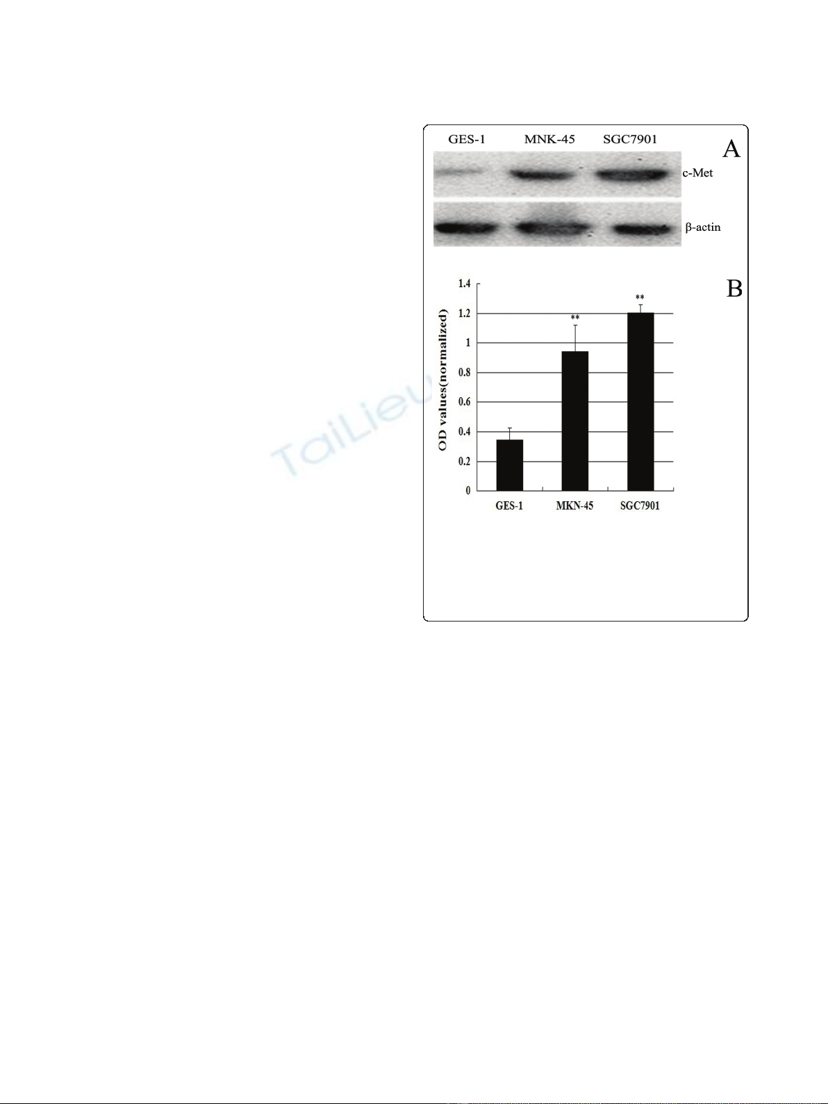
Increased c-Met expression in MKN-45 and SGC7901 cells
To determine the c-Met protein expression levels in GC,
we used western blotting to examine c-Met protein in
two GC cells (MKN-45 and SGC7901) and one normal
gastricmucosacellsGES-1(Figure1A).c-Metproteins
is 3-4 fold higher in MKN-45 and SGC7901cells than
GES-1 cells. SGC7901 cells express slightly more c-Met
than MKN-45 cells (Figure 1B). The optical densities
(OD’s) of the Western blot bands were measured using
ImageJ. The OD for each band was normalized to b-
actin. MKN-45 and SGC7901 had a 0.94 and 1.27 fold
increase in the expression of c-Met over the control, but
only 0.34 fold increased in GES-1.
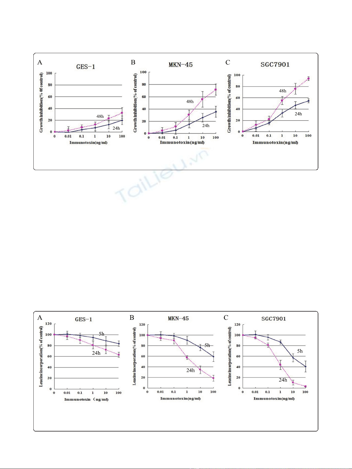
IT anti-c-Met/PE38KDEL inhibited cell proliferation and
protein synthesis
GC cells have significantly higher c-Met protein levels
than normal gastric mucosa cells, therefore we tried to
determine if IT anti-c-Met/PE38KDEL has GC-specific
effects. The anti-proliferative effect of IT anti-c-Met/
PE38KDEL on GES-1, MKN-45 and SGC7901 cells
was measured using CCK8 kit. Cells were harvested at
24 or 48 hr after IT treatment. As shown in Figure 2,
IT inhibited GC cell growth in a time- and dose-
dependent manner. 1, 10 and 100 ng/ml of IT caused
a dramatic growth inhibition in MKN-45 and
SGC7901 cells (P< 0.01). 48 hr of IT treatment (100
ng/ml) resulted in a growth inhibition of 30% in GES-
1 cells (Figure 2A). However, inhibitions of 75% and
95%wereobservedinMKN-45andSGC7901cells
(Figure 2B and 2C), respectively. Further, we found
that there is a strong correlation between c-Met
expression and in vitro immunotoxin efficacy.
Given the high c-MET levels in MKN-45 and
SGC7910 cell lines, we hypothesize that anti-c-Met/
PE38KDEL can attenuate cancer cell growth through
inhibition of protein synthesis via c-Met inhibition.
The effects of anti-c-Met/PE38KDEL on protein
synthesis in GES-1, MKN-45 and SGC7901 cells are
showninFigure3.TheIT’sIC
50
value on GES-1 cells
was approximately 120 ng/ml. However, IT induced
more potent inhibitions of protein synthesis in MKN-
45 and SGC7901 cells, with IC
50
values of 5.34 ng/ml
and 0.83 ng/ml, respectively. Nearly 80% and 100% of
inhibitions were observed with 100 ng/ml of IT treat-
ment in these two GC cells (Figure 3B and 3C). In
contrast, 100 ng/ml of IT only caused a 35% decrease
in protein synthesis in GES-1 cells (Figure 3A). These
results suggested that anti-c-Met/PE38KDEL can
attenuate cell growth through the inhibition of protein
synthesis.
Figure 1 Overexpression of c-Met in castric carcinoma cell
lines. Lysates (80 μg/lane) from normal gastric mucosa cells GES-1
and GC cell lines MKN-45 and SGC7901 were analyzed for c-Met
protein level by western blot using an anti-c-Met antibody and an
anti- b-actin antibody (loading control) (Figure 1A). The optical
densities (OD’s) of the Western blot bands were measured using
Image J (Figure 1B).
Wei et al.Journal of Experimental & Clinical Cancer Research 2011, 30:67
http://www.jeccr.com/content/30/1/67
Page 3 of 7
IT anti-c-Met/PE38KDEL inhibits tumor cell growth
through induction of apoptosis
To determine whether the anti-proliferative effect of IT
was due to cell apoptosis, we used flow cytometric
(FCM)) to further determine if IT induces cell apoptosis.
As shown in Figure 4A and 4B, apoptotic rates in MKN-
45 and SGC7901 cells were increased from 1.89% and
2.4% (0 ng/ml), to 19.19% (P < 0.01) and 27.37% (P <
0.01) (50 ng/ml), respectively. The apoptosis rate of
GES-1 cells is significantly lower than two GC cells
(5.98%, P < 0.01) at the IT dose of 50 ng/ml. These data
indicate that anti-c-Met/PE38KDEL induced apoptosis
in GC cells.
IT anti-c-Met/PE38KDEL activates caspase-3
To determine whether apoptotic pathway is activated by
IT in GC cells, we measured caspase-3 and caspase-8
activities following IT treatment. As shown in Figure 5B
and 5C, MKN-45 and SGC7901 cells showed 3.70 and
5.02 fold of increases in caspase-3 enzyme activity as
compared to untreated controls after 24 hr IT treatment
(P < 0.01). GES-1 exhibited a 2.03-fold increase in cas-
pase-3 enzyme activity (P < 0.05) (Figure 5A). Caspase-8
enzyme activity in two GC cell lines also increased (P <
0.05), suggesting caspase-3 activation mediates IT anti-
c-Met/PE38KDEL-induced biological effects.
The caspases are synthesized as inactive precursors
(zymogens) that are proteolytically processed to generate
active subunits by cleaving specific aspartic acid residues
[18], and are essential for the execution process of apopto-
sis as effector proteases [19]. In the process of IT-inducd
apoptosis, caspase-3 appeared to play a role. We investi-
gated whether caspase-3 is regulated in anti-c-Met/
PE38KDEL-induced cell death. As shown in Figure 6,
Figure 3 Anti-c-Met/PE38KDEL induced inhibition of protein synthesis. The ability of IT to inhibit protein synthesis in GES-1, MKN-45 and
SGC7901 cells were evaluated by using the [
3
H]-leucine incorporation assay. [
3
H]-leucine incorporation for protein synthesis as a function of
varying concentration of IT (expressed as a percentage of untreated cells), Normal cell GES-1 (A), GC cells MKN-45 (B) and SGC7901 (C) were
treated with varying concentration of IT for 24 hr and 48 hr.
Figure 2 IT anti-c-Met/PE38KDEL induced inhibition of cell proliferation. Cell growth inhibition as a function of varying concentrations of IT
(expressed as a percentage of untreated cells), Normal cell GES-1 (A), GC cells MKN-45 (B) and SGC7901 (C) were treated with various
concentrations of IT for 24 hr and 48 hr.
Wei et al.Journal of Experimental & Clinical Cancer Research 2011, 30:67
http://www.jeccr.com/content/30/1/67
Page 4 of 7
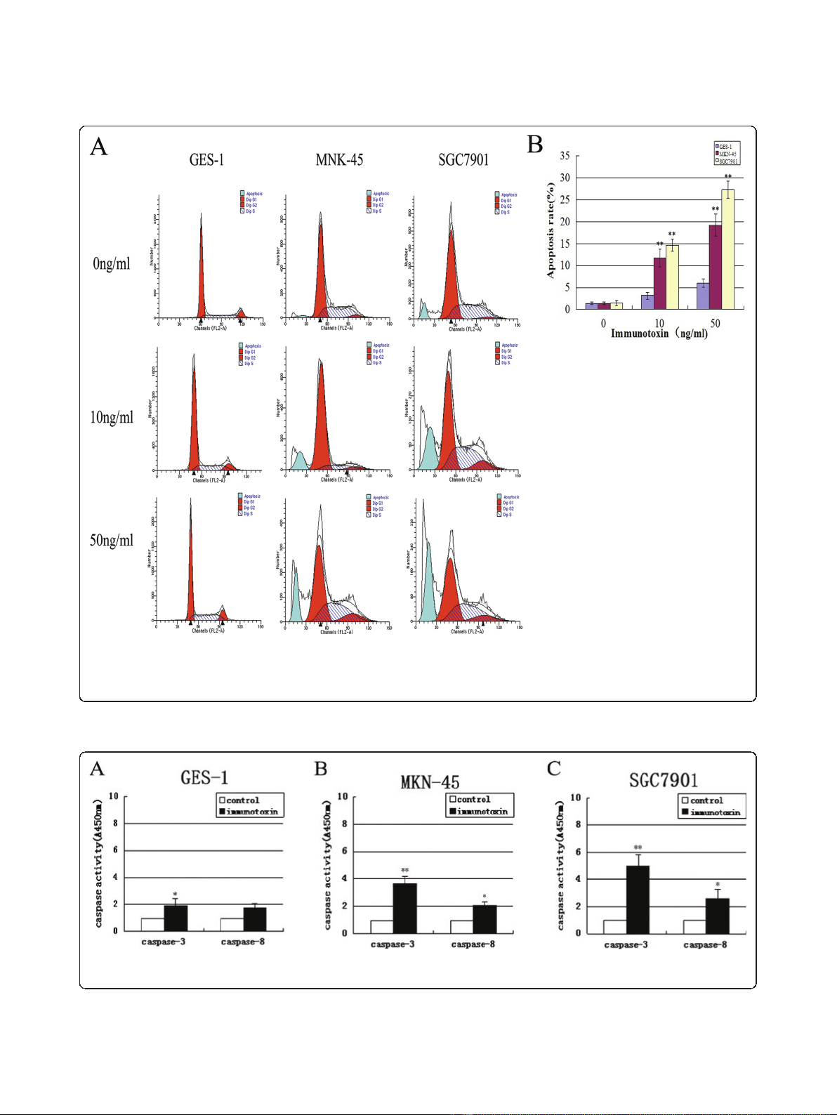
Figure 4 IT anti-c-Met/PE38KDEL inhibited tumor cell growth through induction of apoptosis. To measure the dose response effect of IT
on cell apoptosis rate of GES-1, MKN-45 and SGC7901, cells were treated with different concentrations of anti-c-Met/PE38KDEL. Cells were
incubated with IT at 0, 10 and 50 ng/ml for 24 hr, and the percentage of cell apoptosis was determined by flow cytometry. IT induced apoptosis
for its anticancer effect.
Figure 5 IT anti-c-Met/PE38KDEL mainly activates caspase-3. Caspase-3 and caspase-8 activities in GES-1 (A), MKN-45 (B) and SGC7901 (C)
cells were measured in control or IT-treated cells (immunotoxin) (24 hr) using the Caspase colorimetric assay kit. * P < 0.05, **P < 0.01.
Wei et al.Journal of Experimental & Clinical Cancer Research 2011, 30:67
http://www.jeccr.com/content/30/1/67
Page 5 of 7


![Vaccine và ứng dụng: Bài tiểu luận [chuẩn SEO]](https://cdn.tailieu.vn/images/document/thumbnail/2016/20160519/3008140018/135x160/652005293.jpg)


