11/1/2014 1
CHAÅN ÑOAÙN HÌNH AÛNH
BEÄNH TIM MAÉC PHAÛI KHAÙC
BS.NGUYEÃN QUYÙ KHOAÙNG
BS.NGUYEÃN QUANG TROÏNG
11/1/2014 2
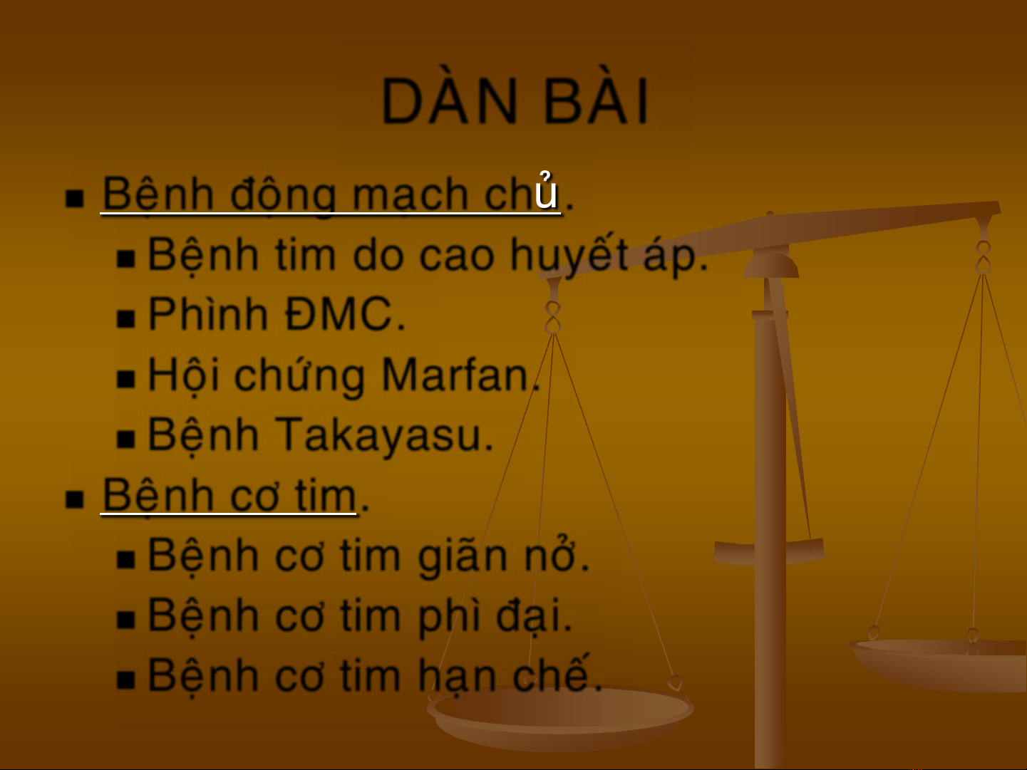
DAØN BAØI
Beänh ñoäng maïch chuû.
Beänh tim do cao huyeát aùp.
Phình ÑMC.
Hoäi chöùng Marfan.
Beänh Takayasu.
Beänh cô tim.
Beänh cô tim giaõn nôû.
Beänh cô tim phì ñaïi.
Beänh cô tim haïn cheá.
11/1/2014 3
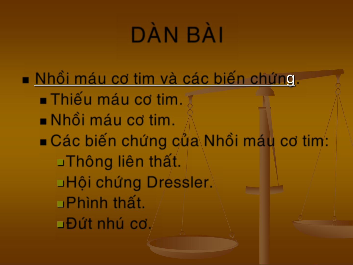
DAØN BAØI
Nhoài maùu cô tim vaø caùc bieán chöùng.
Thieáu maùu cô tim.
Nhoài maùu cô tim.
Caùc bieán chöùng cuûa Nhoài maùu cô tim:
Thoâng lieân thaát.
Hoäi chöùng Dressler.
Phình thaát.
Ñöùt nhuù cô.
11/1/2014 4
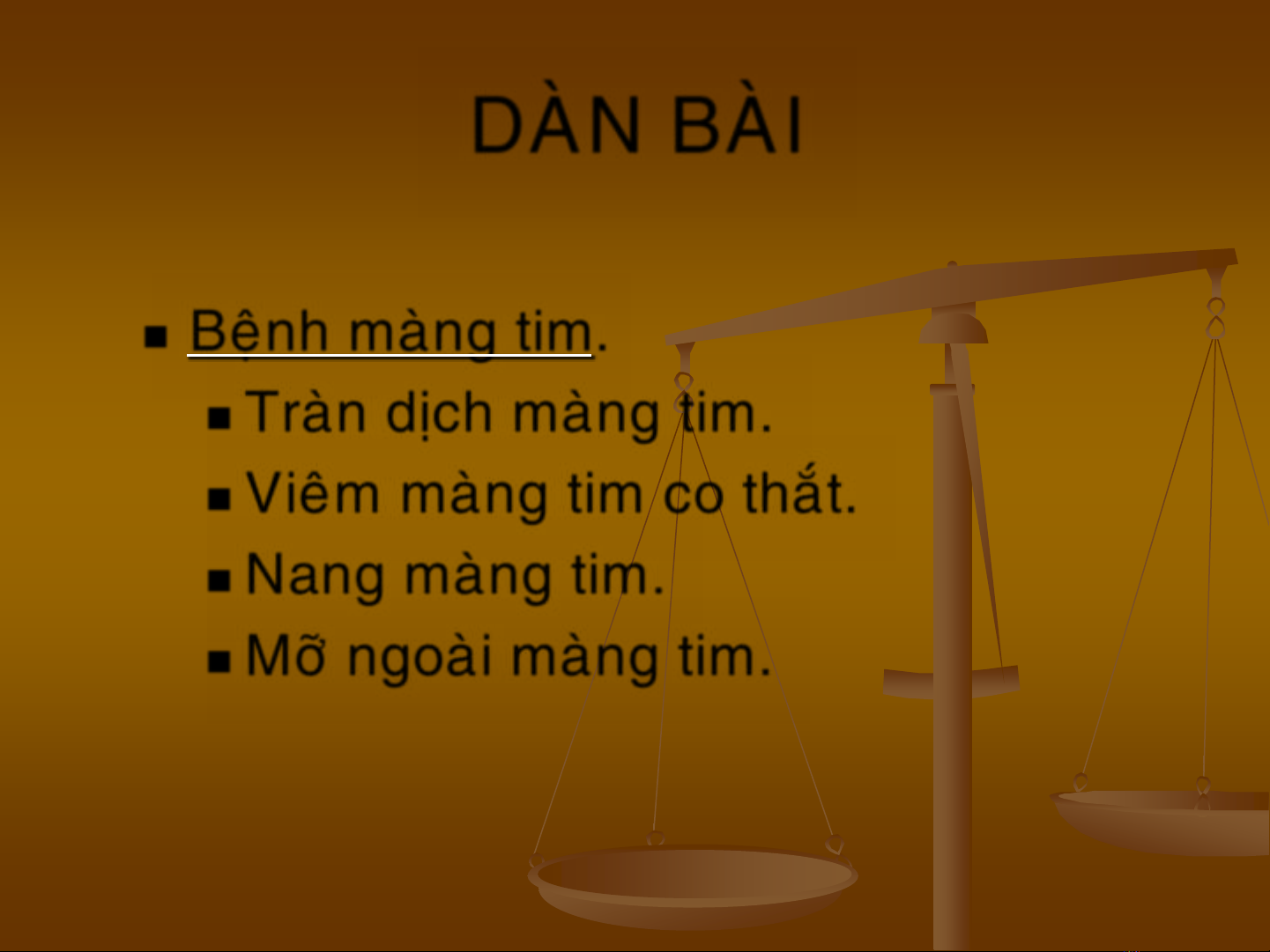
DAØN BAØI
Beänh maøng tim.
Traøn dòch maøng tim.
Vieâm maøng tim co thaét.
Nang maøng tim.
Môõ ngoaøi maøng tim.
11/1/2014 5
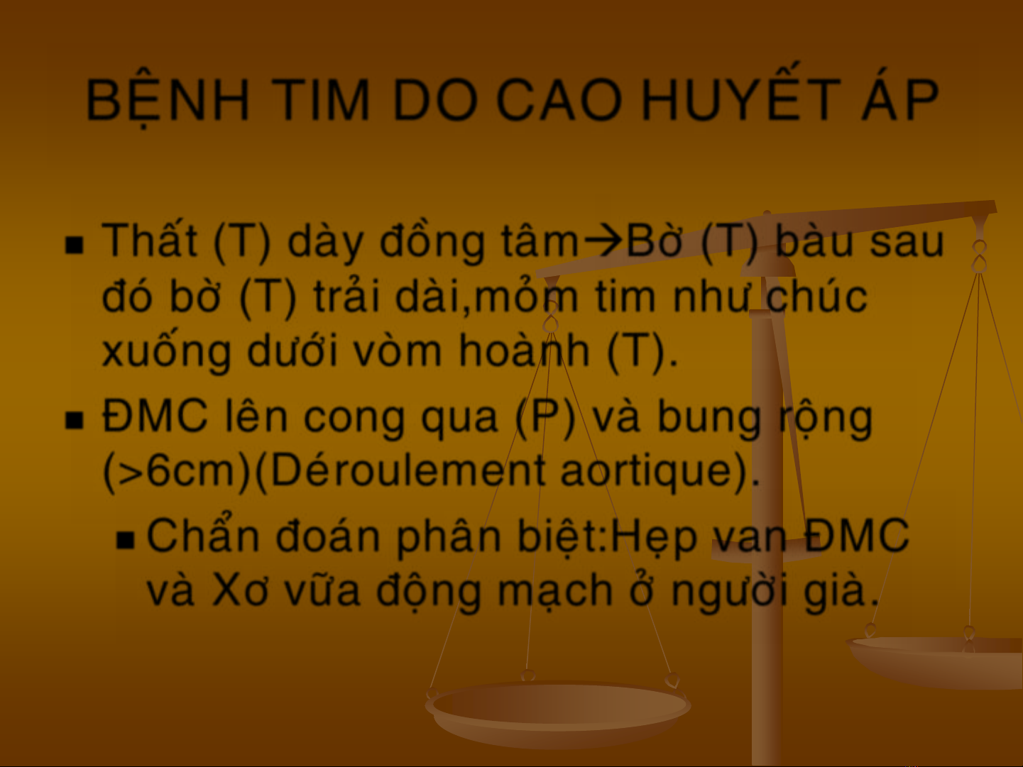
BEÄNH TIM DO CAO HUYEÁT AÙP
Thaát (T) daøy ñoàng taâmBôø (T) baøu sau
ñoù bôø (T) traûi daøi,moûm tim nhö chuùc
xuoáng döôùi voøm hoaønh (T).
ÑMC leân cong qua (P) vaø bung roäng
(>6cm)(Deùroulement aortique).
Chaån ñoaùn phaân bieät:Heïp van ÑMC
vaø Xô vöõa ñoäng maïch ôû ngöôøi giaø.


![Giáo trình Chẩn đoán hình ảnh [chuẩn nhất]](https://cdn.tailieu.vn/images/document/thumbnail/2025/20251019/syan5050@gmail.com/135x160/15031761021299.jpg)


![Tài liệu về Hội chứng chèn ép khoang [chuẩn nhất]](https://cdn.tailieu.vn/images/document/thumbnail/2025/20250806/tuhaolg/135x160/27201754535086.jpg)