
ATP N-glycosidase
A novel ATP-converting activity from a marine sponge
Axinella polypoides
To
˜nu Reintamm, Annika Lopp, Anne Kuusksalu, To
˜nis Pehk and Merike Kelve
Laboratory of Molecular Genetics, National Institute of Chemical Physics and Biophysics, Tallinn, Estonia
A novel nucleosidase enzymatic activity was discovered in
the marine sponge Axinella polypoides. This enzyme, desig-
nated as ATP N-glycosidase, converts adenosine-5¢-tri-
phosphate into adenine and ribose-5-triphosphate. The
crude extract of A. polypoides was capable of hydrolysing
25 lmol ATPÆmin
)1
per g wet weight of sponge. The cata-
lytic activity of a sponge crude extract per mg total protein is
comparable with specific activities of purified plant adeno-
sine and bacterial AMP nucleosidases. The preferred sub-
strate for the novel enzyme is ATP but any compound
containing adenosine-5¢-diphosphoryl fragment is also
cleaved. The biochemical properties (K
m
,K
ip
, environmental
requirements) of ATP N-glycosidase show similarities with
previously described adenine-specific nucleosidases; how-
ever, the pattern of its biochemical characteristics does not
match with that of any of those enzymes.
Keywords: adenosine nucleotide metabolism; ATP; Axinella
polypoides; marine sponge; nucleosidase.
Most of the biological and chemical literature concerning
marine sponges is primarily dedicated to the isolation and
characterization of exotic secondary metabolites and studies
of their biological activity (antibacterial, antifungal, anti-
cancer, etc.) [1]. These works have been rooted and inspired
by the discovery of unusual nucleosides in Cryptotethya
crypta-arabinothymidine and -uridine [2] which have led to
the development of pharmaceuticals with antiviral and
anticancer action. We have shown the presence of 2¢,5¢-
oligoadenylates (2-5A) in a marine sponge Geodia cydonium
[3]. The synthesis of 2-5A from ATP in sponges proceeds
independently from dsRNA [4], in contrast with higher
animals (birds and mammals) [5]. There is an evolutionary
gap in occurrence of this signal molecule between the
sponges and birds, as no 2-5A synthetase genes have been
found in completed insect, worm and fish genomes [6,7].
In the present study, a completely novel and unexpected
ATP-utilizing activity in Axinella polypoides was found. The
enzymatic activity, cleaving the most abundant high-energy
nucleotide (ATP) into a free nucleobase without touching
the energy–charge-carrying triphosphate moiety, seems to
be in conflict with the current understanding of nucleotide
utilization, salvage and catabolism in nature.
The capacity of the A. polypoides crude extract to utilize
ATP in yet an undescribed direction is impressive. Its rate
could be compared with the rate of ATP turnover in human
muscle [8] and it masks any other ATP-utilizing activity
potentially present in natural crude extracts. Such a
fortunate circumstance enabled us to characterize the novel
activity enzymatically without purification or enrichment of
the crude extract. Substrate preferences and factors deter-
mining the reaction rate in the physiological concentration
rangewerestudied.
Whether the newly discovered enzyme, ATP N-glycosi-
dase, participates in the purine nucleotide salvage pathway,
regulation of cellular adenylate levels, signalling, or other
mechanisms, remains to be established.
Materials and methods
Reagents and enzymes
Reagents and enzymes were purchased from commercial
suppliers (Sigma, Fluka, Reanal, Fermentas, USB Cor-
poration), except for those mentioned below. pppA2¢p5¢A
was enzymatically synthesized by Geodia cydonium
2¢,5¢-oligoadenylate synthetase [4]. c-P-(4-amino-n-butyl-
amido)adenosine-5¢-triphosphate (DAB-ATP) and (5¢,5¢¢)-
diadenosine(a,x)-oligophosphates (A5¢p
n
5¢A, n¼2–5)
were chemically synthesized according to the published
Correspondence to M. Kelve, Laboratory of Molecular Genetics,
National Institute of Chemical Physics and Biophysics, Akadeemia tee
23, 12618 Tallinn, Estonia. Fax: +372 6398382. Tel.: +372 6398352,
E-mail: merike@kbfi.ee
Abbreviations: DAB-ATP, c-P-(4-amino-n-butylamido)adenosine-
5¢-triphosphate; cADPR, cyclic ADP-ribose; cADPRP, cyclic ADP-
ribose 2¢-phosphate; ADPR, b-P-(5-ribosyl) adenosine-5¢-diphosphate
(ADP-Ribose); ATPR, c-P-(5-ribosyl) adenosine-5¢-triphosphate;
ATePR, d-P-(5-ribosyl) adenosine-5¢-tetraphosphate; APPR,
e-P-(5-ribosyl)adenosine-5¢-pentaphosphate; FDPR, b-P-(ribosyl)-
lactoflavin-5¢-diphosphate; MTA, 5¢-methylthio-5¢-deoxyadenosine;
SAH, S-adenosylhomocysteine; Ado, adenosine; 2–5 A, 5¢-tri
(di-, mono-)phosphorylated (2¢,5¢)oligoadenylates; (2¢,5¢)p
3
A
n
,
5¢-triphospho(2¢,5¢)oligoadenylates; (2¢,5¢)A
n
,(2¢,5¢)oligoadenylates;
A5¢p
n
5¢A, P
1
,P
n
-bis(5¢-adenosyl)oligophosphates; NDPR,
b-P-(5-ribosyl)-1-b-
D
-ribofuranosylnicotinamide)5¢-diphosphate.
Enzymes: snake venom phosphodiesterase (EC 3.1.15.1); alkaline
phosphatase (EC 3.1.3.1); ribonuclease U2 (EC 3.1.27.4); purine
nucleosidase (EC 3.2.2.1); 5¢-methylthioadenosine/S-adenosylhomo-
cysteine (MTA/SAH) nucleosidase (EC 3.2.2.9, EC 3.2.2.16); AMP
nucleosidase (EC 3.2.2.4); adenosine nucleosidase (EC 3.2.2.9);
ADP ribosyl cyclase (EC 3.2.2.5).
(Received 13 June 2003, revised 18 August 2003,
accepted 26 August 2003)
Eur. J. Biochem. 270, 4122–4132 (2003) FEBS 2003 doi:10.1046/j.1432-1033.2003.03805.x

methods [9,10]. Phosphodiesterase from the snake venom
(Vipera lebetina) was a gift from J. Siigur (National Institute
of Chemical Physics and Biophysics, Tallinn, Estonia).
Natural sponge material
The marine sponges A. polypoides (Porifera, Demospong-
iae, Ceractinomorpha, Halicondrida, Axinellidae) were
collected near the Kalymnos Island (Greece). The material
was kept in natural seawater during the transportation
(< 24 h). Then it was frozen in liquid nitrogen and stored
at )70 C. All experiments, if not otherwise stated, were
performed using this material.
The alternative sample of A. polypoides was generously
provided by W.E.G. Mu
¨ller (Johannes Gutenberg-Universita
¨t,
Mainz, Germany) from his sponge collection (stored at
)70 C). The air-dried powder of A. polypoides was provided
by W. Schatton (Klinipharm GmbH, Frankfurt, Germany).
Preparation of sponge extracts and their characterization
The sponge material, which had been mechanically pow-
dered and thoroughly mixed at liquid nitrogen temperature,
was used for the extraction of total RNA, the low molecular
weight nucleotides and enzymes. The total RNA from a
sample of A. polypoides was prepared and analysed by the
Chomczynski method [11]. Low molecular mass nucleotides
were extracted with 5% trichloroacetic acid (7 mLÆg
sponge
)1
). The appropriately diluted trichloroacetic acid
extract (5%) was analysed by HPLC and the ATP content
was measured by the luciferase assay [12].
An extract with a maximal yield of ATP N-glycosidase
activity and stable in storage was obtained using an
extraction buffer, containing ‡100 m
M
KCl. All of the
experiments described in the current work were performed
using the single extract (hereafter referred to as crude
extract), which was prepared as follows. Two-hundred
milligrams of the sponge powder (made from frozen sponge
pieces from different body parts of several individuals
collected from the same geographical location; each piece
0.5 g, total mass 5 g) was extracted with 0.1
M
Mops
pH 6.7, containing 0.1
M
KCl (1200 lL) at room tempera-
ture for 30 min. The insoluble material was removed by
centrifugation and 1100 lL of solution was collected. The
protein content was estimated by the Bradford method [13].
The crude extract was kept unfrozen at 4 C. The specific
activity of the crude extract quantified by standard assay in
parallel to each kinetic series yielded average deviation of
7.5%. No statistically significant decrease in the specific
activity of this preparation was found throughout the
biochemical characterization period (2 months).
HPLC analysis
All HPLC analyses were performed, using the C18
HPLC column (5 lm, 4.6 ·250 mm, Supelco, USA) and
the Waters Model 600 chromatograph with a tunable
wavelength detector (Model 486), controlled by the
MILLE-
NIUM
32 software (Waters, USA). Eluent A was 50 m
M
ammonium phosphate pH 7.0 and eluent B was 50%
methanol in water. The flow rate was 1 mLÆmin
)1
and
the column temperature was 40 C. The products were
separated and analysed in a linear gradient of eluent B (1–
60%, 30 min); the column was equilibriated with 1% eluent
B before the next injection (10 min). Fast isocratic separa-
tions (8 or 20% of eluent B, 15 or 10 min) were used in
the routine kinetic point analysis in appropriate cases.
Retention times (min) of the adenosine nucleotide deriva-
tives are listed in an ascending order: cADPRP (2.49),
ADPRP (2.68), NDPR (2.89), unknown cADPR derivate
(3.18), APPR (3.21), ATePR (3.35), ATP (3.60), ATPR
(3.70), ADP (3.8), NADP + (3.84), 5¢-AMP (4.00), cADPR
(4.28), DAB-ATP (4.60), (2¢,5¢)p
3
A
2
(4.6), ADPR (4.80),
dATP (5.58), dADP (6.52), (2¢,5¢)p
3
A
3
(6.60), A5¢p
5
5¢A
(6.70), 3¢-AMP (7.8), A5¢p
4
5¢A (8.0), dAMP (8.2), (2¢,5¢)p
3
A
4
(8.95), A5¢p
3
5¢A (9.1), Ade (9.24), NAD
+
(9.6), nicotin-
amide (10.4), (2¢,5¢)p
3
A
5
(10.46), (2¢,5¢)p
3
A
6
(11.25),
(2¢,5¢)p
3
A
7
(11.75), A5¢p
2
5¢A (12.44), NADH (12.5),
2¢-AMP (12.7), (2¢,3¢)cAMP (14.7), Ado (16.8), (3¢,5¢)cAMP
(17.1), (2¢,5¢)A
5
(18.0), (2¢,5¢)A
4
(18.4), (2¢,5¢)A
2
(18.9),
(2¢,5¢)A
3
(19.0), poly(A) (21.48), (3¢,5¢)A
3
(24.43), FDPR
(26.3), FAD (27.9). The set of adenylate retention times
has been derived from the chromatograms, which were
internally or externally calibrated with ATP (3.6 ± 0.05)
and Ado (16.8 ± 0.3).
Whenever possible, both the substrate and the product
were quantified for the calculation of the reaction yield to
exclude the partial loop filling method related error (10%).
The HPLC raw data were recalculated according to different
molar absorption coefficients of adenine and the substrates.
ATP N-glycosidase assay
Summing up the knowledge obtained during the work, a
simple procedure was developed for the A. polypoides ATP
N-glycosidase quantification.
Fifteen microlitres of 1
M
KCl, 20 lL5m
M
ATP,
pH 7.0 (25 C), 10 lL200m
M
Mes, pH 5.3 (25 C) and
50 lL deionized water were mixed and equilibriated at
37 C. The reaction was started by adding 5 lLofthe
sponge extract, appropriately diluted with deionized water,
to keep the half-decay of the substrate over 10 min. The
reaction was monitored by HPLC with a 10-lL aliquot of
the reaction mixture injected immediately at the time-point
analysed.
A unit of ATP N-glycosidase activity is an amount of the
enzyme which releases adenine at an initial rate of
1lmolÆmin
)1
under standard conditions (1 m
M
ATP,
pH 5.0–5.5, 150–200 m
M
KCl, 37 C). ATP decay by
A. polypoides ATP N-glycosidase proceeds with pseudo-
first order kinetics under the described assay conditions and
the initial rates of the reaction were calculated from the
progress curve of ATP decay, given that the concurrent
reactions of ATP (and adenine) are slow. The accuracy of
the assay was estimated by 10 parallel standard assays
giving the initial rate with average deviation of 1.6%.
The ATP N-glycosidase activity in the A. polypoides
crude extract could be observed under a variety of assay
conditions. The reaction rate is dependent on pH and ionic
strength (which could be adjusted equally with KCl or NaCl
or LiClO
4
). It should be noted that any additional
component in the assay buffer capable of altering pH or
ionic strength may therefore have an indirect influence on
the reaction rate.
FEBS 2003 ATP N-glycosidase (Eur. J. Biochem. 270) 4123
NMR measurements
NMR spectra were recorded with the Bruker spectrometer
AMX500 at room temperature. The
1
H NMR signals are
given, adjusted for the chemical shift of the residual water
peak of 4.82 p.p.m. The
31
P signal chemical shifts were
determined, using 85% H
3
PO
4
as an external standard.
13
C
chemical shifts are given relative to residual acetone
(30.89 p.p.m. [14]), present in the sample NMR-B. Hetero-
nuclear spectra were recorded with
1
H-saturation. The
samples were prepared as follows. NMR-A: A 1-cm
2
piece
of Hybond-N+ filter (Amersham) was soaked in 100 lL
A. polypoides extract for 30 min at room temperature and
washed several times with an excessive amount of deionized
water. The filter was incubated with 1 mL 10 m
M
ATP
pH 7.0 in 100 m
M
KCl at 37 C until no more substrate
could be detected by the HPLC-analysis. NMR-B: 1 mL
42 m
M
ATP pH 7.0 (25 C) in 195 m
M
LiClO
4
was
incubated with 50 lLA. polypoides crude extract at 37 C
for 29 h, monitoring the reaction by HPLC. After 29 h the
HPLC analysis revealed the presence of 8% ATP, 8% ADP
and 84% adenine in the reaction mixture. The phosphate-
containing compounds were precipitated with acetone
(20 vols). The precipitate was washed with acetone, dis-
solved in aqueous 0.5
M
LiClO
4
and the precipitation
procedure was repeated to remove any coprecipitated
adenine. The precipitate was dissolved in 0.5 mL D
2
Oand
the absence of adenine was confirmed by HPLC. The
NMR-B sample contained acetone in trace amounts,
serving as an excellent internal reference for
1
Hand
13
C
spectra (2.22 and 30.89 p.p.m., respectively [14]).
Results
Incubation of ATP with
A. polypoides
extract gives
unexpected UV
254
visible single product identified
as adenine
When a panel of marine sponge extracts was assayed for
their 2-5A synthetase activity [15], a different HPLC profile
of products was obtained with the crude extract from
A. polypoides. The substrate ATP was exhausted quickly,
giving a single UV/visible product with a retention time of
9.24 min. No other peaks in addition to ATP and the
unidentified product were detected in the HPLC profile with
shorter incubation times where the reaction was incomplete.
The HPLC retention time of the product did not match
either that of ADP, AMP and adenosine or any of the 2-5A
derivatives, or any other adenosine derivatives (see Mate-
rials and methods, HPLC analysis).
This peak was collected and its UV spectrum was found
to be identical with that of the unmodified adenine
chromophore (data not shown). This excluded the hypo-
xantine/inosine nucleosides/nucleotides as candidate prod-
ucts, which could be formed due to deaminase activity in the
extract.
Because an apparent loss of the UV/visible material
occurred during the reaction, an oligomeric product was
suspected. The absence of terminal phosphoryl and adeno-
sine-5¢-phosphoryl groups, as well as a 3¢,5¢–internucleotidic
linkage in the structure of unknown product, was shown by
alkaline phosphatase, snake venom phosphodiesterase and
ribonuclease U2 treatments, respectively [15]. The activity of
the enzymes was qualitatively and quantitatively confirmed
in parallel assays with their common substrates added.
The initially most improbable candidate compound,
adenine, was run in HPLC and found to have a retention
time similar to that of the unidentified product from
A. polypoides. An absolute match of adenine and the
A. polypoides product was revealed by the peak shape
analysis in the HPLC profile of a mixed probe.
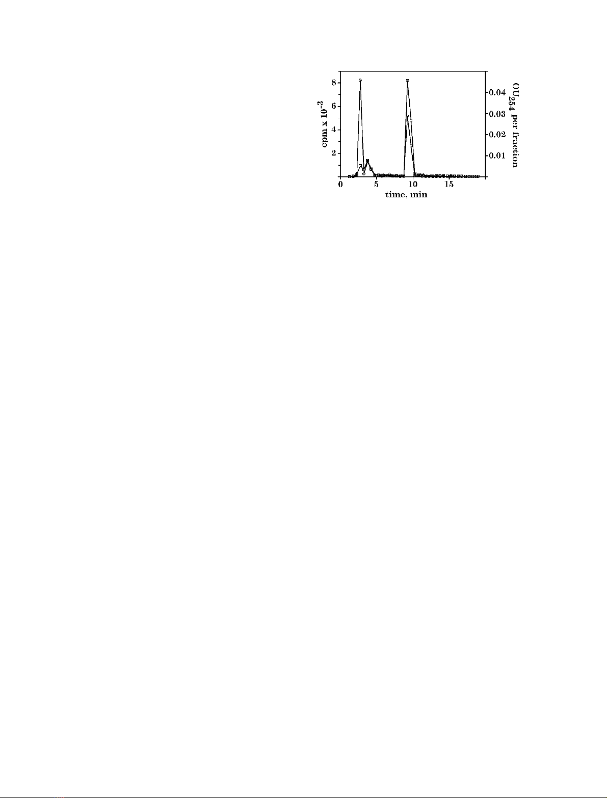
Finally, ATP together with [U-
14
C]ATP tracer were
treatedwiththeA. polypoides extract and the reaction
mixture was analysed by HPLC (Fig. 1). UV
254
trace
showed two peaks: one at 3.6 min corresponding to residual
ATP and another at 9.24 min corresponding to adenine. In
addition to these two peaks, radioactivity was detected at
2.75 min. The ratio of radioactivity in peaks at 2.75 min
and 9.5 min was 1.05, which approximately corresponds to
the number of carbon atoms in ribose moiety and hetero-
cycle. This experiment proved that ATP had been split into
two molecules – adenine and a yet unidentified derivative of
ribose.
Adenine is not a result of a multistep conversion
of ATP by phosphatases and N-glycosidases
The formation of adenine from ATP could be explained as a
result of multiple known enzymatic activities, first of all by
the combination of a relatively slowly acting phosphatase or
ATPase and a relatively rapidly acting well-known AMP/
adenosine nucleosidase. Thus, adenosine, AMP and ADP
were incubated under the same conditions as ATP with the
A. polypoides extract. Adenosine and AMP were not
digested during the period, which was sufficient for ATP
to be degraded almost completely; the release of adenine
from ADP was significantly slower than that from ATP.
This preliminary result completely excluded the possibility
of the formation of adenine by the way of combined action
of known enzymes. More detailed studies on these sub-
strates will be described below.
Fig. 1. HPLC analysis of products formed by A. polypoides extract
from exogeneous ATP. A Hybond N+ filter, presoaked in A. poly-
poides extract, was incubated in a mixture containing 1 m
M
ATP (with
[U-
14
C]ATP as a tracer), 100 m
M
KCl, pH 7.0 at 37 C. Ten micro-
litres of reaction mixture was subjected to HPLC fractionation. The
radioactivity of the fractions (500 lL) was measured (s). The amount
of the UV-absorbing material (OU
254
) in the fractions (h)wasdeter-
mined by integration of the computer-stored UV
254
-trace.
4124 T. Reintamm et al. (Eur. J. Biochem. 270)FEBS 2003
The second product of ATP degradation
in
A. polypoides
extract is ribose-5-triphosphate
The simplest reaction leading to the release of adenine from
ATP is the hydrolysis of the N-glycosidic bond. If adenine
results from hydrolysis of this bond the second reaction
product has to be ribose-5-triphosphate. Here we show that
the only way to interpret our results is to assign the NMR
signals of the second reaction product to ribose-5-triphos-
phate.
Samples for the NMR analysis were prepared by
treatment of a concentrated ATP solution (10–40 m
M
)with
the crude A. polypoides extract either in solution (named
NMR-B) or on a solid-phase support (Hybond-N+)
(named NMR-A). The reaction rate for these reactions,
performed on a preparative scale, decreased more rapidly
than would be expected from the first-order-kinetics at lower
substrate concentrations (0.1–5 m
M
). Only a small portion
of adenine-releasing activity was adsorbed on the Hybond-
N+ filter; therefore very long incubations (2 weeks for
10 m
M
ATP) were needed for the complete reaction. Still,
the solid-phase approach was useful for NMR samples as
the HPLC analysis revealed no concurrent dephosphoryla-
tion of the substrate in this sample. Presumably the ATP
dephosphorylating enzymes had a lower adsorbing capacity
to the Hybond-N+ than the ATP N-glycosidase, leading to
occasional enrichment of the latter.
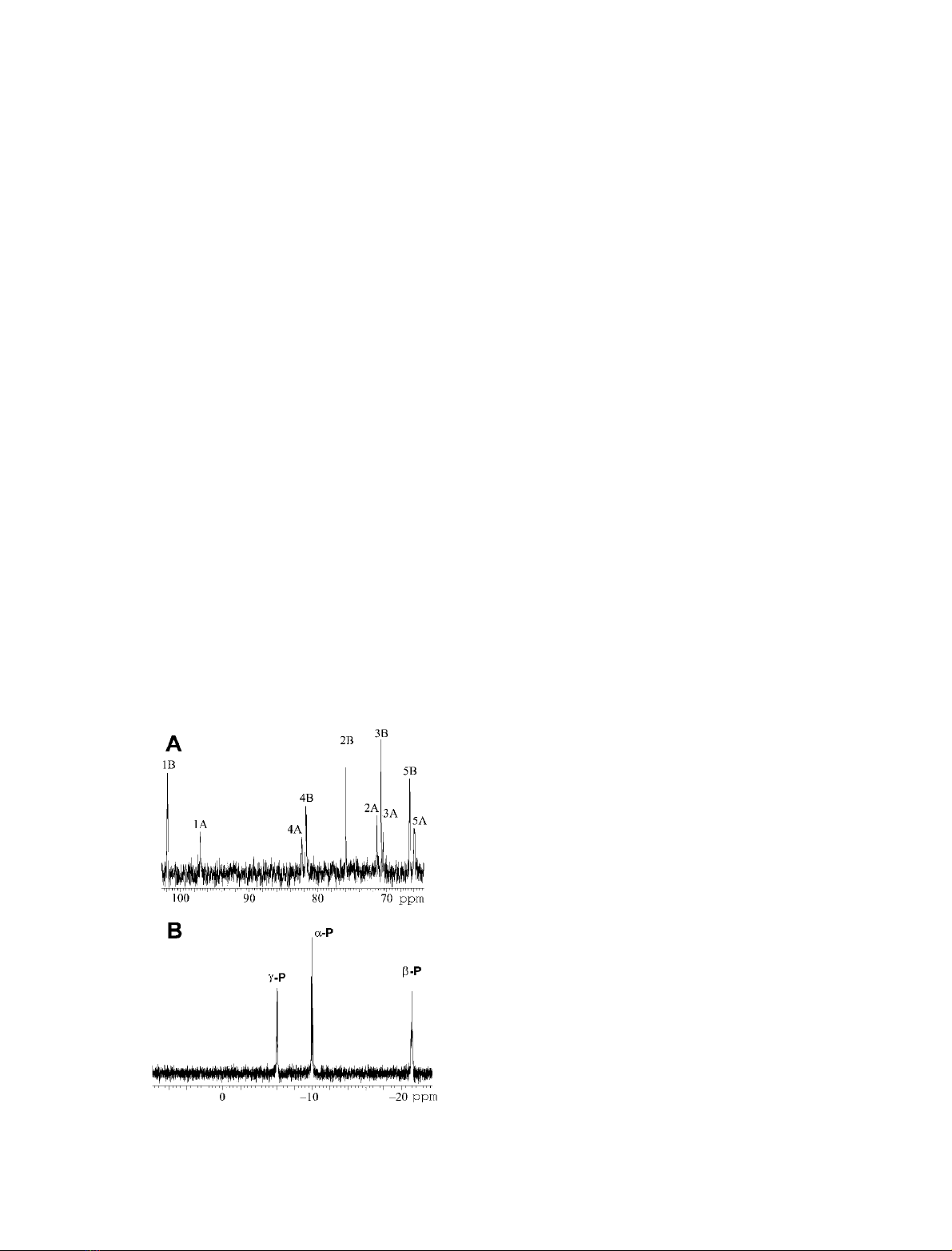
Ten signals were registered in the aliphatic region of
NMR-A
13
C-spectrum (Fig. 2A). The comparison of their
chemical shifts,
31
P–
13
C coupling constants and anomer
distribution (two-thirds of b-anomer) with available data
for the
D
-ribose-5-phosphate [16] revealed that they unam-
bigously belonged to the 5-phosphorylated a-and
b-
D
-ribofuranosides. The
1
H-NMR spectrum of NMR-A
was almost unusable because of the large water signal and
insufficient concentration. Still, the signals belonging to H-1
of ribose and aromatic protons of adenine could be
detected, indicating a 1 : 1 ratio of adenine to
D
-ribose-
5-triphosphates. The
31
P-NMR spectrum of NMR-A had
three groups of multiplets assignable to a-, b-andc-phos-
phates of the triphosphate monoester, while neither inor-
ganic phosphate nor any other additional resolved signals
were detected in NMR-A (Fig. 2B). However, the multiplet
appeared to be more complex than expected from a single
triphoshpate-containing compound.
The complete
13
C,
1
Hand
31
Pdataforthe
D
-ribose-
5-triphosphate were obtained with the sample NMR-B
(Table 1). The NMR-B sample contained a mixture of
a-andb-
D
-ribofuranoside-5-triphosphates as the main
product. The minor components (ATP, ADP and
D
-ribose-
5-diphosphates, inorganic phosphate) were identified and
quantified by one- and two-dimensional
31
P-NMR. It
should be noted that no
13
C-NMR signal was resolved for
the 5-diphosphorylated ribose. This indicates that the
differences up to 1 p.p.m. (Table 1) between the reported
13
C-NMR data of the ribose-5-monophosphate and our
data were probably caused by environmental differences in
the spectra registration rather than by the influence of the
number of phosphate groups.
1
H-NMR signals of a-and
b-anomers of phosphorylated ribose were resolved by two-
dimensional NMR. A small resolution between the
1
H
signals of diphosphorylated and triphosphorylated com-
pounds was evident, but these weak signals could not be
assigned to particular positions in particular isomers
because of the overall complexity of the spectrum.
It was possible to derive almost complete NMR data for
ATP/ADP from the NMR-B spectra. The spectral charac-
teristics of ATP and ADP obtained from NMR-B (Table 1)
are included in Table 1 because they serve as fine-tuning
internal standards for the ribose-5-triphosphate.
Thus, we can conclude that the second product formed by
A. polypoides extract is the
D
-ribose-5-triphosphate (as a
mixture of a-andb-anomers 1 : 2).
Preliminary kinetic studies of the hydrolysis of the
N-glycosidic bond in ATP by the
A. polypoides
ATP
N-glycosidase
Based on the results of product identification described
above, the novel enzyme catalyses the reaction of hydrolysis
of the N-glycosidic bond in ATP. This novel enzyme was
named the ATP N-glycosidase.
The conversion of ATP catalysed by the ATP
N-glycosidase present in the A. polypoides extract followed
the exponential-like kinetics at the 1 m
M
substrate concen-
tration (Fig. 3). Similar progress curves were registered
within the whole range of substrate concentrations used for
K
m
determination (0.1–4 m
M
ATP). The K
m
values
(KpH7
m¼0.16 m
M
and KpH5
m¼0.10 m
M
)calculatedfrom
the initial rates were found to be smaller than the substrate
concentration used (Fig. 4). The exponential form of
progress curves at [S] > K
m
could not be explained by
enzyme degradation during the reaction, because no change
in its activity was determined during the preincubation of
the extract up to 4 h under assay conditions before the
substrate was added (data not shown).
Fig. 2. NMR spectra of
D
-ribose-5-triphosphate. (A)
13
C-NMR spec-
trum of NMR-A. The assignment of signals in a-andb-anomers is
shown. (B)
31
P-NMR spectrum of NMR-A.
FEBS 2003 ATP N-glycosidase (Eur. J. Biochem. 270) 4125

Competitive inhibition by a product with K
ip
K
m
[17]
predicts pseudo-first order kinetics at substrate concentra-
tions above K
m
. The inhibition of the ATP N-glycosidase by
adenine was examined. K
ip
for adenine, estimated from the
decrease of the initial reaction rate by addition of adenine to
1m
M
ATP at pH ¼7.0, appeared to be close to the K
m
value
(Fig. 5). The progress curves obtained in the assays for K
m
determination (Fig. 4, pH 7) and for adenine inhibitory
effect (Fig. 5) were analysed together, using the procedure
described in [17]. Similar values of K
m
(0.15 m
M
)andK
ip
(0.15 m
M
) were obtained for the ATP N-glycosidase.
At very high substrate concentrations (> 10 m
M
ATP)
the kinetic model K
m
K
ip
was incomplete to simulate the
progress curves, as the reaction rate decreased even faster
than predicted by this model. Thus the kinetics of ATP
glycohydrolysis by the A. polypoides enzyme is actually
more complex than described by the relatively simple
KATP
mKAde
ip scheme.
The reaction rate was cross-dependent on ionic
strength and pH. The optimal pH was about 5 and
the optimal salt concentration was 100–250 m
M
(Fig. 6).
Alteration of the environmental condition did not lead to
a drastic change of the KATP
mand KAde
ip ratio,asfarasit
could be judged by progress curve shapes. The enzyme
activity was not substantially altered by the presence of
10 m
M
EDTA, 140 m
M
mercaptoethanol or the inorganic
phosphate.
The enzyme appeared to be relatively stable. The
temperature dependence of the reaction (Fig. 7) showed
that the denaturation of the enzyme started above 60 C.
The reaction catalysed by the ATP N-glycosidase
was described by a single activation energy (DH
a
)of
11.6 kcalÆmol
)1
in the temperature range 10–60 C.
Heating of the extract for 10 min at 92 Cresultedin
Table 1.
1
H,
13
Cand
31
P-NMR data of the NMR-B sample. The differences in chemical shifts from those of the
D
-ribose-5-phosphate [16] are shown
in brackets. The resolved and assigned signals are separated by slashes, signals unassigned to a particular molecule are separated by commas. NA,
Not applicable; ND, not detected.
Nucleus
b-
D
-ribose-5-triphosphate/
b-
D
-ribose-5-diphosphate
a-
D
-ribose-5-triphosphate/
a-
D
-ribose-5-diphosphate ATP/ADP/P
i
Chemical shift Coupling constants Chemical shift Coupling constants Chemical shift Coupling constants
1
H 1H 5.23
3
J
HH
¼1.6 5.40
3
J
HH
¼4.70 6.13 J
HH
¼5.33
2H 4.04 4.17 4.78, 4.74
3H 4.37 4.26 4.58
4H 4.1 4.08 4.37
5H (4.15,4.02) (4.15,4.02) 4.21, 4.27
13
C 1C 101.79 [)0.61] 97.07 [-0.43] 87.67, 87.34
2C 75.81 [)0.59] 71.35 [-0.55] 74.94, 74.86
3C 70.84 [)0.86] 70.48 [-0.82] 70.90, 70.60
4C 81.76 [)0.74] J
CP
¼8.9 82.40 [-1.20] J
CP
¼8.3 84.56, 84.38 J
CP
¼9.5, 9.9
5C 66.74 [0.14] J
CP
¼6.2 66.05 [0.25] J
CP
¼5.3 65.76/ND J
CP
¼5.0/ND
31
PaP)9.82/)8.92 J
PP
¼18.5/20.7 )9.88/)9.03 J
PP
¼18.5/18.4 )10.11/)9.23 J
PP
¼18.6/20.6
bP)20.1/)5.73 )20.1/)5.81 )20.1/)5.78
cP)5.52/NA J
PP
¼18.6 )5.55/NA J
PP
¼18.5 )5.46/NA J
PP
¼18.5
p
i
1.86
Fig. 3. Progress curves of ATP degradation by A. polypoides crude
extract. ATP (1 m
M
), KCl (100 m
M
), pH 7.0, 37 C, dilution of the
crude extract 1 : 100. The almost perfectly fitted exponential line
through the experimental points is shown.
Fig. 4. Lineweaver–Burk plots of A. polypoides ATP N-glycosidase
activity on ATP and ADP. The initial rates of each reaction containing
A. polypoides crude extract in a dilution of 1 : 100 were found from the
progress curves, assuming pseudo first-order kinetics. ATP was
investigated at two pH values: at pH 7. ± 0.1 (100 m
M
KCl, 37 C,
K
m
¼0.158 m
M
,v
max
¼0.031 m
M
Æmin
)1
,s) and at pH 5.3 ± 0.1
(20 m
M
Mes, 170 m
M
KCl, 37 C, K
m
¼0.102 m
M
,v
max
¼
0.044 m
M
Æmin
)1
,h). ADP was assayed at pH 5.1 ± 0.2 (20 m
M
Mes,
170 m
M
KCl, 37 C, K
m
¼0.122 m
M
,v
max
¼0.027 m
M
Æmin
)1
,m).
pH for each reaction mixture at the assay temperature was determined.
4126 T. Reintamm et al. (Eur. J. Biochem. 270)FEBS 2003


