
BioMed Central
Page 1 of 15
(page number not for citation purposes)
Journal of Nanobiotechnology
Open Access
Research
Capillary electrophoresis for the characterization of quantum dots
after non-selective or selective bioconjugation with antibodies for
immunoassay
Mark Pereira and Edward PC Lai*
Address: Department of Chemistry, Ottawa-Carleton Chemistry Institute, Carleton University, Ottawa, ON K1S 5B6, Canada
Email: Mark Pereira - mpereir2@connect.carleton.ca; Edward PC Lai* - edward_lai@carleton.ca
* Corresponding author
Abstract
Capillary electrophoresis coupled with laser-induced fluorescence was used for the
characterization of quantum dots and their conjugates to biological molecules. The CE-LIF was
laboratory-built and capable of injection (hydrodynamic and electrokinetic) from sample volumes
as low as 4 μL via the use of a modified micro-fluidic chip platform. Commercially available quantum
dots were bioconjugated to proteins and immunoglobulins through the use of established
techniques (non-selective and selective). Non-selective techniques involved the use of EDCHCl/
sulfo-NHS for the conjugation of BSA and myoglobin to carboxylic acid-functionalized quantum
dots. Selective techniques involved 1) the use of heterobifunctional crosslinker, sulfo-SMCC, for
the conjugation of partially reduced IgG to amine-functionalized quantum dots, and 2) the
conjugation of periodate-oxidized IgGs to hydrazide-functionalized quantum dots. The migration
times of these conjugates were determined in comparison to their non-conjugated QD relatives
based upon their charge-to-size ratio values. The performance of capillary electrophoresis in
characterizing immunoconjugates of quantum dot-labeled IgGs was also evaluated. Together, both
QDs and CE-LIF can be applied as a sensitive technique for the detection of biological molecules.
This work will contribute to the advancements in applying nanotechnology for molecular diagnosis
in medical field.
Background
Quantum dots (QDs) are fluorescent nanoparticles that
receive increasing recognition as a viable alternative (to
conventional organic fluorophores) for molecular labe-
ling. Their quantum mechanical and electronic character-
istics give QDs unique optical properties that are
advantageous in the fields of bioanalytical, biomedical
and biophotonic research. Such optical properties include
size-tunable emission wavelengths, broad excitation
wavelengths, long fluorescence lifetimes, large Stokes
shifts, and high quantum yields [1-3]. Other advanta-
geous properties include resistance to photo- and chemi-
cal- degradation and their capability for performing
multiplexing experiments [3]. QDs are relatively large par-
ticles, with typical diameters ranging from 1–10 nm [1].
The inorganic core (typically a semiconductor) is respon-
sible for their fluorescent properties. This core is typically
surrounded by a shell (ZnS is common) for protection
from chemical- and photo-oxidation [2]. The shell also
provides a means of functionalizing the QD with carbox-
Published: 1 October 2008
Journal of Nanobiotechnology 2008, 6:10 doi:10.1186/1477-3155-6-10
Received: 3 May 2008
Accepted: 1 October 2008
This article is available from: http://www.jnanobiotechnology.com/content/6/1/10
© 2008 Pereira and Lai; licensee BioMed Central Ltd.
This is an Open Access article distributed under the terms of the Creative Commons Attribution License (http://creativecommons.org/licenses/by/2.0),
which permits unrestricted use, distribution, and reproduction in any medium, provided the original work is properly cited.
Journal of Nanobiotechnology 2008, 6:10 http://www.jnanobiotechnology.com/content/6/1/10
Page 2 of 15
(page number not for citation purposes)
ylic acids or primary amines, for good solubility in aque-
ous solutions and relative ease of specific labeling
reactions [1].
QDs, often applied for the labeling of biological mole-
cules (proteins, peptides, antibodies, etc.), require specific
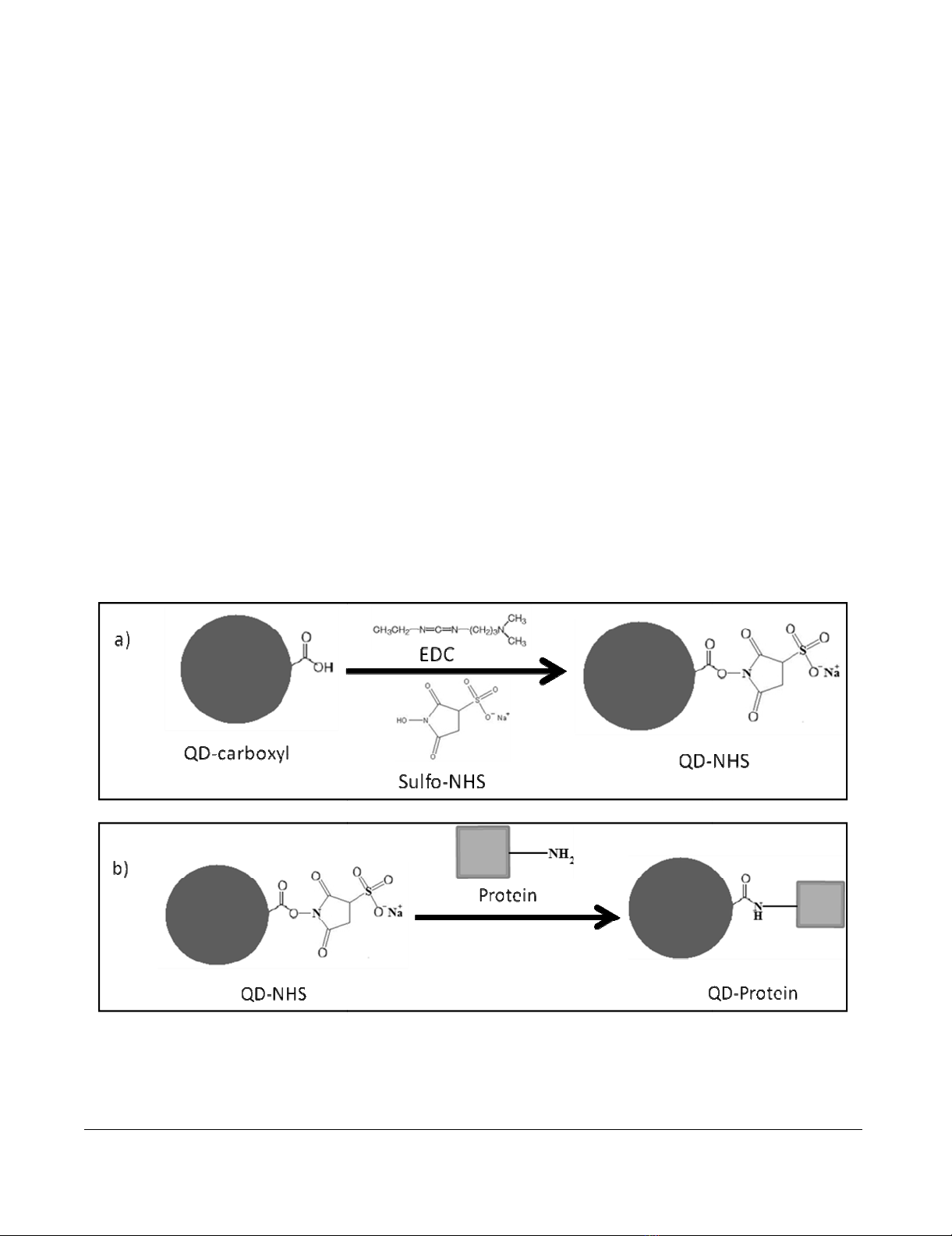
techniques for their conjugation [4-7]. The most popular
bioconjugation technique involves the use of a zero-
length crosslinker, 1-ethyl-3- [3-dimethylaminopro-
pyl]carbodiimide hydrochloride (EDCHCl) [1-4,6,7], in
the presence of a hydrophilic active group, N-hydroxysul-
fosuccinimide (sulfo-NHS) [8], for the formation of a sta-
ble amide bond between carboxylic acid-functionalized
QDs (QD-COOH) and any biomolecules containing a
primary amine [9] (Figure 1).
This method, while proven to yield exclusively QD-pro-
tein conjugates in a controlled manner, randomizes the
location on a protein to which conjugation can occur,
resulting in a non-selective bioconjugation [9]. Despite
high bioconjugation efficiencies, this can be detrimental
in the case where an immunoassay is to be performed
next. For instance, a labeled protein serving as an antigen
might lose its antigenicity (ability to bind an antibody)
when conjugated to a large QD. A similar concern can be
conveyed if an antibody were conjugated in a region close
to the antigen-binding site (the hypervariable region).
Either one of these variations can significantly reduce the
efficiency of immunoassay applications [9].
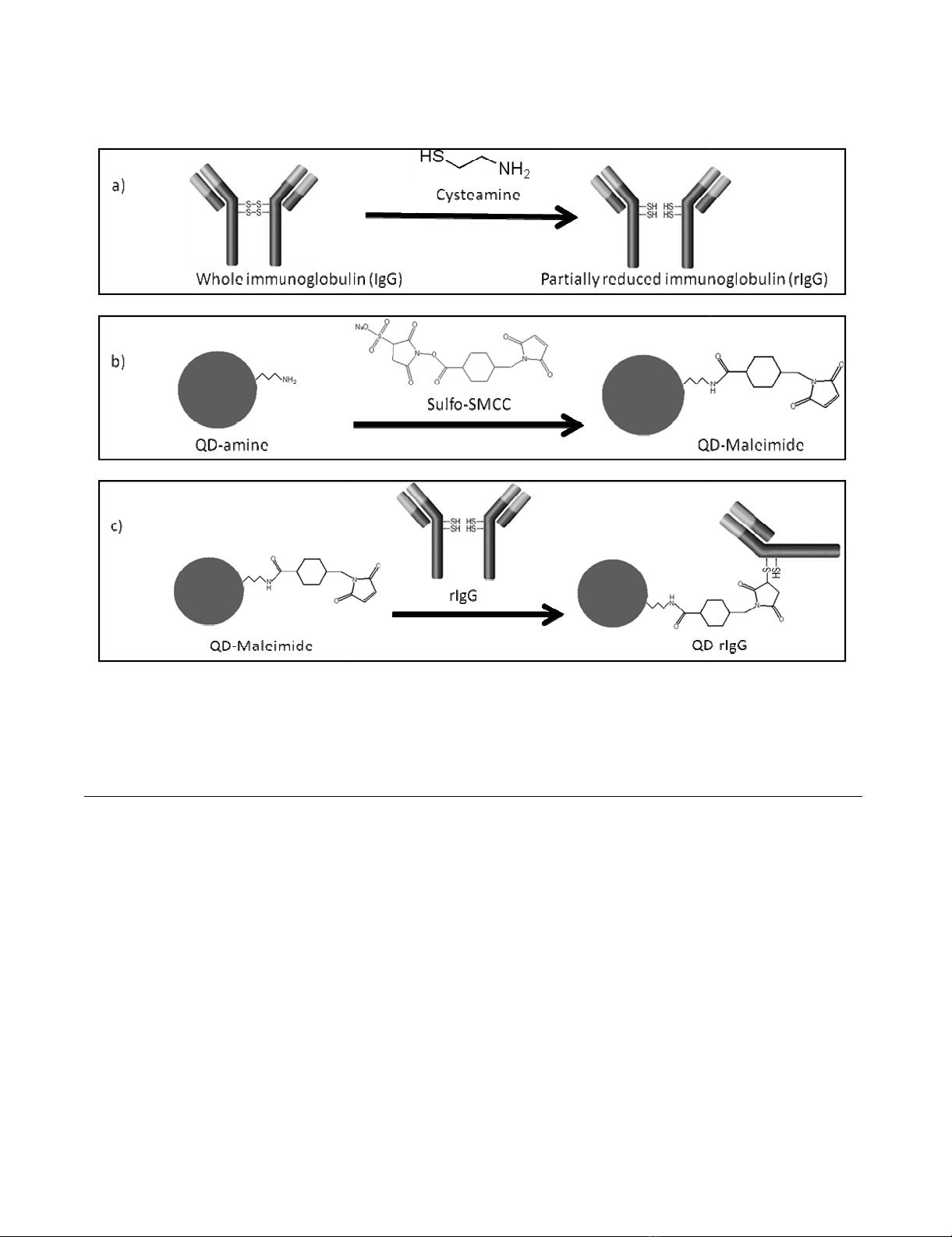
Other techniques make effective use of selective bioconju-
gation, targeting specific sites on the protein. These
include the use of a heterobifunctional crosslinker such as
sulfosuccinimidyl-4-(N-maleimidomethyl)cyclohexane-
1-carboxylate (sulfo-SMCC) [9-11]. In the case for anti-
bodies, as shown in Figure 2 below, sulfo-SMCC can form
stable amide bonds to amine-functionalized QDs (QD-
NH2) [9]. The resultant QDs, through sulfo-SMCC's male-
imide region, can next form stable a thioether bond with
a sulfhydryl-exposed antibody [9]. Mild reducing reagents
such as cysteamineHCl (or DTT) can selectively cleave the
disulfide bonds (hinge region) connecting the IgG heavy
chains, while leaving the other disulfide bonds that make
up the antigen binding site (hypervariable region) unaf-
fected, thus producing a partially reduced IgG (rIgG) [12].
In addition, the resulting exposed sulfhydryls (hinge
region) are sufficiently far away (from the hypervariable
region) for QD-bioconjugation to occur. The resulting
Non-selective bioconjugation reaction scheme of carboxylated QDs (QD-COOH) to amine-containing proteinsFigure 1
Non-selective bioconjugation reaction scheme of carboxylated QDs (QD-COOH) to amine-containing pro-
teins. This two-step reaction involves a) the activation of QD-COOH with EDC/sulfo-NHS, resulting in a semi-stable active
ester (QD-NHS), and b) the nucleophilic reaction between the QD-NHS and amine-containing protein, forming a QD-protein
conjugate via a stable amide bond.
Journal of Nanobiotechnology 2008, 6:10 http://www.jnanobiotechnology.com/content/6/1/10
Page 3 of 15
(page number not for citation purposes)
quantum dot-conjugated half antibody (QD-rIgG) will
allow an immunoreaction to proceed readily.
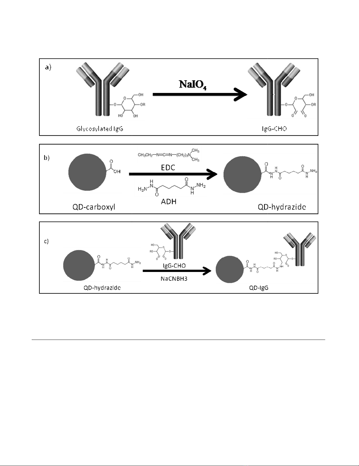
Reductive amination is a bioconjugation technique popu-
lar in the labeling of glycoproteins. Taking advantage of
the polysaccharide chains within the Fc region of an anti-
body, it can allow bioconjugation to occur relatively far
away from the antigen binding site. Through oxidation
(using sodium periodate) of the carbohydrate hydroxyls,
the aldehydes formed are highly reactive toward primary
amines and hydrazides [9]. This makes QD-NH2 or QD-
COOH (derivatized with adipic acid dihydrazide (ADH))
suitable candidates for conjugation [9]. In addition, selec-
tive bioconjugation can occur without a proceeding
reduction reaction, thus retaining the integrity of the anti-
body (Figure 3).
Capillary electrophoresis (CE) has seen increasing use in
the separation and characterization of inorganic nanopar-
ticles (Ag, Au, TiO2, Al2O3, Fe2O3) [13-17], polystyrene
microspheres [18], biomolecules (proteins, peptides) [19-
30], QDs [31], QD-conjugates with bovine serum albu-
min (BSA) and horse radish peroxidase (HRP) [7], and
QD-conjugates with Ulex europaeus (UEA-1) and anti-von
Willebrand factor (anti-vWF) [32]. CE has also been used
for immunoassays involving hepatitis B, prion protein,
alpha-fetoprotein, etc [24-30]. Recently, a CE-based
immunoassay involving QDs conjugated to anti-IgM anti-
bodies followed by immuno-conjugation to its compli-
mentary antigen IgG was performed with satisfactory
results [33]. Another recent advancement involved the
CE-characterization of QDs (of differing emission wave-
lengths) exclusively conjugated to biotin and streptavidin
Selective bioconjugation reaction scheme of amino QDs (QD-amine) to free sulhydryl-containing IgG antibodiesFigure 2
Selective bioconjugation reaction scheme of amino QDs (QD-amine) to free sulhydryl-containing IgG antibod-
ies. The reaction involves a) the mild reduction of IgG with cysteamine to yield partially reduced IgG antibody fragments
(rIgG); b) the activation of QD-NH2 by nucleophilic reaction with NHS-moiety of sulfo-SMCC, resulting in maleimide-function-
alized quantum dot (QD-maleimide); and c) the rIgG and QD-maleimide conjugation (QD-rIgG) via the formation of a
thioether bond.
Journal of Nanobiotechnology 2008, 6:10 http://www.jnanobiotechnology.com/content/6/1/10
Page 4 of 15
(page number not for citation purposes)
[34]. Their work followed the characterization of the con-
jugates' affinity to each other via strong biotin-streptavi-
din interactions. However, present publications reporting
the use of QDs in CE-based immunoassays are very pre-
liminary, due in part to a QD-biomolecule conjugate's
(and immunoconjuagte's) complex charge-to-size ratio.
Thus, more research is required in its development as a
fast and efficient method for performing immunoassays.
In this paper, we report more preliminary results of cova-
lently bioconjugating QDs to various biomolecules (pro-
teins and immunoglobulins). These QD-conjugated
biomolecules are characterized via a laboratory-built cap-
illary electrophoresis instrument with laser-induced fluo-
rescence detection (CE-LIF) [35]. The instrumental
capabilities (comparable to commercial CE-LIF systems)
include the use of a micro-sample injection platform that
can load sample volumes as low as 4 μL [35]. We also dis-
cuss some of the challenges faced when performing bio-
conjugation through the various schemes described
above. The purpose is to validate a fast, selective, and
reproducible CE-LIF analysis method that can be efficient
Selective bioconjugation reaction scheme of hydrazide QDs (QD-hydrazide) to aldehyde-containing IgG antibodies (IgG-CHO)Figure 3
Selective bioconjugation reaction scheme of hydrazide QDs (QD-hydrazide) to aldehyde-containing IgG anti-
bodies (IgG-CHO). The reaction involves a) mild periodate oxidation of glycosylated IgG, yielding IgG-CHO; b) synthesis of
QD-hydrazide via derivatization of QD-COOH with EDC/ADH; and c) conjugation of QD-hydrazide with IgG-CHO via for-
mation of hydrazone linkage to yield QD-IgG.

Journal of Nanobiotechnology 2008, 6:10 http://www.jnanobiotechnology.com/content/6/1/10
Page 5 of 15
(page number not for citation purposes)
and robust. This work will evolve to perform QD-based
immunoassays using CE-LIF as an effective separation and
sensitive detection technique. The aim is to apply this
research in the area of infectious biological materials that
are generally present in relatively low concentrations and
small volumes.
Methods
Chemicals and reagents
Boric acid (certified A.C.S.), sodium meta-periodate (crys-
talline, A.C.S. grade), sodium hydroxide (reagent grade)
were purchased from Fisher Scientific (Ottawa, Ontario,
Canada). CdSe/ZnS carboxy-terminated QDs (Maple Red-
Orange, 620 nm) and CdSe/ZnS amine-terminated QDs
(Maple Red-Orange, 620 nm) were purchased from Evi-
dent Technologies (Troy, NY, USA). EDCHCl, Sulfo-NHS,
lysozyme (Lys), and MES buffered saline packs were pur-
chased from Pierce Biotechnology. Sodium acetate (rea-
gent grade) and hydroxylamine hydrochloride (reagent
grade) was purchased from Anachemia. EDTA (0.1 M vol-
umetric standard), ADH (= 98%), sulfo-SMCC (= 98%),
DL-DTT (1 M in water solution), anti-human albumin
(polyclonal IgG produced in rabbit), human serum albu-
min (HSA), cysteamine hydrochloride (Purum = 97.0%),
2-mercaptoethanol (14 M), 10× PBS concentrate, bovine
serum albumin (BSA), horse myoglobin (Myo) cyto-
chrome c (CytC), ethanolamine, and sodium cyanoboro-
hydride (5 M in 1 M sodium hydroxide) were purchased
from Sigma Aldrich. Coumarin 521 was purchased from
Exciton (Dayton, Ohio, USA). Micro-centrifuge tubes (50
kDa and 100 kDa MWCO) were purchased from Fisher
Scientific.
Preparation of buffer solutions and stock solutions
All buffer solutions were prepared and pH-adjusted using
sodium hydroxide (10 M, 5 M, and 1 M) and hydrochloric
acid (1 M and 0.5 M). All CE separation buffers were fil-
tered through a 0.45 μm membrane filter (Pall Corpora-
tion, Ann Arbor, MI, USA).
Carboxy- and amine- terminated QDs were used from
supply stock (11 μM) without any prior treatment.
Stock solutions of EDCHCl (20 mM) and sulfo-NHS (50
mM) were prepared by dissolution of dry reagents in 0.1
M MES (pH 5.2) buffered saline and used immediately
after preparation. Stock solutions of 2-mercaptoethanol
(1 M) and hydroxylamine hydrochloride (1 M) were pre-
pared and stored at room temperature.
Stock solutions of cysteamineHCl (100 mM) were pre-
pared by dissolution of dry reagent in 1× PBS (pH 7.2), 10
mM EDTA and used immediately after preparation. Stock
solutions of DTT (100 mM) were prepared by dilution of
a 1 M DTT stock solution and used within 3 days of prep-
aration.
Stock solutions of NaIO4 (100 mM) were prepared by dis-
solution of dry reagents in 0.1 M sodium acetate (pH 5.5)
buffered saline. Preparation and storage was performed in
minimal lighting and used immediately after use. Sodium
cyanoborohydride (5 M in 1 N NaOH) was used as pre-
pared from supplier. Stock solution of ethanolamine (1
M) was prepared by dissolution of dry reagent in distilled
deionized water (ddw) and pH adjusted to 9.6.
Stock solutions (1 mg/mL) of bovine serum albumin
(BSA), myoglobin (Myo), cytochrome c (CytC), and lys-
ozyme (Lys), were prepared in 1× PBS (pH 7.2). Human
serum albumin (HSA) was prepared in ddw (11 mg/mL).
Anti-human albumin IgG (4 mg/mL) was prepared in 1×
PBS (pH 7.2).
Non-specific bioconjugation of whole IgG using EDCHCl/
sulfo-NHS
A mixture containing 2 mM EDCHCl, 5 mM sulfo-NHS,
and 1.1 μM carboxy-terminated QDs (QD-carboxyl) was
prepared in 0.1 M MES, pH 6.0 and incubated for 15 min-
utes at room temperature. The remaining unreacted EDC
was quenched with the addition of 2-mercaptoethanol (1
M) to a final concentration of approximately 20 mM and
the mixture was left to stand for 10 minutes. The activated
QDs were purified of unreacted reagents and byproducts
by dialysis using 100 kDa MWCO microcentrifuge tubes
and re-suspended in 1× PBS (pH 7.2) containing dis-
solved protein. The reaction proceeded for 2 hours with
gentle mixing. The reaction was quenched with addition
of hydroxylamine hydrochloride (1 M) to a final concen-
tration of approximately 10 mM. The bioconjugation mix-
ture was left to stand for 10 minutes at room temperature
prior to purification by dialysis using 100 kDa MWCO
microcentrifuge tubes. The mixture was analyzed by CE-
LIF and stored at 4°C.
Selective bioconjugation of reduced IgG (rIgG) using
cysteamineHCl or DTT and sulfo-SMCC
A mixture containing approximately 1 mg/mL rabbit anti-
human albumin IgG and cysteamineHCl (concentration
ranging from 0.1 mM to 100 mM) was incubated at 37°C
for 90 minutes in 0.1 M sodium phosphate (pH 7.0), 0.15
M, 0.01 M EDTA. The resulting partially reduced antibody
(rIgG) was purified of byproducts and unreacted com-
pounds via dialysis using a 50 kDa MWCO microcentri-
fuge tube with successive washings of 0.1 M sodium
phosphate (pH 6.8), 0.15 M NaCl, 0.01 M EDTA buffer.
The rIgG was temporarily stored at 4°C until use for QD
coupling.


