
Cyclosporin A-induced oxidative stress is not the
consequence of an increase in mitochondrial membrane
potential
Marco van der Toorn
1
, Henk F. Kauffman
2
, Margaretha van der Deen
3
, Dirk-Jan Slebos
1
,
Gerard H. Koe
¨ter
1
, Rijk O. B. Gans
1
and Stephan J. L. Bakker
1
1 Department of Internal Medicine, University Medical Center Groningen, University of Groningen, the Netherlands
2 Groningen University Institute for Drug Exploration, University Medical Center Groningen, University of Groningen, the Netherlands
3 Department of Medical Oncology, University Medical Center Groningen, University of Groningen, the Netherlands
Keywords
cyclosporin A; mitochondria; mitochondrial
membrane potential; mitochondrial
permeability transition; reactive oxygen
species
Correspondence
S. J. L. Bakker, Department of Internal
Medicine, University Medical Center
Groningen, PO Box 30001, 9700 RB
Groningen, the Netherlands
Fax: +31 503 619069
Tel: +31 503 613677
E-mail: s.j.l.bakker@int.umcg.nl
(Received 7 December 2006, revised 6 April
2007, accepted 11 April 2007)
doi:10.1111/j.1742-4658.2007.05827.x
Cyclosporin A induces closure of the mitochondrial permeability transition
pore. We aimed to investigate whether this closure results in concomitant
increases in mitochondrial membrane potential (DW
m
) and the produc-
tion of reactive oxygen species. Fluorescent probes were used to assess
DW
m
(JC-1, 5,5¢,6,6¢-tetrachloro-1,1¢,3,3¢-tetraethyl-benzimidazolyl-carbo-
cyanine iodide), reactive oxygen species [DCF, 5- (and 6)-chloromethyl-
2¢,7¢-dichlorodihydrofluorescein diacetate, acetyl ester] and [Ca
2+
] [Fluo-3,
glycine N-[4-[6-[(acetyloxy)methoxy]-2,7-dichloro-3-oxo-3H-xanthen-9-yl]-
2-[2-[2-[bis[2-[(acetyloxy)methoxy]-2-oxyethyl]amino]-5-methylphenoxy]eth-
oxy]phenyl]-N-[2-[(acetyloxy)methoxy]-2-oxyethyl]-(acetyloxy)methyl ester]
in human kidney cells (HK-2 cells) and in a line of human small cell
carcinoma cells (GLC4 cells), because these do not express cyclospo-
rin A-sensitive P-glycoprotein. We used transfected GLC4 cells expressing
P-glycoprotein as control for GLC4 cells. NIM811 (N-methyl-4-isoleucine-
cyclosporin) and PSC833 (SDZ-PSC833) were applied as selective
mitochondrial permeability transition pore and P-glycoprotein blockers,
respectively. To study the effect of cyclosporin A on mitochondrial func-
tion, we isolated mitochondria from fresh pig livers. Cyclosporin A and
PSC833 induced a more than two-fold increase in JC-1 fluorescence in
HK-2 cells, whereas NIM811 had no effect. None of the three substances
induced a significant increase in JC-1 fluorescence in GLC4 cells. Despite
this, cyclosporin A, NIM811 and PSC833 induced a 1.5-fold increase in
DCF fluorescence (P<0.05) and a two-fold increase in Fluo-3 fluores-
cence (P<0.05). Studies in isolated mitochondria showed that blockage
of mitochondrial permeability transition pores by cyclosporin A affected
neither DW
m
, ATP synthesis, nor respiration rate. The mitochondrial per-
meability transition pore blockers cyclosporin A and NIM811, but also the
non-mitochondrial permeability transition pore blocker PSC833, induced
comparable degrees of reactive oxygen species production and cytosolic
[Ca
2+
]. Neither mitochondria, effects on P-glycoprotein nor inhibition of
Abbreviations
CsA, cyclosporin A; DCF, 5- (and 6)-chloromethyl-2¢,7¢-dichlorodihydrofluorescein diacetate, acetyl ester; DNP, 2,4-dinitrophenol; DW
m
,
mitochondrial membrane potential; Fluo-3, glycine N-[4-[6-[(acetyloxy)methoxy]-2,7-dichloro-3-oxo-3H-xanthen-9-yl]-2-[2-[2-[bis[2-
[(acetyloxy)methoxy]-2-oxyethyl]amino]-5-methylphenoxy]ethoxy]phenyl]-N-[2-[(acetyloxy)methoxy]-2-oxyethyl]-(acetyloxy)methyl ester; GLC4,
human small cell carcinoma; HK-2, human kidney; JC-1, 5,5¢,6,6¢-tetrachloro-1,1¢,3,3¢-tetraethyl-benzimidazolyl-carbocyanine iodide; MPTP,
mitochondrial permeability transition pore; NIM811, N-methyl-4-isoleucine-cyclosporin; PSC833, SDZ-PSC833; ROS, reactive oxygen species.
FEBS Journal 274 (2007) 3003–3012 ª2007 The Authors Journal compilation ª2007 FEBS 3003
Immunosuppressive treatment with cyclosporin A
(CsA) is accompanied by accelerated atherosclerosis
and fibrosis, which contribute to the development of
chronic transplant dysfunction [1]. It has been sugges-
ted that reactive oxygen species (ROS) play an import-
ant underlying role [2–4]. Different studies have shown
that CsA is able to increase levels of superoxide anion
(O
2
Æ–
), hydrogen peroxide, malondialdehyde, and thio-
barbituric acid reactive substances [5,6]. Mitochondrial
enzymes with antioxidative properties, including super-
oxide dismutase, catalase, and glutathione peroxidase,
become upregulated upon exposure to CsA [7]. It is
evident that CsA induces oxidative stress, but its origin
remains speculative.
Mitochondria represent a major source of intracellu-
lar ROS, and play a crucial role in cellular Ca
2+
homeostasis, which affects various cell signaling path-
ways [8]. The primary function of mitochondria is pro-
duction of ATP, a process linked to the action of the
electron transfer chain. Normally, electrons supplied
by metabolic fuel (NADH and FADH
2
) are trans-
ferred along the electron transfer chain to oxygen.
Optimally, the terminal enzyme of the electron transfer
chain, cytochrome coxidase, binds oxygen until it has
accepted four electrons, when it is released as water.
Most of the energy released during the transfer of
these electrons is used to pump protons from the mito-
chondrial matrix towards the inner membrane space,
thereby creating a proton gradient. The energy stored
in the proton gradient is used to drive the process of
oxidative phosphorylation of ADP to ATP. When the
intramitochondrial ADP concentration drops (e.g.
under conditions of low energy demand), the proton
gradient will rise as a consequence of decreased con-
sumption [9–12]. This increased proton gradient
impairs the flow of electrons along the electron trans-
fer chain, which results in accumulation of electrons
along the electron transfer chain [13]. This results
in an increased likelihood of leakage of electrons
from the chain, with increased ROS production as a
consequence [14].
One mechanism by which the mitochondrial mem-
brane potential (DW
m
) can decrease is through opening
of the mitochondrial permeability transition pore
(MPTP) [15–17]. CsA is well known as an inhibitor of
calcineurin and P-glycoprotein, but it is also a strong
inhibitor of the MPTP [18,19]. Indeed, it has been sug-
gested that in several cell types CsA prevents opening
of the MPTP, thereby leading to an increased DW
m
[17,20]. The CsA analog N-methyl-4-isoleucine-cyclos-
porin (NIM811) is also known as an inhibitor of
MPTP, and to lead to an increase in DW
m
[21]. Fluor-
escent probes used to assess DW
m
are pumped out of
cells by P-glycoprotein [22]. Thus, probe accumulation
caused by CsA may result from effects on P-glycopro-
tein as well as effects on MPTP. The CsA analog
SDZ-PSC833 (PSC833) may serve as a useful control
substance in this context, because it is an inhibitor of
P-glycoprotein rather than MPTP, and is devoid of
calcineurin-inhibiting properties [23].
We hypothesized that an increase in steady-state
DW
m
underlies increased ROS production in associ-
ation with CsA exposure. We set out to investigate the
effects of CsA on DW
m
in relation to the production of
ROS, with NIM811 and PSC833 as controls.
Results
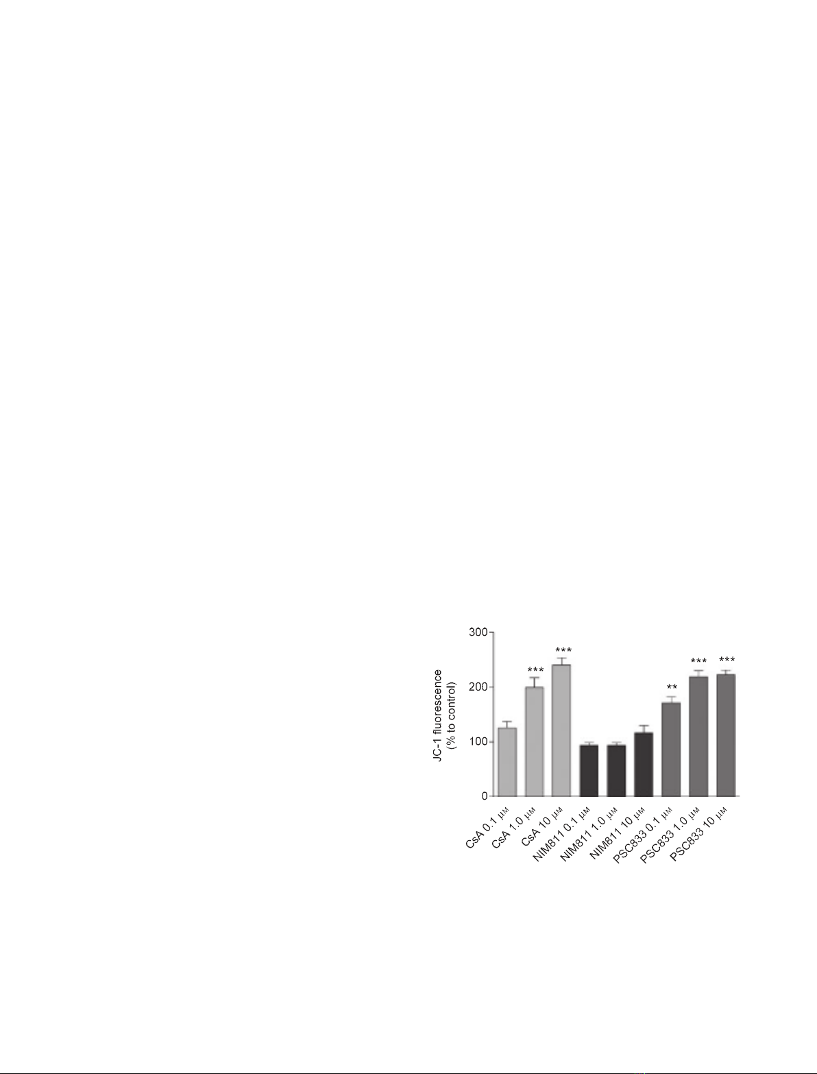
Closure of the MPTP and DW
m
Human kidney (HK-2) cells are known to express
P-glycoprotein [24,25]. Both CsA and PSC833 induced
a dose-dependent increase in 5,5¢,6,6¢-tetrachloro-1,
1¢,3,3¢-tetraethyl-benzimidazolyl-carbocyanine iodide
(JC-1) fluorescence in these cells (Fig. 1). NIM811,
calcineurin therefore play a role in cyclosporin A-induced oxidative stress
and disturbed Ca
2+
homeostasis.
Fig. 1. Effect of CsA and its analogs on mitochondrial membrane
potential in HK-2 cells. JC-1 probe (5 lgÆmL
)1
) was used to study
mitochondrial membrane potential. Data are expressed as mean
value ± SEM, and refer to three experiments. *P< 0.05 versus
control, **P< 0.01 versus control, ***P< 0.001 versus control by
Newman–Keuls multiple comparison test.
Cyclosporin A-induced oxidative stress M. van der Toorn et al.
3004 FEBS Journal 274 (2007) 3003–3012 ª2007 The Authors Journal compilation ª2007 FEBS
however, did not induce a significant increase in JC-1
fluorescence.
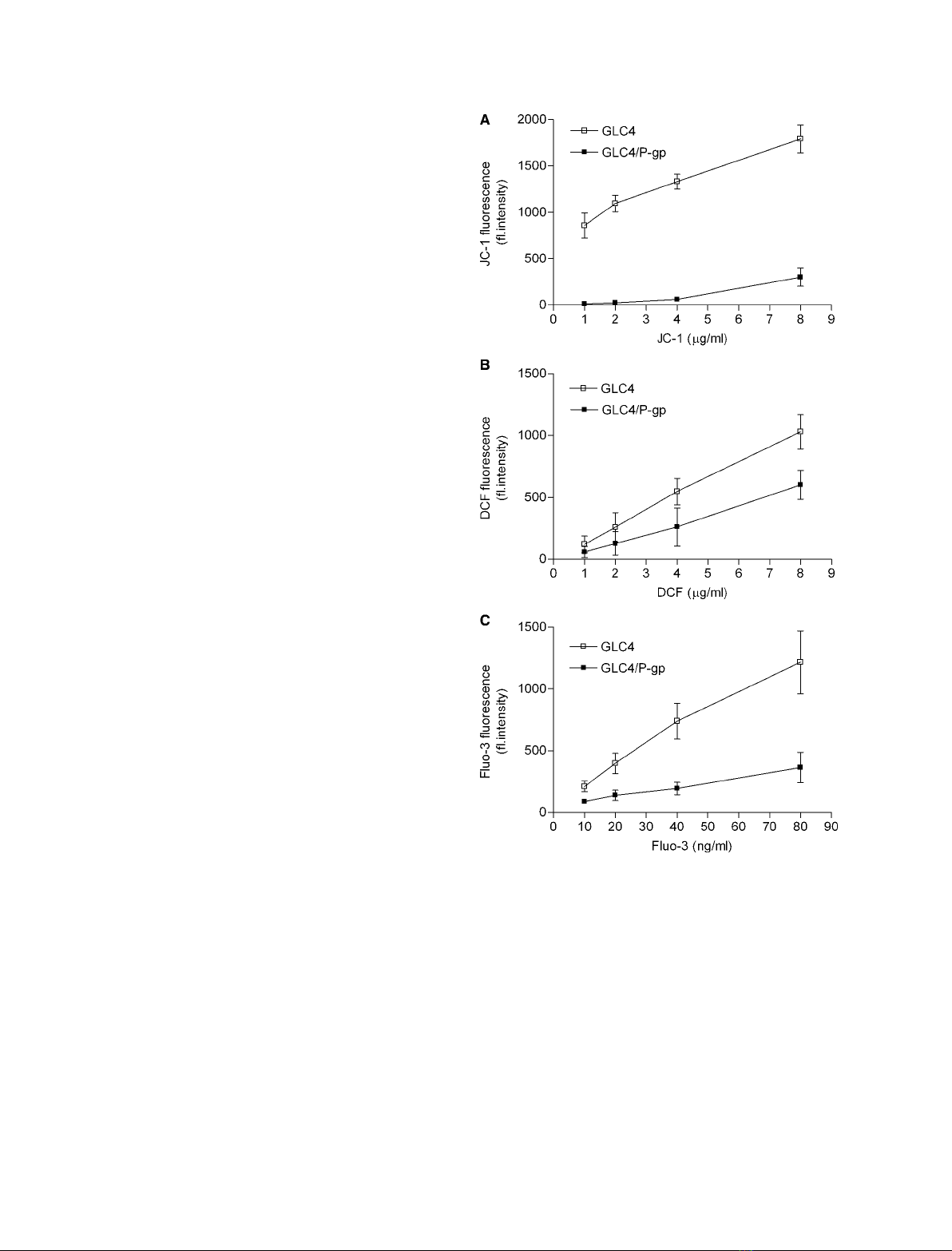
We subsequently investigated whether, and to what
extent, P-glycoprotein expression affects intracellular
accumulation of three different fluorescent probes.
Expression of P-glycoprotein resulted in significant
decreases in fluorescence intensity as compared to non-
P-glycoprotein-expressing cells [effect of P-glycoprotein
presence: JC-1, P< 0.0001; 5- (and 6)-chloromethyl-
2¢,7¢-dichlorodihydrofluorescein diacetate, acetyl ester
(DCF), P< 0.05; glycine N-[4-[6-[(acetyloxy)methoxy]-
2,7-dichloro-3-oxo-3H-xanthen-9-yl]-2-[2-[2-[bis[2-[(ace-
tyloxy)methoxy]-2-oxyethyl]amino]-5-methylphenoxy]
ethoxy]phenyl]-N-[2-[(acetyloxy)methoxy]-2-oxyethyl]-
(acetyloxy)methyl ester (Fluo-3), P< 0.0001 by two-
way ANOVA] (Fig. 2).
We used human small cell carcinoma (GLC4) cells
and GLC4 ⁄P-glycoprotein cells to investigate the
effects of CsA and its analogs on DW
m
. There were no
significant increases in JC-1 fluorescence in response to
either CsA or its analogs in GLC4 cells. Inhibition of
P-glycoprotein by CsA and its analogs, including
NIM811, resulted in significant increases in JC-1 fluor-
escence as compared to GLC4 ⁄P-glycoprotein control
cells untreated with CsA and its analogs (Fig. 3A).
We also used GLC4 cells to investigate CsA and its
analogs in the absence of disturbing effects mediated
by inhibition of P-glycoprotein pumps. Analyses with
DCF as probe for assessment of ROS production
showed, for all three analogs, a significant, more than
1.5-fold, increase in fluorescence (Fig. 3B). Treatment
with the antioxidant vitamin E blunted these increases
in DCF fluorescence. The Fluo-3 measurements pre-
sented in Fig. 3C suggest increases in cytosolic [Ca
2+
]
in response to CsA and its analogs. Both the intra-
cellular Ca
2+
chelator BAPTA and the extracellular
Ca
2+
chelator EGTA caused significant attenua-
tion of the effects of CsA and its analogs on Fluo-3
fluorescence.
Effects of CsA and its analogs on mitochondrial
function
We concluded that experiments in isolated mitochon-
dria were necessary to discern whether mitochondria
could be a source of increased ROS production,
because we observed ROS production with CsA and
both of its analogs even in GLC4 cells that were devoid
of P-glycoprotein. To perform these experiments, we
used mitochondria that were isolated from fresh
liver obtained from pigs. We first confirmed that CsA
and NIM811 actually inhibit the MPTP, using the
mitochondrial swelling assay. As shown in Fig. 4, iso-
lated mitochondria undergo large-amplitude swelling
that is dependent on Ca
2+
, which is a classical inducer
of MPTP opening. Pretreatment of mitochondria with
Fig. 2. Probe accumulation in GLC4 cells without expression of
P-glycoprotein (GLC4) and GLC4 cells with expression of P-glyco-
protein (GLC4 ⁄P-gp). After loading of cells with probes and subse-
quent washing, they were kept in culture medium for 1 h, and then
measured by flow cytometry. (A) Dose–response curve of JC-1
(mitochondrial membrane potential). (B) Dose–response curve of
DCF (intracellular levels of ROS). (C) Dose–response curve of Fluo-3
(intracellular levels of Ca
2+
). The data presented are from at least
three independent experiments, and represent the mean value ±
SEM. If no error bar appears, it is hidden by the marker for the
mean value.
M. van der Toorn et al. Cyclosporin A-induced oxidative stress
FEBS Journal 274 (2007) 3003–3012 ª2007 The Authors Journal compilation ª2007 FEBS 3005
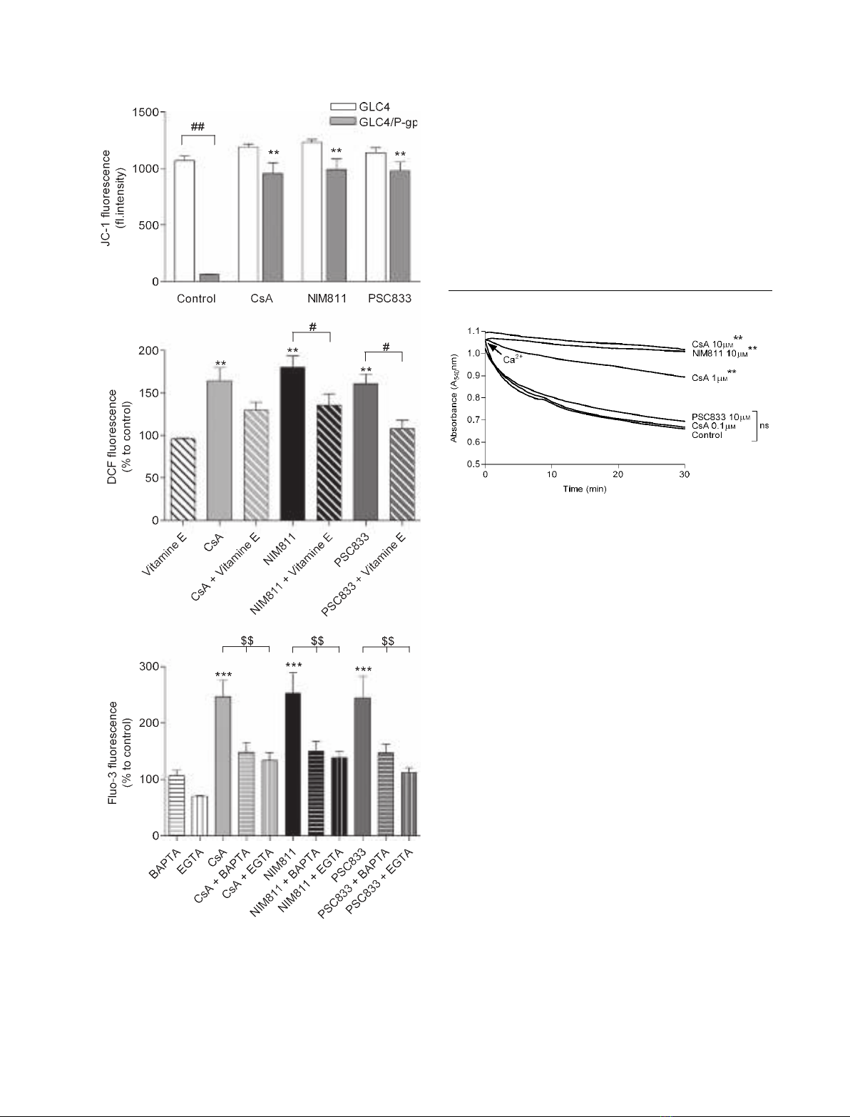
CsA (1 and 10 lm) and NIM811 (10 lm) significantly
reduced mitochondrial swelling, whereas CsA (0.1 lm)
and PSC833 (10 lm) did not.
Isolated mitochondria
To further examine whether closure of the MPTP
results in an increase in DW
m
, isolated mitochondria
were loaded with JC-1. After addition of succinate and
ADP, state III respiration was reached. Figure 5A
shows that CsA did not result in an increase in JC-1
fluorescence. In response to induction of state IV res-
piration, however, JC-1 fluorescence increased by
13.5 ± 2.8%. The protonophore 2,4-dinitrophenol
(DNP), which dissipates DW
m
, resulted in a significant
(50.7 ± 12.9%, P< 0.001) decrease.
Mitochondrial ATP levels were monitored during
state III respiration. CsA did not result in an increase
in ATP production (Fig. 5A). State IV respiration and
DNP were used as negative controls. State IV respir-
ation could not result in ATP production, because
there was no supply of ADP. Addition of DNP, an
established uncoupler of oxidative phosphorylation,
resulted in a decrease in ATP to 28.9 ± 4.5%
(P< 0.01) as compared to state III.
Fig. 4. Effects of different concentrations of CsA and its analogs
on the Ca
2+
-dependent induction of opening of the MPTP. The data
are representative of four experiments. A concentration of 1 mM
Ca
2+
was used to induce opening of the MPTP. CsA (1 and 10 lM)
and NIM811 (10 lM) caused significant inhibition of mitochondrial
swelling. **P< 0.01 versus control; ns, not significant by two-way
ANOVA.
A
B
C
Fig. 3. Effects of CsA (10 lM), NIM811 (10 lM) and PSC833
(10 lM) in GLC4 cells without expression of P-glycoprotein (GLC4)
and GLC4 cells expressing P-glycoprotein (GLC4 ⁄P-gp). (A) JC-1
(5 lgÆmL
)1
) was used to assess mitochondrial membrane potential.
(B) DCF (5 lgÆmL
)1
) was used to detect the generation of ROS. (C)
Fluo-3 (50 ngÆmL
)1
) was used to determine Ca
2+
levels. The data
presented are from four independent experiments, and represent
the mean value ± SEM. (A)
##
P< 0.01 versus GLC4; **P< 0.01
versus control. (B) **P< 0.01 versus control;
#
P< 0.05 versus
vitamin E (200 lM) treatment. (C) ***P< 0.001 versus control;
$$P< 0.01 versus BAPTA (10 lM) or EGTA (0.1 mM). P-values are
according to the Newman–Keuls multiple comparison test.
Cyclosporin A-induced oxidative stress M. van der Toorn et al.
3006 FEBS Journal 274 (2007) 3003–3012 ª2007 The Authors Journal compilation ª2007 FEBS
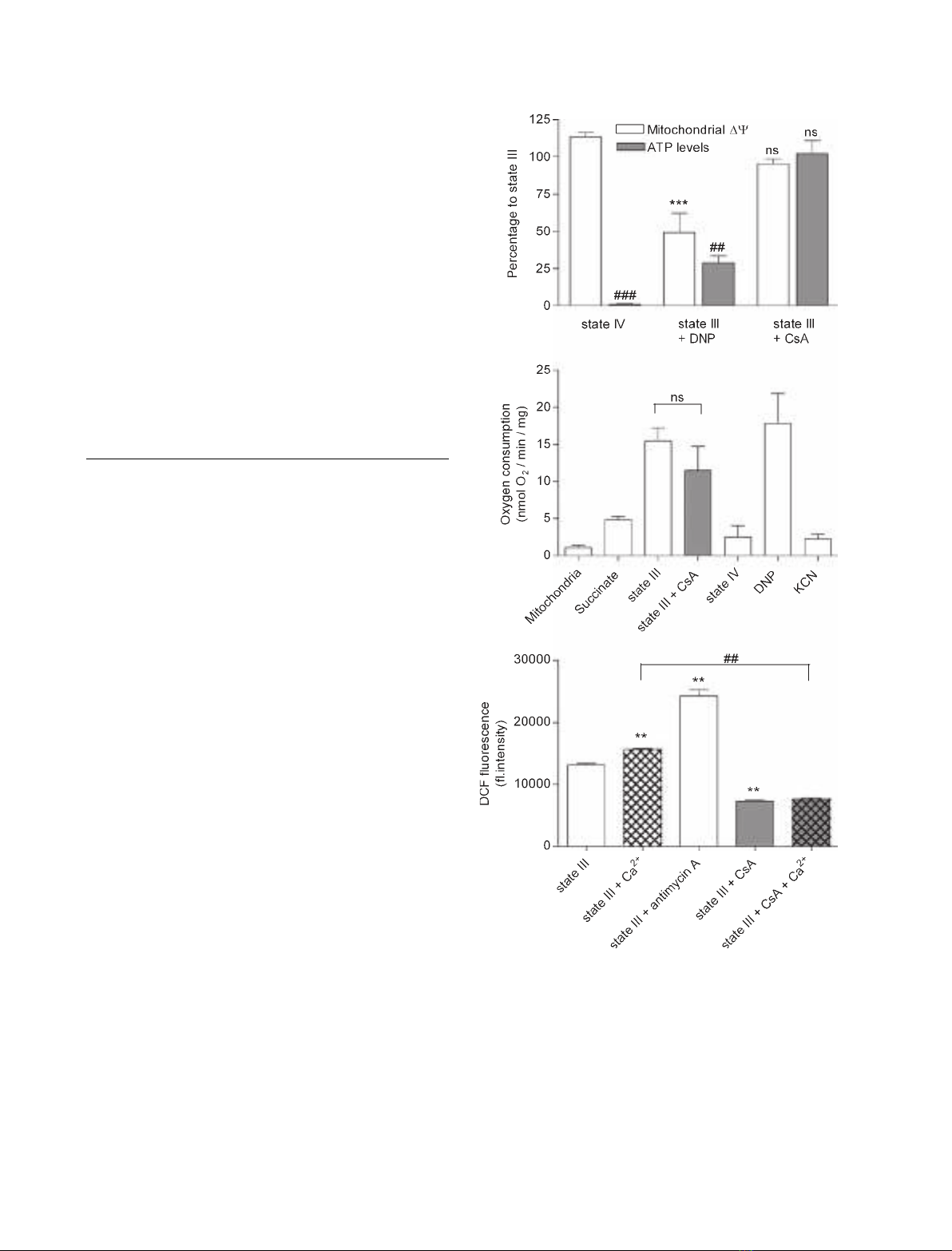
Oxygen consumption was monitored with sequential
addition of succinate, ADP (to induce state III respir-
ation) and CsA, until state IV respiration was reached
again, when all ADP was converted to ATP. DNP
was then added, followed by KCN (Fig. 5B). Isolated
mitochondria were incubated in an oxygraph sample
chamber with air-saturated respiration buffer in these
experiments. After addition of succinate as metabolic
substrate, mitochondria start to respire (4.8 ± 0.5
nmol O
2
Æmin
)1
Æmg
)1
). Addition of ADP causes a burst
of oxygen uptake (15.4 ± 1.8 nmol O
2
Æmin
)1
Æmg
)1
).
The respiratory control index was 3.2 ± 0.3. Addition
of CsA during state III respiration did not cause a sig-
nificant change in oxygen consumption as compared to
state III control. DNP was used as positive control.
Uncoupling of the mitochondria caused a burst of oxy-
gen uptake (17.8 ± 4.0 nmol O
2
Æmin
)1
Æmg
)1
). KCN,
a blocker of complex IV, was used as negative con-
trol. Addition of KCN acutely blocked respiration
of the uncoupled mitochondria (2.2 ± 6.8 nmol O
2
Æ
min
)1
Æmg
)1
).
Finally, we examined whether CsA exposure induces
changes in ROS production during state III respiration
in the presence and absence of 1 mmCa
2+
. Mitoch-
ondrial ROS production was monitored with DCF in
these experiments. Figure 5C shows that addition of
Ca
2+
results in a significant increase in DCF fluores-
cence. Antimycin A, a blocker of complex III and a
well-known inducer of ROS production [26], was used
as positive control. Addition of CsA resulted in signifi-
cant attenuation of DCF fluorescence during state III
respiration, both in the absence and in the presence of
Ca
2+
, with no significant difference between the latter
two conditions.
Discussion
In this study, we found that CsA induces increases in
the production of ROS and in cytosolic [Ca
2+
]. In
contrast to expectations, we found that these increases
A
B
C
Fig. 5. Effects of 10 lMCsA in isolated liver mitochondria. (A) Mit-
ochondrial membrane potential (DW
m
) and ATP levels. (B) Respir-
ation rate. (C) ROS. Measurements for assessment of DW
m
, ATP
levels and ROS were performed under different conditions. Meas-
urements of oxygen consumption for assessment of respiration
rate represent four experiments in which isolated mitochondria
were subsequently exposed to different conditions, starting with
respiration medium with mitochondria alone (indicated as mitochon-
dria) and ending with addition of KCN (indicated as KCN). JC-1
(0.2 lgÆmL
)1
) probe was used to monitor mitochondrial membrane
potential. Mitochondrial ATP levels were quantified by using a
chemiluminescent ATP assay. Mitochondrial respiration rate was
measured using an oxygraph. DCF (1 lgÆmL
)1
) was used to quan-
tify ROS. Data are expressed as mean value ± SEM and are repre-
sentative of four experiments. (A) (mitochondrial DW
m
)
***P< 0.001 versus state III; ns, not significant. (A) (ATP levels)
##
P< 0.01 versus state III;
###
P< 0.001 versus state III; ns, not
significant. (B) ns, not significant; (C) **P< 0.01 versus state III;
##
P< 0.01 versus state III + Ca
2+
.P-values are according to the
Newman–Keuls multiple comparison test.
M. van der Toorn et al. Cyclosporin A-induced oxidative stress
FEBS Journal 274 (2007) 3003–3012 ª2007 The Authors Journal compilation ª2007 FEBS 3007


