
Differentiation stage-dependent preferred uptake of
basolateral (systemic) glutamine into Caco-2 cells results
in its accumulation in proteins with a role in cell–cell
interaction
Kaatje Lenaerts, Edwin Mariman, Freek Bouwman and Johan Renes
Maastricht Proteomics Center, Nutrition and Toxicology Research Institute Maastricht (NUTRIM), Department of Human Biology,
Maastricht University, the Netherlands
Glutamine has an important function in the small
intestine with respect to maintaining the gut epithelial
barrier in critically ill patients [1,2]. Several studies
performed in different experimental settings reveal
that it serves as an important metabolic fuel for
enterocytes [3], and as a precursor for nucleotides,
amino sugars, proteins and several other molecules
such as glutathione [4,5]. In vitro cell culture studies
demonstrate that glutamine specifically protects intes-
tinal epithelial cells against apoptosis [6,7], has
trophic effects on the intestinal mucosa [8] and pre-
vents tumour necrosis factor (TNF)-alpha induced
bacterial translocation [9]. In experimental models
of critical illness, glutamine was able to attenuate
Keywords
apical and basolateral; barrier function;
clinical nutrition; intestinal cells; protein
turnover
Correspondence
K. Lenaerts, Maastricht Proteomics Center,
Nutrition and Toxicology Research Institute
Maastricht (NUTRIM), Department of
Human Biology, Maastricht University,
PO Box 616, 6200MD, Maastricht,
the Netherlands
Fax: +31 43 3670976
Tel: +31 43 3881509
E-mail: K.Lenaerts@HB.unimaas.nl
(Received 4 February 2005, revised 22 April
2005, accepted 3 May 2005)
doi:10.1111/j.1742-4658.2005.04750.x
Glutamine is an essential amino acid for enterocytes, especially in states of
critical illness and injury. In several studies it has been speculated that the
beneficial effects of glutamine are dependent on the route of supply (lumi-
nal or systemic). The aim of this study was to investigate the relevance of
both routes of glutamine delivery to in vitro intestinal cells and to explore
the molecular basis for proposed beneficial glutamine effects: (a) by deter-
mining the relative uptake of radiolabelled glutamine in Caco-2 cells;
(b) by assessing the effect of glutamine on the proteome of Caco-2 cells
using a 2D gel electrophoresis approach; and (c) by examining glutamine
incorporation into cellular proteins using a new mass spectrometry-based
method with stable isotope labelled glutamine. Results of this study show
that exogenous glutamine is taken up by Caco-2 cells from both the apical
and the basolateral side. Basolateral uptake consistently exceeds apical
uptake and this phenomenon is more pronounced in 5-day-differentiated
cells than in 15-day-differentiated cells. No effect of exogenous glutamine
supply on the proteome was detected. However, we demonstrated that exo-
genous glutamine is incorporated into newly synthesized proteins and this
occurred at a faster rate from basolateral glutamine, which is in line with
the uptake rates. Interestingly, a large number of rapidly labelled proteins
is involved in establishing cell–cell interactions. In this respect, our data
may point to a molecular basis for observed beneficial effects of glutamine
on intestinal cells and support results from studies with critically ill patients
where parenteral glutamine supplementation is preferred over luminal sup-
plementation.
Abbreviations
CBB, Coomassie brilliant blue; DMEM, Dulbecco’s modified Eagle’s medium; FBS, fetal bovine serum; IPG, immobilized pH gradient;
LI-cadherin, liver-intestine cadherin; PTFE, polytetrafluoroethylene.
3350 FEBS Journal 272 (2005) 3350–3364 ª2005 FEBS

proinflammatory cytokine expression and to improve
gut barrier function [1,10–12].
The intestinal cells obtain glutamine through exo-
genous and endogenous routes. The exogenous gluta-
mine comes from uptake of the amino acid itself or
of glutamine-containing peptides from the intestinal
lumen via transporters in their apical brush border
membranes [13], and from the bloodstream via their
basolateral membranes [14]. The endogenous glutamine
arises from conversion of glutamate and ammonia by
glutamine synthetase [15]. However, in human and rat,
intestinal glutamine synthetase activity is very low
[16,17]. This suggests that enterocytes strongly depend
on the external glutamine supply, either from the diet
or from the blood circulation.
In many studies it has been proposed that the bene-
ficial effect of glutamine is dependent on the dose and
route of supplementation. Data from a meta-analysis
suggested that glutamine supplementation in critically
ill patients may be associated with a decrease in com-
plications and mortality rate, particularly when deliv-
ered parenterally at high dose [18]. Panigrahi et al.
demonstrated that especially apical deprivation of glu-
tamine in Caco-2 cells resulted in a significant rise of
bacterial transcytosis [19]. Similar results were found
in HT-29 cells, where apical delivery of glutamine
decreased transepithelial permeability [20]. Le Bacquer
et al. reported that, regardless of its route of delivery,
glutamine is able to restore protein synthesis in cells
submitted to apical fasting [21]. Another study showed
that glutamine is utilized by the rat small intestine to
a similar extent when given by luminal or systemic
routes [22]. Hence, these studies indicate that both
luminal and systemic routes can be used interchange-
ably to supply the enterocytes with glutamine. Alto-
gether, these data do not allow a conclusion on the
preferred side of glutamine supplementation.
Although the uptake rate of lumen-derived and
blood-derived glutamine by the rat small intestine
ex vivo and in vivo has been reported [22,23], the relat-
ive uptake from each glutamine source in in vitro cell
culture systems is unknown. Another area that remains
unexplored is the overall influence of glutamine on
gene expression of intestinal cells, which may reveal
the underlying mechanism for the so-called ‘health’
effect of glutamine. In this respect, it is important to
know whether glutamine taken up by the cells from
the apical or basolateral side enters a common meta-
bolic pool.
The purpose of this study was to investigate the rele-
vance of the route of glutamine delivery to in vitro
intestinal cells and to explore a molecular basis for
the proposed beneficial effects of glutamine; (a) by
determining the relative uptake of glutamine; (b) by
searching for changes in the intestinal proteome; and
(c) by examining glutamine incorporation into cellular
proteins. The Caco-2 cell line was used for this study.
Although originally derived from a human colon
adenocarcinoma, the cells undergo spontaneous entero-
cytic differentiation and share many characteristics
with human small intestinal cells in their differentiated
state. Caco-2 cells form a polarized monolayer with
junctional complexes and a well-developed brush bor-
der with associated hydrolases [24–26]. This cell line is
commonly used in a Transwell system, which enables
an effective separation of the apical or ‘luminal’ and
the basolateral or ‘systemic’ compartment, similar to
the intestinal barrier in in vivo situations [27,28].
Results
Uptake of glutamine by differentiating Caco-2
cells
To determine whether the glutamine uptake is depend-
ent on the differentiation stage of Caco-2 cells, mono-
layers were exposed to radiolabelled glutamine for 1 h
at several time points after the formation of tight junc-
tions (from day 1 to day 15 after reaching confluence).
Three different concentrations of glutamine (0.1, 2.0
and 8.0 mm) were tested, administered from either the
apical or the basolateral side. Higher glutamine con-
centrations in the medium resulted in higher glutamine
uptake by the cells (Fig. 1A). Uptake of apically and
basolaterally administered glutamine was significantly
different at every time point, for each concentration
used. Basolateral exposure of the monolayers to gluta-
mine-containing medium for 1 h resulted in 15.3 ± 3.2
to 4.3 ± 0.7 times higher glutamine uptake compared
to apical exposure. The difference between apical and
basolateral glutamine uptake was smaller at the end of
the differentiation period. This originated from the fact
that basolateral l-[
3
H]glutamine uptake decreased con-
siderably during differentiation of the cells, especially
from day 6 postconfluence. Comparing day 1 with day
15, we observed a 2.0 ± 0.6, 1.8 ± 0.5 and 1.4 ± 0.3-
fold decrease, for, respectively, 0.1, 2.0 and 8.0 mm
basolateral glutamine, and only a 1.3 ± 0.2, 1.1 ± 0.2
and 1.3 ± 0.2-fold decrease for apical glutamine.
Time course of glutamine uptake in Caco-2 cells,
at two stages of differentiation
To investigate the influence of exogenous glutamine on
protein metabolism of Caco-2 cells, longer exposure
times are required. To see whether exogenously added
K. Lenaerts et al. Glutamine incorporation in Caco-2 proteins
FEBS Journal 272 (2005) 3350–3364 ª2005 FEBS 3351
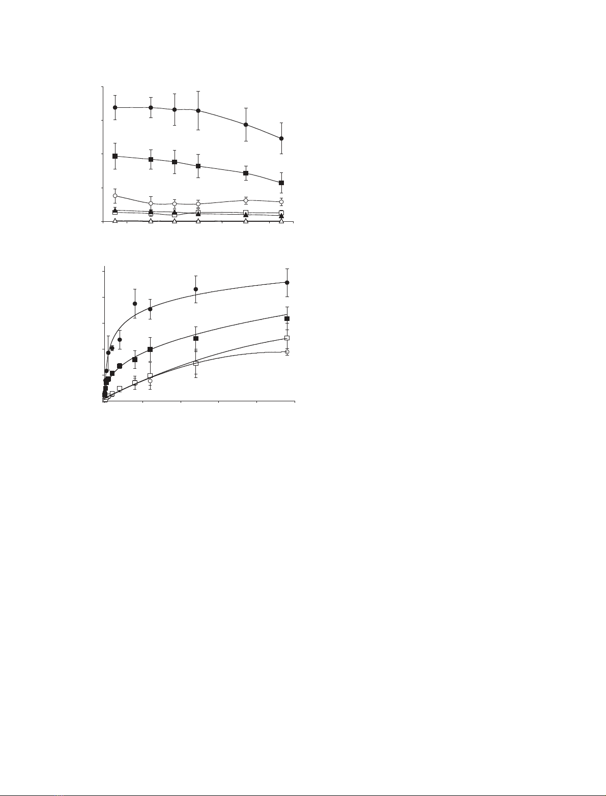
glutamine still contributed to the total glutamine pool
in a side-dependent way after prolonged supplementa-
tion, cells were exposed to 2.0 mmglutamine for 5 min
to 48 h. At day 5 (Fig. 1B, circles), basolaterally
administered glutamine led to a time-dependent
increase of label in the cells with a maximum at 24 h,
after which a steady state level was reached. Remark-
ably, an increase of radioactivity was observed at the
apical compartment of the Transwell system when
monolayers were exposed to radiolabelled glutamine
from the basolateral side, and vice versa (data not
shown). This was not due to leakage as paracellular
diffusion of phenol red was not observed. Therefore,
Caco-2 cells appeared not only to take up, but also to
expel or secrete (metabolized) glutamine. With apically
administered glutamine the accumulated label gradu-
ally increased till 48 h. At day 15 of differentiation
(Fig. 1B, squares) the absolute level of labelled gluta-
mine in the cells again remained higher when adminis-
tered from the basolateral side, but steady-state levels
were not yet reached.
Short exposure times (5 min to 30 min) did not
result in a significantly different basolateral ⁄apical
uptake ratio compared to the ratio obtained at 1 h
(data not shown). At 30 min the basolateral ⁄apical
uptake ratio was 9.1 ± 3.7 and 5.2 ± 0.3 for 5-day-
and 15-day-differentiated cells, respectively. At 24 h
the basolateral ⁄apical uptake ratio was 3.0 ± 0.6 and
1.7 ± 0.3 for 5-day and 15-day-differentiated cells,
respectively. This indicates that the basolateral ⁄apical
uptake ratio depends on the differentiation state of
Caco-2 cells. From these results, exogenous glutamine
supply to 5-day-differentiated cells for 24 h was selec-
ted as the optimal condition for further studies.
Effects of glutamine availability on protein
expression profiles of Caco-2 cells
To detect differences in protein expression related to
glutamine addition to the Caco-2 cells, proteins were
isolated from 5-day-differentiated cells exposed for
24 h to experimental medium containing 0.1, 2.0 and
8.0 mmglutamine from apical or basolateral side, and
separated by 2D gel electrophoresis. Approximately
1600 spots were detected per gel within a pH range
of 3–10, and a molecular mass range of 10–100 kDa.
When comparing spot intensities after different
glutamine treatment, none of them showed a signifi-
cant up- or down-regulation (data not shown).
Accumulation of L-[
2
H
5
]glutamine in proteins
of Caco-2 cells
We further investigated whether the supplied gluta-
mine was incorporated into proteins and whether this
was dependent on the delivery site. We examined this
using our newly developed method [29] based on
mass spectrometric detection of incorporated stable
isotope labelled amino acids into proteins. After incu-
bating Caco-2 monolayers for 0, 24, 48 and 72 h with
medium containing l-[
2
H
5
]glutamine from the apical
or the basolateral side, proteins were isolated from
0
40
80
120
160
0246810121416
0
50
100
150
200
250
0 1020304050
Uptake gln (nmol·mg protein-1)
Days after confluence
Uptake gln (nmol·mg protein-1)
Time (h)
A
B
Fig. 1. (A) Glutamine uptake in Caco-2 monolayers across the apical
(open symbols) and basolateral (closed symbols) membrane surface
at various stages of differentiation (at day 1, 4, 6, 8, 12 and day 15
postconfluence). Uptake was measured after exposing cells to
medium containing 0.1 mM(triangles), 2.0 mM(squares) and
8.0 mM(circles) glutamine, trace-labelled with 28.5 kBqÆmL
)1
L-[
3
H]glutamine for 1 h. Data represent mean ± SD for three mono-
layers. (B) Time course of apical and basolateral glutamine uptake
in Caco-2 monolayers. Apical (open symbols) and basolateral
(closed symbols) uptake was measured after exposing cells to
medium containing 2.0 mMglutamine, trace-labelled with 28.5
kBqÆmL
)1
L-[
3
H]glutamine, from apical or basolateral side for up to
48 h, at day 5 (circles) and day 15 postconfluence (squares). Data
represent mean ± SD for three monolayers.
Glutamine incorporation in Caco-2 proteins K. Lenaerts et al.
3352 FEBS Journal 272 (2005) 3350–3364 ª2005 FEBS
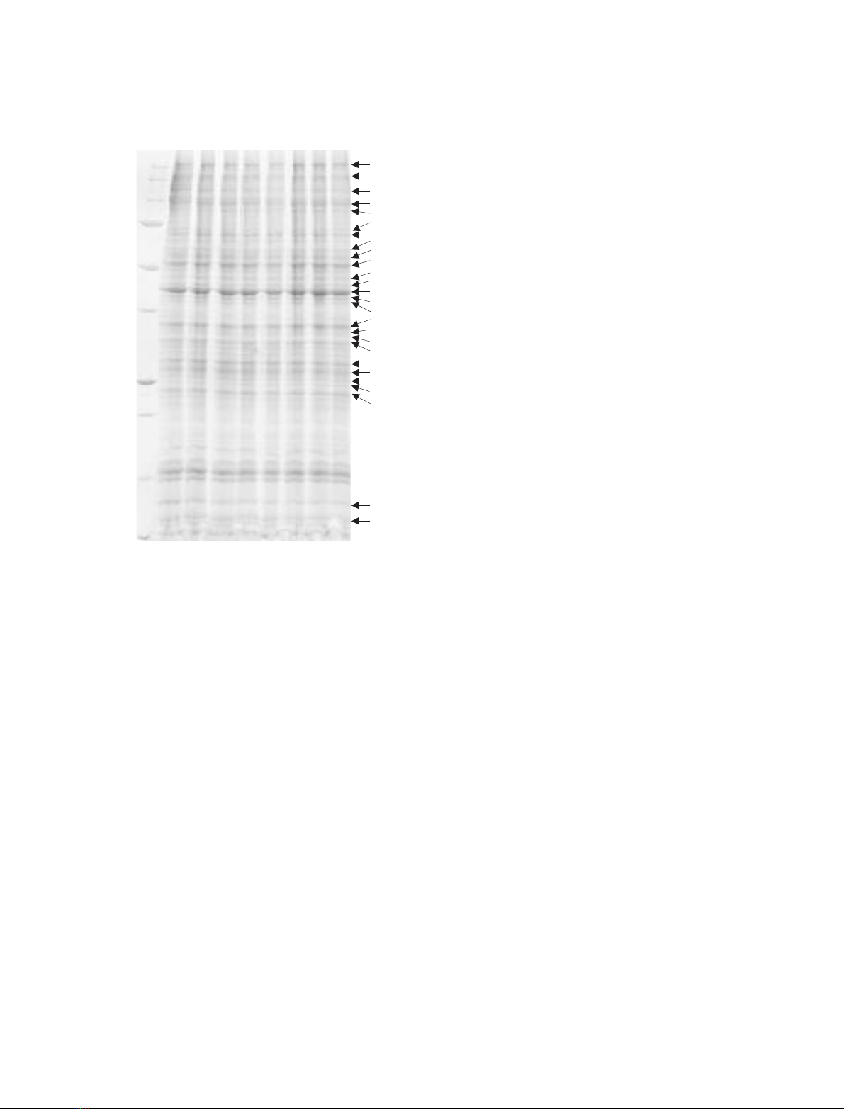
the cells and separated in one dimension by
SDS ⁄PAGE (Fig. 2). MALDI-TOF MS analysis of
36 clearly visible protein bands covering the entire
molecular mass range of the 1D gel led to the identi-
fication of 33 distinct proteins in 26 bands by search-
ing the Swiss-Prot database. This discrepancy is
explained by the fact that one band in the gel can
contain a mixture of several different proteins. Twelve
of those 33 proteins showed label incorporation
(Table 1). In addition, protein samples of Caco-2 cells
labelled with l-[
2
H
5
]glutamine for 0 and 72 h from
the apical or the basolateral side were separated by
2D electrophoresis. An example of a 2D gel is shown
in Fig. 3. From each gel, 120 protein spots were sub-
jected to MALDI-TOF MS analysis. This resulted in
the identification of 80 distinct proteins represented
by 114 spots in the gel, as some proteins were present
as more than one spot due to protein processing or
modification. In total, 20 proteins showed label incor-
poration (Table 2), from which eight proteins were
also detected as labelled in the 1D electrophoresis
experiment.
As an example the spectra and coverage maps of
actin and galectin-3, respectively, band 13 and 20 in
Table 1, are depicted in Fig. 4. Tryptic peptides that
were matched with peaks in the spectrum are boxed in
the amino acid sequence of the protein. A glutamine-
containing spectrum peak of actin at m⁄z1790 corres-
ponds to the tryptic peptide SYELPDGQVIT
IGNER, and was analyzed at high resolution. No sig-
nificant isotopomer peak (M+5) could be detected
after labelling with l-[
2
H
5
]glutamine for up to 72 h,
from either the apical or the basolateral side
(Fig. 5A,B). Hence this protein did not incorporate
labelled glutamine significantly during this time period.
On the contrary, analysis of such a peak of galectin-3
at m⁄z1650, which corresponds to the tryptic peptide
VAVNDAHLLQYNHR, clearly shows the appearance
of an isotopomer peak (M+5) after 24 h of labelling
(Fig. 5C,D). According to our criteria, labelling was
only significant after 48 h incubation with l-[
2
H
5
]gluta-
mine at the basolateral side. The isotopomer peak
appearing upon basolateral exposure to labelled gluta-
mine for 72 h is 57.9% of the original mass peak,
while the apical isotopomer peak is only 23.3% of the
original peak. These data demonstrate incorporation
of labelled glutamine into the protein galectin-3. Sim-
ilar results were obtained for 11 other proteins of the
1D gel (Table 1), and for 20 proteins of the 2D gel
(Table 2). This indicates that glutamine incorporates
into a common pool of proteins independent from the
site of application. The only difference is their rate of
labelling which is for most of the proteins at least
twice as high for basolaterally administered glutamine
compared to apically administered glutamine.
Discussion
Essential in this study is that the gut epithelial lining
utilizes glutamine from two sources, i.e. from the lumi-
nal and the systemic side. By using an in vitro cell
study approach, in which polarized human intesti-
nal Caco-2 cells cultured on Transwell inserts are
exposed to external glutamine from the apical or the
basolateral side, we were able to investigate the influ-
ence of the polarity on cellular glutamine uptake and
glutamine incorporation into proteins.
We demonstrated that compared to the apical side
the overall glutamine uptake from the basolateral side
is consistently higher. It is known that uptake of gluta-
mine across the apical (brush border) membrane of
Caco-2 cells is mainly dependent on three mechanisms
(a) Na
+
-dependent and (b) Na
+
-independent saturable
0 h AP
0 h BL
48 h AP
72 h AP
24 h BL
48 h BL
72 h BL
250
MW(kDa)
150
100
75
50
37
25
20
15
10
1
Band
2
3
4
5
6
7
8
9
10
11
12
13
14
15
16
17
18
19
20
21
22
23
24
25
26
24 h AP
Fig. 2. 1D pattern of proteins extracted from Caco-2 cells after
exposure to stable isotope labelled glutamine for 0, 24, 48 and 72 h,
apical (AP) and basolateral (BL). Protein bands were made visible by
Coomassie brilliant blue staining. The 26 indicated protein bands
were identified by MALDI-TOF MS and are depicted in Table 1.
K. Lenaerts et al. Glutamine incorporation in Caco-2 proteins
FEBS Journal 272 (2005) 3350–3364 ª2005 FEBS 3353

transport processes as well as (c) passive diffusion,
which even exceeds Na
+
-independent uptake at high
concentrations of glutamine (> 3.0 mm) [30–32]. The
Na
+
-dependent uptake of glutamine occurs mainly via
the Na
+
-dependent neutral amino acid transporter B
0
(ATB
0
), which is also expressed in Caco-2 cells [33]
and was found to mediate the majority of total gluta-
mine uptake across the apical membrane. Na
+
-inde-
pendent glutamine uptake in Caco-2 cells occurs
largely through system L [31]. Although it is suggested
that systemic (basolateral) glutamine plays an import-
ant role in enterocyte homeostasis and function [34],
also in intestinal injury [35], few data are available on
the uptake mechanisms of glutamine by the basolateral
membrane of Caco-2 cells. As mentioned above, sys-
tem L plays a role in glutamine uptake across the
brush border membrane of Caco-2 cells and it is sug-
gested that especially LAT-1, the first isoform of sys-
tem L, is responsible for that [36]. A second isoform of
this system, known as LAT-2, is prominently expressed
in the basolateral membranes of epithelial cells in the
villi of the mouse intestine [37]. A study performed in
Caco2-BBE cells also showed a basolateral localization
of LAT-2 [38]. As the Caco2-BBE cell line is a clone
isolated from the cell line Caco-2 [39], it is most likely
that the LAT-2 protein has a similar distribution pat-
tern in the cells used in this study. In addition, experi-
ments with rodent and human LAT isoforms revealed
Table 1. List of identified proteins from bands of the 1D gel. Thirty-three proteins from 26 bands (see Fig. 2) were identified by MALDI-TOF
MS and semiquantitative analysis of glutamine-containing peptides and the corresponding isotopomer peaks at high resolution revealed signi-
ficant labelling of 12 proteins, which are indicate in bold. NQ, No glutamine-containing peptides in spectrum peaks.
Band
Accession
number Protein name
Peak ratio ( ·100%) Peak ratio ( ·100%)
m⁄z24 h AP 48 h AP 72 h AP 24 h BL 48 h BL 72 h BL
1 O15061 Desmulin 1608 7.7 12.3 13.7 11.4 21.0 18.8
2 Q00610 Clathrin heavy chain 1 NQ
3 Q12864 Cadherin-17 [precursor] 1547 4.3 20.1 23.9 21.1 – 73.2
4 O43707 Alpha-actinin 4 1174 7.8 14.3 20.9 12.1 33.4 75.1
P14625 Endoplasmin [precursor] 1081 5.3 15.2 18.4 4.4 13.8 24.1
5 P08238 Heat shock protein HSP 90-beta 2257 12.2 12.2 14.4 2.6 10.3 24.0
P09327 Villin 1 NQ
6 P38646 Stress-70 protein, mitochondrial [precursor] 1695 11.5 18.7 19.5 10.2 21.8 33.7
7 P31040 Succinate dehydrogenase [ubiquinone]
flavoprotein subunit, mitochondrial [precursor]
1268 0.0 2.0 11.0 3.6 3.8 14.0
8 P10809 60 kDa heat shock protein, mitochondrial [precursor] 1919 5.6 8.6 23.2 2.1 4.7 10.2
9 P30101 Protein disulfide-isomerase A3 [precursor] 1515 9.5 10.0 21.2 14.1 29.3 42.2
P07237 Protein disulfide-isomerase [precursor] 1834 5.7 14.8 21.2 15.4 27.8 47.9
10 P05787 Keratin, type II cytoskeletal 8 1079 0.0 0.0 3.6 3.3 3.3 7.3
P00367 Glutamate dehydrogenase 1, mitochondrial [precursor] 1738 5.0 6.9 9.0 1.6 2.6 8.2
11 P50454 Collagen-binding protein 2 [precursor] 1293 16.7 22.5 34.9 11.3 23.7 58.4
12 P04181 Ornithine aminotransferase, mitochondrial [precursor] 1811 – 21.3 16.5 19.5 53.2 64.0
13 P60709 Actin, cytoplasmic 1 1791 2.5 6.0 9.4 1.6 3.2 6.3
14 P08727 Keratin, type I cytoskeletal 19 1675 4.6 9.1 14.6 10.5 17.7 30.3
15 P00505 Aspartate aminotransferase, mitochondrial [precursor] 1449 0.0 2.3 2.5 11.0 18.0 21.6
16 P07355 Annexin A2 1111 1.9 5.0 10.1 14.1 26.1 36.8
P22626 Heterogeneous nuclear ribonucleoprotein A2 ⁄B1 1087 1.5 5.4 5.6 3.4 4.9 8.3
17 P09651 Heterogeneous nuclear ribonucleoprotein A1 1049 15.4 3.0 5.2 0.0 0.0 10.2
Q07955 Splicing factor, arginine ⁄serine-rich 1 NQ
18 P09651 Heterogeneous nuclear ribonucleoprotein A1 1628 7.8 8.2 12.1 9.0 13.4 15.4
19 P09525 Annexin A4 1118 1.2 9.7 14.1 13.3 30.5 49.0
20 P17931 Galectin-3 1650 10.5 16.9 23.3 25.4 38.8 57.9
P35232 Prohibitin 1396 4.0 5.9 9.6 5.0 7.1 11.8
21 P30084 Enoyl-CoA hydratase, mitochondrial [precursor] 1467 2.5 8.2 16.5 3.7 14.2 21.4
22 P60174 Triosephosphate isomerase 1458 0.1 2.0 6.9 3.3 5.1 10.4
23 P09211 Glutathione S-transferase P 1883 5.7 9.9 13.4 13.9 25.3 37.3
24 P62820 Ras-related protein Rab-1 A 1316 3.3 8.4 14.7 8.16 19.0 32.7
P51149 Ras-related protein Rab7 1187 13.6 24.4 36.2 36.0 76.3 97.5
25 P61604 10 kDa heat shock protein, mitochondrial 1325 2.1 2.4 4.6 4.3 5.7 3.7
26 P62805 Histone H4 NQ
Glutamine incorporation in Caco-2 proteins K. Lenaerts et al.
3354 FEBS Journal 272 (2005) 3350–3364 ª2005 FEBS


![Báo cáo seminar chuyên ngành Công nghệ hóa học và thực phẩm [Mới nhất]](https://cdn.tailieu.vn/images/document/thumbnail/2025/20250711/hienkelvinzoi@gmail.com/135x160/47051752458701.jpg)


