
BioMed Central
Page 1 of 7
(page number not for citation purposes)
Journal of Negative Results in
BioMedicine
Open Access
Research
Failure of E. coli bacteria to induce preterm delivery in the rat
Emmet Hirsch*1,2, Yana Filipovich1 and Roberto Romero3
Address: 1Department of Obstetrics and Gynecology, NorthShore University HealthSystem, Evanston, IL, USA, 2Department of Obstetrics and
Gynecology, Feinberg School of Medicine, Northwestern University, Chicago, IL, USA and 3Perinatology Research Branch, Eunice Kennedy Shriver
National Institute of Child Health and Human Development, National Institutes of Health, Department of Health and Human Services, Bethesda,
MD and Detroit, MI, USA
Email: Emmet Hirsch* - ehirsch@northshore.org; Yana Filipovich - yfilipovich@netscape.net; Roberto Romero - prbchiefstaff@med.wayne.edu
* Corresponding author
Abstract
Background: We sought to develop a model of bacterially induced preterm delivery in rats to
parallel similar models in mice.
Methods: Female Sprague-Dawley rats on day 17 of gestation (normal term = 21–22 days) were
inoculated into the uterus with either 2 × 10⁹ – 7 × 10¹⁰ killed E. coli organisms, 1 – 4 × 10⁸ live E.
coli or sterile solution. These inoculations were made either via trans-cervical catheter or by direct
intrauterine injection at laparotomy. Animals were then observed for delivery for variable periods
up to term. Necropsies were performed and fetal viability was assessed.
Results: No rats delivered prematurely after bacterial exposure (27 animals observed for at least
48 hours), and all animals followed to term (n = 3) delivered live pups. No dams exhibited signs of
systemic illness. There was a statistically significant but small negative effect of killed E. coli on fetal
viability (100% of 80 fetuses from 6 control pregnancies and 93% of 182 fetuses from 14 bacterially-
treated pregnancies were alive at necropsy, p = 0.014). Live bacteria had a larger effect on fetal
viability, with only 64% of 14 fetuses, 47% of 28 fetuses and 32% of 31 fetuses surviving after trans-
cervical administration of 7 × 10⁷, 2 × 10⁸ and 4 × 10⁸ E. coli, respectively.
Conclusion: Unlike mice, it has proven difficult to induce preterm labor in the rat using E. coli as
a stimulating agent. The relevant literature is reviewed and hypotheses are offered to explain this
phenomenon.
Background
Preterm birth is the major cause of neonatal morbidity
and mortality in the developed world [1]. Infection within
the gestational compartment is thought to be the primary
cause of a large proportion of cases of preterm labor,
accounting for as many as 50% or more of premature
deliveries, especially at very early gestational ages. Given
the complexity inherent in the processes of parturition
and the obstacles to conducting properly controlled and
prospective studies in human subjects, animal models
have proven helpful in developing an understanding of
the physiology and pathophysiology of parturition [2].
Novel insights have been generated in animal models of
infection-induced preterm labor in mice using a wide
range of inflammatory stimuli from a large number of
labs (small representative sample found in references [3-
9]), including our own. Other well established models
exist in rabbits [10-13] and non-human primates [14-16].
Published: 4 January 2009
Journal of Negative Results in BioMedicine 2009, 8:1 doi:10.1186/1477-5751-8-1
Received: 8 August 2008
Accepted: 4 January 2009
This article is available from: http://www.jnrbm.com/content/8/1/1
© 2009 Hirsch et al; licensee BioMed Central Ltd.
This is an Open Access article distributed under the terms of the Creative Commons Attribution License (http://creativecommons.org/licenses/by/2.0),
which permits unrestricted use, distribution, and reproduction in any medium, provided the original work is properly cited.

Journal of Negative Results in BioMedicine 2009, 8:1 http://www.jnrbm.com/content/8/1/1
Page 2 of 7
(page number not for citation purposes)
In order to capitalize on certain advantages afforded by
the rat over the mouse (such as larger tissue samples and
more easily conducted invasive monitoring), we sought to
develop a model of infection-induced preterm labor in
the rat. In this paper we report on the failure of this
attempt, offer hypotheses for the causes of this failure and
suggest possible directions for future study. It is notable
that although there are abundant studies of rat parturition
induced by progesterone antagonism [17-19] only two
groups have reported preterm delivery with an infectious/
inflammatory stimulus in rats in a total of three publica-
tions [20-22].
Methods
Rats
All procedures involving animals were approved by the
Institutional Animal Care and Use Committee of North-
Shore University HealthSystem. Timed pregnant Sprague-
Dawley rats (254 – 380 gm) were obtained from Harlan
(Indianapolis, IN) on day 15 of pregnancy and allowed to
acclimate in the animal facility until the experiment was
performed. In most cases and unless otherwise stated,
experiments occurred on day 17 of a 21–22 day rat preg-
nancy.
Bacterial preparation
Bacteria (a pathogenic strain of E. coli originally isolated
from a patient with urosepsis and obtained from the
American Type Culture Collection (ATCC #12014)) were
stored in 50% glycerol at -80°C and processed as previ-
ously described [4,23]. Live and heat-killed bacteria from
this strain have been used repeatedly by our group to
induced preterm delivery in mice.
For experiments with killed bacteria, a small chip of the
stock culture was thawed in Luria-Bertani (LB) medium,
passaged 4 times prior to overnight culture in 4 L of
medium and concentrated by centrifugation. The bacteria
were then washed three times in phosphate-buffered
saline (PBS) and resuspended in 8 ml of PBS, yielding a
concentration of 1.7 × 10¹¹ bacteria per ml. This dense live
bacterial prep was quantitated by plating serial dilutions
for overnight culture just prior to the remaining stock
being killed by boiling for 5 minutes. Non-viability of this
boiled preparation was confirmed by overnight incuba-
tion of both plate and broth cultures. The stock of killed
E. coli was aliquoted and frozen at -80°C. Prior to each
experiment using killed bacteria, an aliquot was thawed
and diluted in PBS to the desired concentration.
For experiments with live bacteria, E. coli were taken either
from the frozen stock or from fresh plates and passaged
twice, including one overnight culture in 3 ml of LB
medium. At 10 AM, 100 μl of the overnight culture was
added to 5 ml of sterile medium and grown at 37°C for 4
hours, when surgeries were performed. This bacterial prep
was used undiluted and was kept at room temperature
during surgery. The culture was quantified by plating
before the first procedure and after the last procedure of
the day, and the pre- and post-procedure counts were
averaged to determine the inoculum used.
Surgical procedures
Rats were anesthetized with a mixture of 10 mg/kg Keta-
mine and 1 mg/kg Xylazine intraperitoneally. They were
then immobilized in the supine position with the external
genitalia slightly overhanging the edge of the lab bench. A
3 mm lacrimal duct scope attached to an external light
source was advanced into the vagina. Polyethylene tubing
(Becton Dickinson #427416, I.D. 0.76 mm (.030") O.D.
1.22 mm (.048")) was joined to the barrel of the scope
through a narrow channel created from strips of tape. The
tubing could be advanced or withdrawn for cannulation
of the cervix under direct visualization. The cannula
(primed with injectate to remove air) was advanced for a
distance of approximately 1 cm through the cervical open-
ing prior to injection of 100 – 1000 μl of solution. Various
infusion volumes were used to assess their potential dif-
ferential effects. After an average of 30 seconds (to allow
for fluid distribution through the uterine horn while min-
imizing egress through the cervix), the cannula was with-
drawn and visualization continued for another 30
seconds to assess whether fluid leakage occurred. The rat
was then returned to her cage and allowed to awaken from
anesthesia.
Original attempts using both an external pressurized air
supply and a hand-operated bulb to distend the vagina to
ease visualization of the cervix were abandoned due to air
entry into the uterine horns. Thereafter, we were easily
able to visualize and catheterize the cervical orifice with-
out any source of pressurized air.
In general, animals tolerated these procedures well. Four
rats died within 20 hours of surgery (presumably from
anesthetic complications, as no specific findings were
seen on necropsy). An additional 2 animals developed a
severe shock-like syndrome, again with no findings on
necropsy. These 6 subjects were excluded from the analy-
sis.
In some cases, direct intrauterine injection of bacteria was
performed at laparotomy to assess the effects of route of
administration. These abdominal procedures were fash-
ioned after our existing mouse model [4,23]. A 2 cm mid-
line incision was made in the lower abdomen and the
right uterine horn was exposed. Bacteria or sterile medium
were then injected in volumes of 100 – 1000 μl into the
midsection of the right uterine horn at a site between two
adjacent fetuses. The peritoneal cavity was then closed

Journal of Negative Results in BioMedicine 2009, 8:1 http://www.jnrbm.com/content/8/1/1
Page 3 of 7
(page number not for citation purposes)
with interrupted sutures and the skin was closed with sta-
ples.
Surgical procedures lasted 10 – 15 minutes. Animals
recovered in individual, clean cages in the animal facility
and underwent twice-daily observations. The number of
live-born or dead pups was noted. Preterm delivery was
defined as the finding of at least one fetus in the cage or in
the lower vagina within 48 hours of surgery.
In some cases, early euthanasia and necropsy were per-
formed to assess catheter placement, dye distribution or
fetal viability. Fetal viability was assessed by color, spon-
taneous movement and visible vascular pulsations in
either the chest cavity or the vessels within fetal mem-
branes. Other animals were observed for various periods
of time through one week after delivery to record other
observations, such as time to delivery and viability of
delivered pups. Animals were euthanized by CO2 inhala-
tion plus thoracotomy.
Statistics
Statistical analysis of categorical values was by chi-square
or contingency tables, with Fisher exact correction as
needed.
Results
Development of a trans-cervical uterine infusion model in
the rat
A total of 9 rats on days 14–18 of pregnancy underwent
catheterization and immediate euthanasia to assess cathe-
ter position. A separate group of 14 pregnant rats on ges-
tational days 17–19 (7 rats on day 17, 6 rats on day 18 and
1 rat on day 19) was instilled with PBS with or without
sterile bromphenol blue dye in a total volume of 100–500
μl and observed for up to 70 hours to characterize the dis-
tribution of dye and any fetal effects.
After an initial learning period, we were able consistently
to catheterize one uterine horn to the desired distance of
1 cm and infuse it with the dye solution with retention of
dye in the uterine horn, continued viability of fetuses (ver-
ified by both immediate and delayed necropsy) and min-
imal leakage out the cervix and vagina. No control
animals delivered within 48 hours, the previously deter-
mined cutoff for preterm delivery (n = 14), and 11 of 12
rats followed to term delivered healthy litters (averaging
11.1 ± 2.7 pups per litter). The 12th rat had vaginal bleed-
ing 20 hours after surgery and upon necropsy was found
to have 6 absorbing conceptuses in one uterine horn
along with 10 empty implantation sites distributed in
both horns, suggestive of prior delivery from these sites.
Therefore, we concluded that the trans-cervical infusion of
various volumes of sterile solution was feasible and rarely
affected the course of pregnancy.
Trans-cervical intrauterine infusion of a pathogenic strain
of killed E. coli fails to induce preterm delivery
The trans-cervical infusion method was used to instill 2 ×
109 – 8 × 10¹⁰ killed E. coli organisms into the uteri of 14
pregnant rats at 17 days of gestation (Table 1). None of
these animals delivered within 48 hours. Among these
pregnancies, 6 were euthanized and necropsied at 48
hours to assess fetal status (see results below). The remain-
ing 8 rats were observed for 70 hours without any preterm
deliveries. No animals exhibited signs of systemic illness
(such as piloerection, decreased mobility or decreased
feces production).
Intrauterine infusion of live E. coli fails to induce preterm
delivery
Our existing mouse model is characterized by a dramatic
difference in potency of killed versus live bacteria for
induction of preterm delivery (live bacteria are more
potent by 5–6 orders of magnitude, with as few as 2000
organisms leading to preterm delivery) [4]. Therefore, we
also experimented with live bacteria in an attempt to max-
imize the potential for inducing preterm delivery in rats.
Eight pregnant rats were inoculated trans-cervically with 1
– 4 × 10⁸ live E. coli organisms. None of these animals
delivered prematurely, including 3 animals that were fol-
lowed to term, each of whom delivered live pups. None of
the rats treated with live bacteria showed signs of systemic
illness.
Finally, in order to investigate the effect of method of
inoculation, 5 pregnant females were inoculated with 2 –
4 × 10⁸ live E. coli organisms by direct injection into the
Table 1: Effect of intrauterine E. coli inoculation on preterm delivery.
Treatment Number of E. coli organisms Number of animals Preterm delivery rate (%)
Trans-cervical infusion
Control N/A 16 0
Killed E. coli 0.2 – 8 × 10¹⁰ 14 0
Live E. coli 1 – 4 × 10880
Infusion at laparotomy
Control N/A 1 0
Live E. coli 2–7 × 10850
'Preterm' was defined as 48 hours after surgery, however some animals were observed for longer periods, up to term (see text).
Journal of Negative Results in BioMedicine 2009, 8:1 http://www.jnrbm.com/content/8/1/1
Page 4 of 7
(page number not for citation purposes)
right uterine horn at laparotomy. None of these 5 rats
delivered prematurely.
Fetal effects of intrauterine bacterial infusion
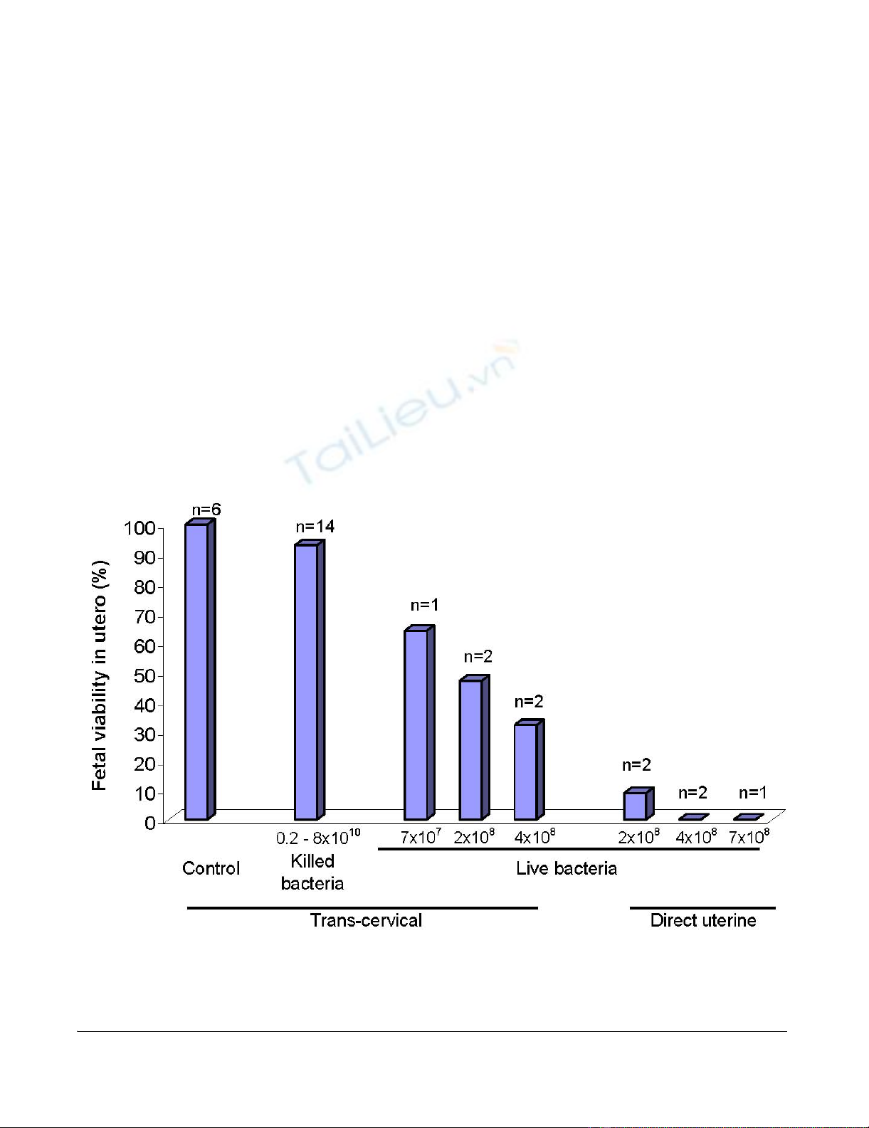
The effect of intrauterine bacterial infusion on fetal viabil-
ity was assessed by timed necropsy after surgery. One hun-
dred percent of 80 fetuses from 6 mothers who received
control (non-bacterial) injections and were euthanized 24
– 70 hours later were still alive at the time of necropsy.
Also, as noted above, dams followed to term after control
injections delivered large, healthy litters. In 14 litters
treated with 2 × 10⁹ – 8 × 10¹⁰ killed bacteria, 93% of 182
fetuses were alive at necropsy 48 – 70 hours later (p =
0.014, comparing control to killed bacterial injection).
There were no significant trends associating inoculum size
with fetal survival. Thus, killed E. coli has a statistically sig-
nificant though small impact on fetal viability.
In contrast, live bacteria had a large effect on fetal viability
that was dependent upon route of administration. Trans-
cervical inoculation with live E. coli was associated with
survival at necropsy 48 hours later in 64% of 14 fetuses,
47% of 28 fetuses and 32% of 31 fetuses from 5 pregnant
rats administered 7 × 10⁷, 2 × 10⁸ or 4 × 10⁸ live E. coli,
respectively (p = 0.13) (Figure 1). Direct administration of
live bacteria by intrauterine injection at laparotomy pro-
duced even more profound effects on fetal survival, with
only 3.4% of 63 fetuses from 5 pregnant dams still alive at
necropsy 48 hours later (p = 0.0006 for comparison of the
trans-cervical and direct injection routes of administration
of 2 × 10⁸ bacteria and p = 0.003 for comparison of the
two routes of administration of 4 × 10⁸ bacteria). Finally,
each of 3 pregnant dams who received 1 – 2 × 10⁸ live E.
coli trans-cervically and were followed through spontane-
ous delivery produced live pups at term, but their number
was diminished (average 3 per litter), compared with 10.2
pups per litter in control animals (p = 0.001).
Discussion
The present study describes the inability to induce pre-
term delivery in pregnant rats using live or killed E. coli.
This experience stands in sharp contrast to published
Effect of intrauterine treatment on fetal viabilityFigure 1
Effect of intrauterine treatment on fetal viability. Female rats on day 17 of pregnancy were inoculated with either ster-
ile solution (control), killed E. coli or live E. coli bacteria, by either trans-cervical infusion or direct intrauterine injection at
laparotomy. Animals were euthanized 24–70 hours later and fetal viability was determined. Displayed on the Y-axis is the per-
centage of fetuses alive at the time of autopsy. The number of treated dams in each group is also displayed.

Journal of Negative Results in BioMedicine 2009, 8:1 http://www.jnrbm.com/content/8/1/1
Page 5 of 7
(page number not for citation purposes)
experience with mice, in which live or killed E. coli as well
as multiple other inflammatory/infectious stimuli pro-
duced preterm delivery [3,4,6-9,24,25]. These stimuli
include a variety of bacteria, pathogen extracts (e.g. LPS,
peptidoglycan, lipoteichoic acid, poly inosinic:cytidylic
acid), cytokines (e.g. IL-1, TNF), and others. The strain of
E. coli used in the present study has been used successfully
in models of preterm birth not only by our group but by
others also in mice [8,26] and rabbits [12,27].
We could identify in the literature only two laboratories
that reported induction of preterm delivery using an infec-
tious/inflammatory stimulus in rats [20-22]. In work from
the first group, Sprague Dawley rats underwent intrauter-
ine implantation of catheters on day 15 or 16 followed on
day 17 of pregnancy by infusion of 25 μg or 50 μg of LPS
over 4 hours. Saline treatment was followed by delivery in
an average of 117 hours, whereas 25 μg and 50 μg of LPS
produced delivery in 82 and 63 hours, respectively,
together with increased production of prostaglandin F2α
metabolite in placentas of LPS-exposed animals [20]. In a
separate study from the same group, a 50 μg infusion of
LPS produced preterm delivery in an average of 92 hours,
which was approximately one day before delivery in
saline-treated rats [22]. Treatment with IL-10 restored
delivery to term. It is not clear why there was a discrepancy
of nearly 30 hours in the interval to delivery between
these two studies in rats treated with 50 μg LPS. In a com-
ment, the authors note that slow infusion over 4 hours
appears to be important for 'optimal results' [20]. For our
studies we chose 48 hours as a reasonable cut-off for 'pre-
term delivery' to avoid approaching term and because this
cutoff is practical in the mouse (in which delivery in sim-
ilar models usually occurs in less than 24 hours). When
delivery did not occur in this time frame, we extended our
observations to term and still did not observe early deliv-
ery.
In work from the second group, Wistar rats were treated
with 25 μg/kg of E. coli LPS intraperitoneally on day 16 of
pregnancy [21]. Although no animals in this study were
treated with control injections, the average time to deliv-
ery was approximately 22 hours. It should be noted that
assuming an average weight of 333 gm per rat, these sub-
jects would have received 8.3 μg of LPS each. Somewhat
surprisingly, intraperitoneal treatment with erythromycin
significantly prolonged the interval to delivery, by up to
50 hours.
Whether the different potencies reflected in the above two
experimental designs is due to route of administration
(intrauterine versus intraperitoneal), rate of infusion, rat
strain (Sprague-Dawley versus Wistar) or some other fac-
tor is not clear. In the present report, Sprague Dawley rats
treated with 2 × 10⁹ – 8 × 10¹⁰ killed E. coli would have
received between 20 μg and 800 μg of LPS each, assuming
that each E. coli bacterial cell contains 9.7 × 10⁻¹⁵ gm LPS
[28].
Far more numerous than the 3 studies cited above that
demonstrate preterm birth after LPS administration are
the studies that use hormonal manipulation (primarily
progesterone antagonism) to induce preterm delivery in
rats (examples to be found in [18,29,30]). This notable
fact is reinforced by another, namely that the literature
contains several examples of experimental LPS exposure
in rats after which observation periods of sufficient length
were conducted to effectively rule out subsequent preterm
delivery. For example, E. coli-derived LPS (400 μg/kg intra-
peritoneally) induced cervical changes in Sprague Dawley
rats when administered on day 14 of pregnancy [30]. Ani-
mals were harvested 2 days after treatment with no cases
of preterm delivery reported. In a study of the effect of pre-
natal LPS on neurobehavioral development, pregnant
Wistar rats were administered 2 mg/kg of E. coli LPS sub-
cutaneously on each day of pregnancy from conception
through delivery [31]. Though maternal LPS treatment
induced significant changes in neonatal neurobehavioral
outcomes as well as dopaminergic neurotransmission and
synaptophysin expression, there was no reported effect on
timing of delivery. A different study in which Sprague
Dawley rats were treated with LPS (100 μg/kg intraperito-
neally) every day from day 14 to day 20 of pregnancy
demonstrated a significant increase in fetal death and
reduced size of surviving fetuses compared with controls
[32]. Within the uteri of treated dams there was increased
apoptosis, a doubling of TNFα levels and a tripling of
nitric oxide and myeloperoxidase levels, however no pre-
term labor was reported. In a study focusing on fetal/neo-
natal brain injury, E. coli LPS treatment (500 μg/kg
intraperitoneally) of pregnant Sprague Dawley rats on day
18 and again on day 19 of pregnancy produced white mat-
ter injury, myelination defects and apoptosis and
increased expression of IL-1β, TNFα and IL-6 in pup
brains 7 days after birth [33]. Again, no preterm delivery
was reported. In a Chinese language publication available
in English in abstract form, treatment of Sprague Dawley
rats with intraamniotic LPS (quantity not provided) on
day 15 of pregnancy produced both histologic changes
and reduction in expression of vascular endothelial
growth factor (VEGF) and its receptors (Flk-1 and Flt-1) in
neonatal lungs, but animals delivered at term [34].
Why has the induction of inflammation-induced preterm
birth been so difficult and variable in the rat, while it is so
readily accomplished in mice and other species? The
results cited above and the fact that in the present study
there were measurable effects on fetal survival suggest that
the pregnant Sprague Dawley strain of rat is responsive to
E. coli. Among the demonstrable effects of LPS in pregnant


![Báo cáo seminar chuyên ngành Công nghệ hóa học và thực phẩm [Mới nhất]](https://cdn.tailieu.vn/images/document/thumbnail/2025/20250711/hienkelvinzoi@gmail.com/135x160/47051752458701.jpg)


