
BioMed Central
Page 1 of 9
(page number not for citation purposes)
World Journal of Surgical Oncology
Open Access
Research
Expression of the metalloproteases MMP-1, MMP-2, MMP-3,
MMP-9, MMP-11, TIMP-1 and TIMP-2 in angiocentric midfacial
lymphomas
Abelardo Meneses-García1, Alejandro Mohar Betancourt1,
Jorge Herrera Abarca2, Adriana Becerril Montes2, Lourdes Suarez Roa1 and
Luz Ruíz-Godoy*3
Address: 1Pathology department, Instituto Nacional de Cancerología, México, 2Pharmacology department, Instituto Politécnico Nacional, México
and 3Basic research, Instituto Nacional de Cancerología, México
Email: Abelardo Meneses-García - aameneses@hotmail.com; Alejandro Mohar Betancourt - moharalejandro@hotmail.com;
Jorge Herrera Abarca - macinves@hotmail.com; Adriana Becerril Montes - abmontes@ipn.mx; Lourdes Suarez Roa - lusuroa@gmail.com;
Luz Ruíz-Godoy* - lruizgodoy@gmail.com
* Corresponding author
Abstract
Background: Extranodal T/NK cell lymphomas possess distinctive clinico-pathological
characteristics: they are angiocentric, exhibit extensive necrosis. Prognosis is poor in the short
term. The objective is to explore the expression of different MMPs in the cells and stroma which
are around of the blood vessels damaged and their correlation with clinico-pathological
parameters.
Patients and methods: Twenty cases of this type of lymphomas were studied and collected
patient clinical data. The expressions of MMP-1, 2, 3, 9, 11, 13 and TIMP-1, 2 were studied by
immunohistochemistry. Ultrastructural studies were performed in two cases. Statistical analysis
was done with Fisher's exact test, Chi2 test.
Results: Of the 20 patients, 13 were men with median age of 43 years. In 13 patients the primary
tumor was localized in the nasal cavity. Treatment was combined chemotherapy and radiotherapy
in 60%. The 55% advanced clinical stages, 70% died from the disease. There were neoplastic cell
and peritumoral fibroblasts positivity to MMP-1 and MMP-11 in most of the cases. The MMPs-2, 3
and 9 were expressed in neoplastic cell between 30 to 65%of the cases. TIMP-1 was presented
mainly in the epithelium and TIMP-2 was poor expressed of the all cases.
Conclusion: There were no statistical significance between the different enzymes used and the
clinical parameters, besides status and survival of the patients. It is necessary to study more
enzymes and focus them to quantify and determine their activity, in order to have a better
correlation with histological features in this type of neoplasm.
Published: 27 October 2008
World Journal of Surgical Oncology 2008, 6:114 doi:10.1186/1477-7819-6-114
Received: 23 April 2008
Accepted: 27 October 2008
This article is available from: http://www.wjso.com/content/6/1/114
© 2008 Meneses-García et al; licensee BioMed Central Ltd.
This is an Open Access article distributed under the terms of the Creative Commons Attribution License (http://creativecommons.org/licenses/by/2.0),
which permits unrestricted use, distribution, and reproduction in any medium, provided the original work is properly cited.

World Journal of Surgical Oncology 2008, 6:114 http://www.wjso.com/content/6/1/114
Page 2 of 9
(page number not for citation purposes)
Background
Nasal-type extranodal T/natural killer (T/NK) cell lym-
phomas represent a distinctive clinico-pathological entity,
characterized by progressive destruction. This type of lym-
phoma generally originates in the nasal cavity, the palate
or midfacial region, and its main characteristic is angioin-
vasion and angiodestruction, accompanied by extensive
areas of necrosis [1-4].
The typical localization of this neoplasm is the nasal cav-
ity; however, it may also appear in the palate, adjacent
anatomic regions and/or distant tissues, such as gastroin-
testinal, testicular, liver, spleen, central nervous system,
and even lymph node tissue [1,5,6]. Nasal type extragan-
glionary T/NK cell lymphomas have a characteristic geo-
graphical distribution. They show predominance in Asian
and Latin American countries, including Mexico [7-10],
and rarely appear in Caucasian countries. This gives them
a distinctive racial pattern [11,12]. This type of geograph-
ical distribution is closely associated to a high incidence of
infection by EBV, as has been extensively reported
[1,9,12,13].
In addition to the high positivity of neoplastic cells to the
immunophenotype of NK (CD 56) cells and cytoplasmic
CD3, the lack of rearrangement of the T-clonal receptor
cells and positivity to CD 56 strongly suggest that this type
of lymphoma derives from T/NK cells [6,14-16] This par-
ticular type of lymphoma is different from other lympho-
proliferative varieties by its characteristic destruction of
blood vessels, and the progressive necrosis of soft and
bone tissues. These changes have been associated to
angioinvasion and lysis of the target cells, by the release of
cytotoxic granules such as perforins and granzymes
present in NK cells and in cytotoxic T lymphocytes [16-
18].
Even if the biological information on lymphomas is grow-
ing, the invasive capacity and cell destruction of this neo-
plasm probably due to the participation of proteolytic
enzymes, such as metalloproteases has been scarcely
explored in head and neck carcinomas and non-Hodg-
kin's lymphomas and reactive lymphocytes and peritu-
moral stroma [19-22]. In cancer, in spite of the classical
proteolysis of the basal membrane and the extracellular
matrix, the different MMPs have been involved in other
paths, as the formation of a microenviromment for the
transformation of promoters, mediators in the activation
of growth factors, apoptosis suppression, destruction of
quimocinas and liberation of angiogenic factors. Matrix
metalloproteinases are synthesized as inactive zymogens,
which are then activated predominantly pericellularly
either by other MMPs or by serine proteases. The activity
of MMPs is specifically inhibited by the so-called tissue
inhibitors of metalloproteases (tisullar inhibitors
(TIMPs)). Currently, four different TIMPs are known to
exist: TIMPs 1, 2, 3, and 4. Moreover, the particular case of
T/NK cell lymphomas has been scarcely explored due to
the infrequency of the disease and the difficulty to obtain
representative material due to the extensive necrosis. For
this reason our objective is to explore the expression of
different MMPs in the cells and stroma which are around
of the blood vessels damaged and their possible correla-
tion with some clinico-pathological parameters.
Methods
From 31 cases previously studied, 20 nasal type lympho-
mas were identified as T/NK cells EBV positive from
National Cancer Institute of Mexico and used for this
study. From this series, 13 were men and seven were
women (M:F range 1.8:1) with a median age of 43 (range
form 22 to 93 years). In 13 cases (65%) the primary tumor
was localized in the nasal cavity, in four patients it was
localized in the palate and in three in the nasopharynx
(Fig. 1). In 12 patients the treatment was chemotherapy
followed by radiotherapy; four patients received chemo-
therapy only; in three it was only radiotherapy and one
patient died before any treatment scheme could be
started. Nine patients (45%) presented early disease (clin-
ical stages I and II) and eleven patients (55%), advanced
stages (III and IV). Fourteen patients died from the disease
(70%); six patients are alive, one with tumoral activity
and five of them without it (Table 1). Histopathologically,
all cases showed atypical lymphoid cells with angiocen-
tricity and angiodestruction (Fig. 2). In addition, focal or
confluent coagulative necrosis was observed in all cases.
The morphological spectrum of the atypical lymphoid
cells varied from case to case; most cases showed a mixture
of medium and large-sized cells (17 cases, 85%) (Table 1).
The inflammatory spectrum frequently included plasmo-
cytes, histiocytes, neutrophils and eosinophils. These cells
were localized between the tumor cell nests. Three cases
showed predominance of large cells with vesicleladen
nuclei, apparent nucleoli and frequent mitoses; in these,
the inflammatory component was less obvious.
Immunohistochemistry
Immunohistochemical studies were performed as follows:
immunostaining was conducted using an autostainer
(Dako Corp) according to the company's protocol. After
tissue deparaffinization and slide rehydration, the antigen
retrieval was achieved by boiling the preparations in a
microwave oven with a 0.001 mol/L of citrate buffer, pH
6.0, containing 0.1% Tween 20, by 30 min. The antibody
panel included MMP-1, MMP-2, MMP-3, MMP-9, and
two metalloprotease inhibitors, TIMP-1 and TIMP-2 from
Oncogene Research Products (Boston, MA, USA); MMP-
11 and MMP-13 from Neomarkers, Inc. (Fremont, Cali-
fornia, USA). They were used following the manufac-
turer's recommended protocol for the specific
World Journal of Surgical Oncology 2008, 6:114 http://www.wjso.com/content/6/1/114
Page 3 of 9
(page number not for citation purposes)
monoclonal antibodies. Primary antibodies were diluted
at a concentration of 1:50 and incubated 55 min. A sec-
ondary universal biotin-labeled antibody was used. Later
it was counter dyed with HE, in order to differentiate the
neoplastic versus reactive lymphocytes. The immunos-
tainings were evaluated in tumor, stromal, endothelium
and residual epithelial cells (surface and mucus gland
ductal epithelium). For evaluations a double-headed
microscope was used with a high resolution objective
(40×). Percentage of cellular positive to metalloproteases
(range from 0 to 100%) and intensity (0, +, ++, +++) were
quantified for the IHC studies. Expression was evaluated
in neoplastic, endothelial, stromal and residual epithelial
cells. When immunohistochemical expression was either
absent or weak (0, +) were considered negative. Immuno-
histochemical expression of ++ or +++ was considered
positive.
Table 1: Clinicopathological findings from 20 patients with extra nodal nasal-type T/NK lymphoma.
Case Age Sex Tumour localized Histology CD56 Clinical stage Treatment Follow-up
1 57 M Palate Mixture + III-B Ct+Rt DWD (10 m)
2 30 F Nasal cavity Mixture + I-B Ct+Rt DWD (16 m)
3 42 F Nasal cavity Mixture + IV-B Ct DWD (3 m)
4 32 M Palate Mixture + II-A Ct+Rt DWD (8 m)
5 93 F Nasal cavity Large cells + II-B Rt DWD (1 m)
6 49 M Nasal cavity Mixture + II-B Ct+Rt AWOD (16 m)
7 38 M Nasopharynx Mixture + I-A Ct+Rt AWOD (10 m)
8 23 F Palate Mixture + II-B Ct+Rt AWOD (12 m)
9 23 M Nasopharynx Mixture + III-A Ct+Rt AWOD (60 m)
10 28 F Nasal cavity Mixture + III-B Ct+Rt DWD (4 m)
11 67 F Nasopharynx Mixture + III-B Ct+Rt DWD (36 m)
12 43 M Nasal cavity Mixture + IV-B Ct DWD (1 m)
13 62 M Nasal cavity Mixture + II-B Rt DWD (1 m)
14 66 M Nasal cavity Large cells + II-A Ct+Rt AWOD (60 m)
15 36 M Nasal cavity Mixture + IV-B Ct DWD (1 m)
16 54 M Nasal cavity Mixture + IV-B Ct+Rt DWD (3 m)
17 57 F Nasal cavity Large cells + IV-B Rt DWD (1 m)
18 63 M Nasal cavity Mixture + I-A Ct+Rt AWD (132 m)
19 22 M Nasal cavity Mixture + IV-B None DWD (1 m)
20 43 M Palate Mixture + IV-B Ct DWD (3 m)
Ct, chemotherapy; Rt, radiotherapy; DWD, death with disease; AWOD, alive without disease; AWD, alive with disease.
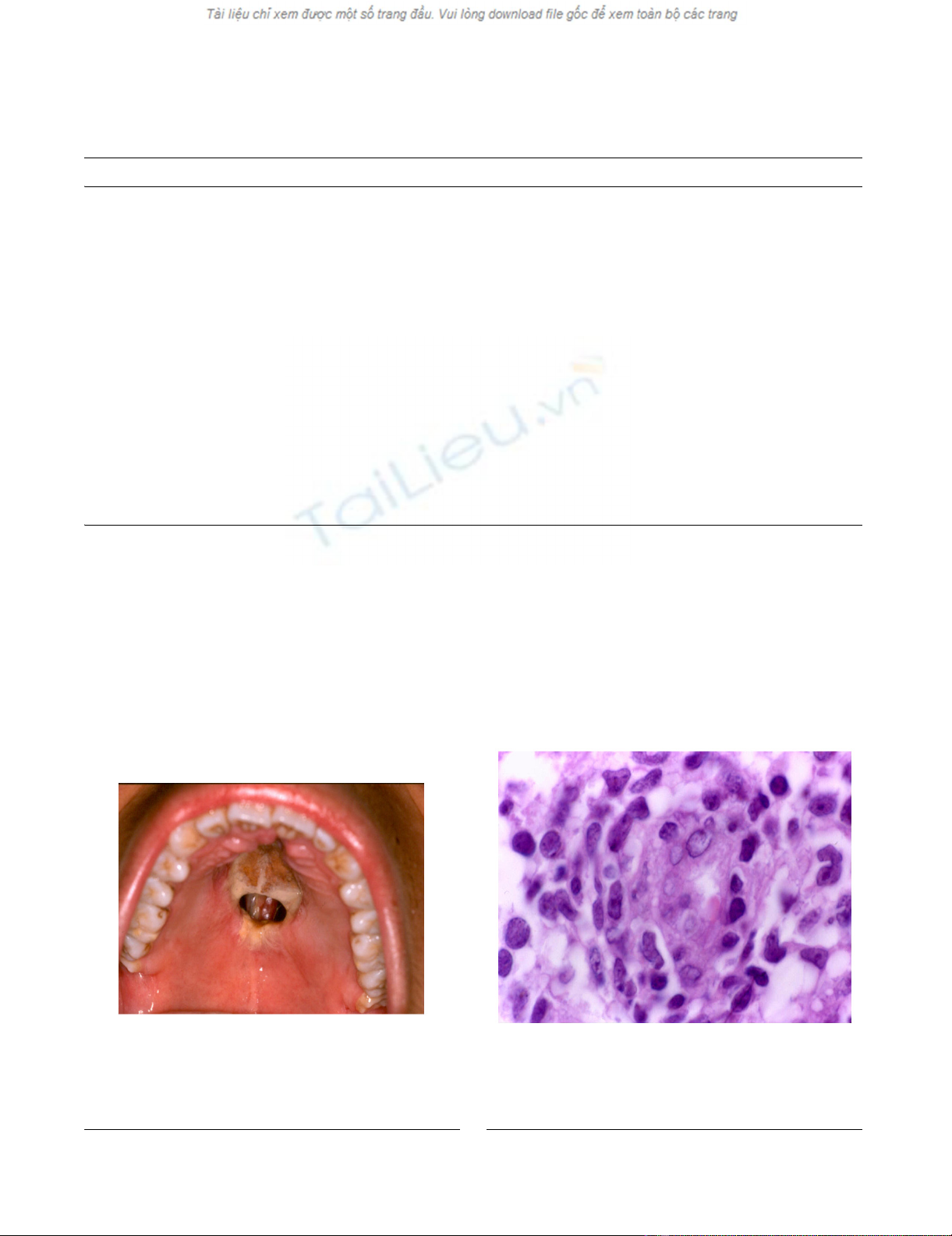
Clinical aspect in patient with T/NK cells angiocentric lym-phoma, which shows destructive ulcerative lesion in hard pal-ate and extensive zones of necrosisFigure 1
Clinical aspect in patient with T/NK cells angiocen-
tric lymphoma, which shows destructive ulcerative
lesion in hard palate and extensive zones of necrosis.
Histological aspect showing polymorphous cell population with malignant lymphoid cellsFigure 2
Histological aspect showing polymorphous cell popu-
lation with malignant lymphoid cells. This picture
shows prominence of endothelial cells as well as invasion of
small and large cleaved neoplastic cells (HE, 400×)

World Journal of Surgical Oncology 2008, 6:114 http://www.wjso.com/content/6/1/114
Page 4 of 9
(page number not for citation purposes)
Statistical analysis
Statistical analysis was based on chi square and Fisher's
exact test and test of Mc Nemar to assess MMP-1, MMP-2,
MMP-3, MMP-9, MMP-11, MMP-13, TIMP-1 y TIMP-2
expression of neoplastic cells, peritumoral fibroblasts,
endothelium cells and epithelium and its relation to clin-
ico-pathological parameters, using the Sigma Stat Ver.
3.00 software (SPSS, USA). A p value < 0.05 was consid-
ered statistically significant. The study received an ethical
waiver from National Institute of Cancer, Mexico.
Results
Expression of MMPs varied from moderate to intense in
the positive cases. The expression of metalloproteases 1, 2,
3, 9, 11, 13 and TIMP-1 and TIMP-2 and their distribution
in tissue is shown in table 2.
The expression in all the cases of MMP-1 thus neoplastic,
fibroblasts and endothelial cells, may indicate a link
action in the degradation of the stroma. The expression of
MMP-2 was present basically in the neoplastic cells. In the
case of MMP-9 most of the cases were negative both
tumoral and reactive cells. The expression of MMP-11 was
seen in the neoplastic cells and fibroblasts, but statistically
the difference in proportions was significant. Regarding
the expression of inhibitors, TIMP-1 was found statisti-
cally significant difference regarding the proportion of
cases that expressed MMP-9, likewise observed for TIMP-
2 regarding MMP-2 (table 2 and 3).
The MMP-2, -3 and -9 were expressed in neoplastic cells
between 30 to 65% of the cases. TIMP-1 was presented
mainly in the epithelium and TIMP-2 was poor expresses
in most of the cells and cases (Figs. 3 and 4). There were
no statistical significance between the different enzymes
used and the clinical pathological parameters included,
besides status and survival of the patients.
From all the studied cases, two of them were analyzed
ultrastructurally, showed prominence of endothelial cells
and infiltration of lymphoid-like cells in capillary vessels.
These cells showed fissures in the nuclear outline and elec-
trodense granules in the cytoplasm. Some granules were
extracellularly located and in contact with subendothelial
collagen fibers (Figs. 5 and 6).
Discussion
The nasal cavity, the palate and in general the midfacial
line are regions continuously stimulated by many extrane-
ous antigens. This stimulus creates a permanent and con-
stant interaction between antigens and cells that
participate in the host's defense. Among these antigens, a
highly antigenic is the EBV. Chronic exposure to this virus
can damage NK cells and T lymphocytes, these cells even-
tually can be transformed with time and generate extran-
odal lymphomas as T/NK nasal type lymphomas [16,23].
Once a neoplastic lesion is established in the nasal cavity
and/or midfacial region, individual tissue modifications
occur in the host. This is histologically translated to
inflammatory infiltrate of macrophages, granulocytes,
lymphoid and plasmatic cells. This reactive tissue
response can mask the disease and make the diagnosis of
lymphoma more difficult [3,7,10].
The chronic exposure of the nasal and palatine mucosa to
EBV is probably added to a genetic and racial predisposi-
tion, which can explain the predominant geographic dis-
tribution of the disease in some countries of Asia and
Latin America, including Mexico, a country which shows
an increasing number of patients with this malignancy [7-
12]. These two factors, EBV and the host's tissue response
can lead to the induction of enzymatic processes that
destroy the extracellular matrix of the blood vessel wall
and thus generate progressive areas of necrosis of soft and
bone tissue.
A previous study performed in Mexico in 23 cases of T/NK
lymphoma showed that 96% of the cases exhibited
expression of cytotoxic granules of TIA-1 and perforins
inside neoplastic cells [16]. Eighteen of these 23 cases
were included in this study.
In addition to the immunohistochemistry results, two of
the cases in the present series were analyzed with electron
microscopy; electrodense granules were observed in the
cytoplasm of the neoplastic cells. Besides, these granules
were identified in the blood vessel wall and were observed
disaggregating subendothelial collagen fibrils, which
strongly suggests a destructive action of the vascular wall
that contributes to angiodestruction. These findings have
also been observed and published by other authors
[23,24].
The phenomena of angiodestruction and necrosis could
be multifactorial and, in addition to the mentioned mech-
anisms, could be potentiated by the action of proteolytic
enzymes such as metalloproteases. These MMPs are a
group of Zn dependent endopeptidases, which break
down a large variety of molecules among them fibronec-
tin, laminin, vitronectin, type IV collagen, thrombospon-
din, elastin, hyaluronic acid, factor VIII, heparan sulfate,
proteoglycans, among others [25]. In addition, they can
activate, and in turn be activated by growth factors, and
thus promote degradation, migration, differentiation and
invasion processes [21,26].
The tumor or stromal expression of these enzymes has
been associated to a more aggressive behavior of malig-

World Journal of Surgical Oncology 2008, 6:114 http://www.wjso.com/content/6/1/114
Page 5 of 9
(page number not for citation purposes)
nant neoplasms; in particular, they have been studied in
head and neck, and colon carcinoma, among others, and
their presence is associated to a poor prognosis [22,26-
28]. In this series, the expression of MMP-1 both in tumor
and in peritumoral fibroblasts, and of MMP-11 in neo-
plastic cells, could explain the phenomenon of break-
down of cell elements related to the blood vessel wall,
such as type IV collagen, laminin and fibronectin. It has
been shown that MMP-1 is actively secreted by tumor
cells. This immunohistochemical study confirms the phe-
Table 2: Relationship between expression of the differents MMPs and TIMPs in the cells.
Factor EvenT frecuency Tes Mc Nemar ≠ Degree of ASSOCIATION
MMP-1
Epithelium (-) vs (+) 9/11 P = 0.004 tumor ≠ epithelium
Tumor (-) vs (+) 0/20 P = 0.000 tumor ≠ endothelium
Stroma (-) vs (+) 3/17
Endothelium (-) vs (+) 18/2
MMP-2
Epithelium (-) vs (+) 9/11 P = 0.031 tumor ≠ stroma
Tumor (-) vs (+) 7/13 P = 0.001 tumor ≠ endothelium
Stroma (-) vs (+) 13/7
Endothelium (-) vs (+) 18/2
MMP-3
Epithelium (-) vs (+) 11/9 P = 0.004 tumor ≠ endothelium
Tumor (-) vs (+) 10/10
Stroma (-) vs (+) 11/9
Endothelium (-) vs (+) 19/1
MMP-9
Epithelium (-) vs (+) 17/3 P = 0.031 tumor ≠ endothelium
Tumor (-) vs (+) 14/6
Stroma (-) vs (+) 16/4
Endothelium (-) vs (+) 20/0
MMP-11
Epithelium (-) vs (+) 8/12 P = 0.039 tumor ≠ stroma
Tumor (-) vs (+) 1/19 P = 0.039 tumor ≠ epithelium
Stroma (-) vs (+) 8/12 P = 0.000 tumor ≠ endothelium
Endothelium (-) vs (+) 16/4
MMP-13
Epithelium (-) vs (+) 12/8 P = 0.031 tumor ≠ endothelium
Tumor (-) vs (+) 14/6
Stroma (-) vs (+) 14/6
Endothelium (-) vs (+) 20/0
TIMP-1
Epithelium (-) vs (+) 8/12 P = 0.002 tumor ≠ stroma
Tumor (-) vs (+) 19/1 P = 0.039 tumor ≠ epithelium
Stroma (-) vs (+) 11/9 P = 0.000 tumor ≠ endothelium
Endothelium (-) vs (+) 16/4
TIMP-2
Epithelium (-) vs (+) 17/3
Tumor (-) vs (+) 18/2
Stroma (-) vs (+) 16/4
Endothelium (-) vs (+) 20/0
MMP-2 vs TIMP-2(+) vs (+) 13/2 P = 0.007
MMP-9 vs TIMP-1(+) vs (+) 6/1 P = 0.001


%20--%3e%3cdefs%3e%3cstyle%3e%20.st0%20{%20fill:%20%23fff;%20}%20.st1%20{%20fill:%20%237800fa;%20}%20%3c/style%3e%3c/defs%3e%3cpath%20class='st1'%20d='M117.78,12.18H43.11c2.9,3.47,4.65,7.94,4.65,12.82,0,5.6-2.3,10.66-6.01,14.29h76.02l7.22-13.56-7.22-13.56Z'/%3e%3cg%3e%3cpath%20class='st0'%20d='M53.58,26.17h-.59v-1.46h.59v-4.96h2.83c1.78,0,2.67.94,2.67,2.82v5.76c0,1.87-.89,2.81-2.67,2.81h-2.83v-4.96ZM55.36,21.37v3.34h1.1v1.46h-1.1v3.34h1.01c.61,0,.91-.37.91-1.1v-5.93c0-.74-.3-1.1-.91-1.1h-1.01Z'/%3e%3cpath%20class='st0'%20d='M65.99,31.14h-1.8l-.31-2.07h-2.19l-.31,2.07h-1.64l1.82-11.39h2.62l1.82,11.39ZM65.28,18.04c-.25.46-.51.77-.75.94-.21.15-.47.22-.79.22-.26,0-.57-.07-.92-.22l-.38-.15c-.14-.05-.26-.07-.37-.07-.3,0-.53.18-.71.54l-.91-.68c.25-.46.51-.77.75-.94.21-.14.48-.21.79-.21.26,0,.57.07.92.21l.38.15c.14.05.26.07.37.07.3,0,.53-.18.71-.54l.91.68ZM61.91,27.52h1.73l-.87-5.76-.87,5.76Z'/%3e%3cpath%20class='st0'%20d='M74.53,26.89v1.52c0,1.91-.89,2.86-2.67,2.86s-2.67-.95-2.67-2.86v-5.93c0-1.91.89-2.86,2.67-2.86s2.67.95,2.67,2.86v1.11h-1.69v-1.22c0-.75-.31-1.12-.93-1.12s-.93.37-.93,1.12v6.15c0,.74.31,1.11.93,1.11s.93-.37.93-1.11v-1.63h1.69Z'/%3e%3cpath%20class='st0'%20d='M81.4,31.14h-1.8l-.31-2.07h-2.19l-.31,2.07h-1.64l1.82-11.39h2.62l1.82,11.39ZM75.9,19.2l1.52-1.91h1.71l1.51,1.91h-1.61l-.76-.95-.75.95h-1.61ZM77.32,27.52h1.73l-.87-5.76-.87,5.76ZM83.1,15.99l-1.76,1.91h-1.26l1.17-1.91h1.86Z'/%3e%3cpath%20class='st0'%20d='M84.86,19.75c1.78,0,2.67.94,2.67,2.82v1.48c0,1.87-.89,2.81-2.67,2.81h-.85v4.28h-1.79v-11.39h2.64ZM84.01,21.37v3.86h.85c.58,0,.87-.36.87-1.08v-1.71c0-.71-.29-1.07-.87-1.07h-.85Z'/%3e%3cpath%20class='st0'%20d='M93.51,19.75c1.78,0,2.67.94,2.67,2.82v1.48c0,1.87-.89,2.81-2.67,2.81h-.85v4.28h-1.79v-11.39h2.64ZM92.66,21.37v3.86h.85c.58,0,.87-.36.87-1.08v-1.71c0-.71-.29-1.07-.87-1.07h-.85Z'/%3e%3cpath%20class='st0'%20d='M98.8,31.14h-1.79v-11.39h1.79v4.88h2.03v-4.88h1.83v11.39h-1.83v-4.88h-2.03v4.88Z'/%3e%3cpath%20class='st0'%20d='M105.36,24.55h2.46v1.62h-2.46v3.34h3.09v1.63h-4.88v-11.39h4.88v1.63h-3.09v3.18ZM108.17,17.29l-1.76,1.91h-1.26l1.17-1.91h1.86Z'/%3e%3cpath%20class='st0'%20d='M112.2,19.75c1.78,0,2.67.94,2.67,2.82v1.48c0,1.87-.89,2.81-2.67,2.81h-.85v4.28h-1.79v-11.39h2.64ZM111.35,21.37v3.86h.85c.58,0,.87-.36.87-1.08v-1.71c0-.71-.29-1.07-.87-1.07h-.85Z'/%3e%3c/g%3e%3ccircle%20class='st1'%20cx='25'%20cy='25'%20r='20'/%3e%3cpath%20class='st0'%20d='M32.78,19.27c2.92,0,4.43,2.55,5.28,5.33l.71,2.17c.14.38-.33.75-.71.75h-5.61c.19-.33.24-.71.09-1.08l-.75-2.45c-.43-1.32-.99-2.64-1.79-3.77.75-.57,1.65-.94,2.78-.94h0ZM25,18.38c3.25,0,4.9,2.78,5.89,5.89l.76,2.45c.14.42-.33.8-.8.8h-11.69c-.42,0-.94-.38-.8-.8l.75-2.45c.99-3.11,2.64-5.89,5.89-5.89h0ZM25,11.35c1.74,0,3.11,1.37,3.11,3.11s-1.37,3.11-3.11,3.11-3.11-1.41-3.11-3.11,1.41-3.11,3.11-3.11h0ZM17.27,19.27c1.08,0,1.98.38,2.73.94-.8,1.13-1.37,2.45-1.74,3.77l-.8,2.45c-.14.38-.05.75.09,1.08h-5.56c-.42,0-.9-.38-.75-.75l.71-2.17c.9-2.78,2.41-5.33,5.33-5.33h0ZM17.27,12.91c1.51,0,2.78,1.27,2.78,2.83s-1.27,2.83-2.78,2.83-2.83-1.27-2.83-2.83,1.27-2.83,2.83-2.83h0ZM32.78,12.91c1.56,0,2.78,1.27,2.78,2.83s-1.23,2.83-2.78,2.83-2.83-1.27-2.83-2.83,1.27-2.83,2.83-2.83h0ZM27.07,28.56v.09c0,.57-.24,1.08-.61,1.46h0v.05c-.38.33-.9.57-1.46.57s-1.08-.24-1.46-.61h0c-.38-.38-.61-.9-.61-1.46v-.09h1.41v.09c0,.19.05.38.19.47v.05c.09.09.28.19.47.19s.38-.09.47-.19v-.05c.14-.09.24-.28.24-.47t-.05-.09h1.41ZM30.99,28.56v.09c0,1.65-.66,3.16-1.74,4.24-1.08,1.08-2.59,1.79-4.24,1.79s-3.16-.71-4.24-1.79l-.05-.05c-1.04-1.08-1.7-2.55-1.7-4.2v-.09h1.41v.09c0,1.27.47,2.4,1.27,3.25h.05c.85.85,1.98,1.37,3.25,1.37s2.4-.52,3.25-1.37c.85-.8,1.37-1.98,1.37-3.25v-.09h1.37ZM34.99,28.56v.09c0,2.78-1.13,5.28-2.92,7.07-1.79,1.79-4.29,2.92-7.07,2.92s-5.23-1.13-7.07-2.92c-1.79-1.79-2.92-4.29-2.92-7.07v-.09h1.41v.09c0,2.4.94,4.53,2.5,6.08,1.56,1.56,3.72,2.5,6.08,2.5s4.52-.94,6.08-2.5c1.56-1.56,2.5-3.68,2.5-6.08v-.09h1.41Z'/%3e%3c/svg%3e)