
Protein tandem repeats – the more perfect, the less
structured
Julien Jorda
1
, Bin Xue
2,3
, Vladimir N. Uversky
2,3,4,5
and Andrey V. Kajava
1
1 Centre de Recherches de Biochimie Macromole
´culaire, CNRS UMR-5237, University of Montpellier 1 and 2, France
2 Center for Computational Biology and Bioinformatics, Indiana University School of Medicine, Indianapolis, IN, USA
3 Institute for Intrinsically Disordered Protein Research, Indiana University School of Medicine, Indianapolis, IN, USA
4 Institute for Biological Instrumentation, Russian Academy of Sciences, Pushchino, Moscow Region, Russia
5 Department of Biochemistry and Molecular Biology, Indiana University School of Medicine, Indianapolis, IN, USA
Introduction
Genome sequencing projects are producing knowledge
about a large number of protein sequences. Under-
standing the biological role of many of these proteins
requires information about their 3D structure as well
as their evolutionary and functional relationships. At
least 14% of all proteins and more than one-third of
human proteins carrying out fundamental functions
contain arrays of tandem repeats (TRs) [1]. The 3D
structures of many of these proteins have already been
determined by X-ray crystallography and NMR
methods. Fibrous proteins with repeats of two to seven
residues (collagen, silk fibroin, keratin, and tropomyo-
sin) were the first objects studied by structural biology
methods [2]. Proteins with repeat lengths from 5 to 50
residues gained special interest in the 1990s, when sev-
eral unusual structural folds, including b-helices [3],
b-rolls [4], the horseshoe-shaped structure of leucine-
rich-repeat proteins [5], b-propellers [6], and a-helical
solenoids [7], were resolved by X-ray crystallography.
Many proteins with repeats longer than 30 residues
have a ‘beads-on-a-string’ organization, with each
repeat being folded into a globular domain, e.g. zinc
Keywords
bioinformatics; disordered conformation;
evolution; protein structure; sequence
analysis
Correspondence
A. V. Kajava, Centre de Recherches de
Biochimie Macromole
´culaire, CNRS, 1919
Route de Mende, 34293 Montpellier,
Cedex 5, France
Fax: +33 4 67 521559
Tel: +33 4 67 61 3364
E-mail: andrey.kajava@crbm.cnrs.fr
(Received 23 February 2010, revised 7 April
2010, accepted 12 April 2010)
doi:10.1111/j.1742-4658.2010.07684.x
We analysed the structural properties of protein regions containing arrays
of perfect and nearly perfect tandem repeats. Naturally occurring proteins
with perfect repeats are practically absent among the proteins with known
3D structures. The great majority of such regions in the Protein Data Bank
are found in the proteins designed de novo. The abundance of natural
structured proteins with tandem repeats is inversely correlated with the
repeat perfection: the chance of finding natural structured proteins in the
Protein Data Bank increases with a decrease in the level of repeat perfec-
tion. Prediction of intrinsic disorder within the tandem repeats in the Swiss-
Prot proteins supports the conclusion that the level of repeat perfection
correlates with their tendency to be unstructured. This correlation is valid
across the various species and subcellular localizations, although the level
of disordered tandem repeats varies significantly between these datasets.
On average, in prokaryotes, tandem repeats of cytoplasmic proteins were
predicted to be the most structured, whereas in eukaryotes, the most struc-
tured portion of the repeats was found in the membrane proteins. Our
study supports the hypothesis that, in general, the repeat perfection is a
sign of recent evolutionary events rather than of exceptional structural and
(or) functional importance of the repeat residues.
Abbreviations
IDP, intrinsically disordered protein; IDR, intrinsically disordered region; PDB, Protein Data Bank; SCA, spinocerebellar ataxia;
TR, tandem repeat.
FEBS Journal 277 (2010) 2673–2682 ª2010 The Authors Journal compilation ª2010 FEBS 2673

finger domains [8], immunoglobulin domains [9], and
human matrix metalloproteinase [10]. It was noticed
that, frequently, proteins with repeats do not have
unique, stable 3D structures [11]. Rough estimates pro-
pose that half of the regions with TRs may be naturally
unfolded [12,13]. Low-complexity regions of eukaryotic
proteins that are enriched in repetitive motifs are rare
among the known 3D structures from the Protein Data
Bank (PDB) [14]. The common structural features,
functions and evolution of proteins with TRs have
been summarized in several reviews [7,11,15–18].
Perfect TRs occupy a special place among protein
repeats, which are usually imperfect because of muta-
tions (substitutions, insertions, and deletions) that have
accumulated during evolution. The high level of perfec-
tion of repeats can indicate substantial structural and
functional importance for each residue in the repeat, as
was observed in collagen molecules and some b-roll
structures [2,19]. It can also indicate recent evolution-
ary events that, for example, in pathogens can allow a
rapid response to environmental changes and can thus
lead to emerging infection threats, and in higher organ-
isms can lead to rapid morphological effects [20].
Perfect and nearly perfect repeats occur in a signifi-
cant portion of proteins. Recently, by using a newly
developed algorithm for ab initio identification of TRs,
we detected this type of repeat in 9% of proteins in
the SwissProt database [21]. To estimate the level of
perfection of the TRs, we used a parameter called P
sim
,
which is based on the calculation of Hamming dis-
tances between the consensus sequence and aligned
repeats of the TR (see Experimental procedures). In
this work, we analysed perfect and nearly perfect TRs
with P
sim
‡0.7.
Specific structural and evolutionary properties of the
perfect repeats pose challenges for the annotation of
genomic data. First, unlike with the aperiodic globular
proteins, prediction of structure–function relationships
by sequence similarity cannot be directly applied to the
perfect or nearly perfect repeats, owing to their
different evolutionary mechanisms. Second, although
ab initio structural prediction for proteins with TRs
generally yields reliable results [11], the very high fidel-
ity of sequence periodicity decreases the accuracy and
reliability of the information obtained from the
sequence alignment of the repeats. Each position of
the perfect repeats is conserved, and this makes it diffi-
cult to distinguish between residues that form the inte-
rior of the structure and those that face the solvent.
TRs are often found in proteins associated with
various human diseases. For example, expansion of
homorepeats is the molecular cause of at least
18 human neurological diseases, including myotonic
dystrophy 1, Huntington’s disease, Kennedy disease
(also known as spinal and bulbar muscular atrophy),
dentatorubral–pallidoluysian atrophy, and a number
of spinocerebellar ataxias (SCAs), such as SCA1,
SCA2, Machado–Joseph disease (SCA3), SCA6,
SCA7, and SCA17 [22,23]. A number of clinical disor-
ders, including prostate cancer, benign prostatic hyper-
plasia, male infertility, and rheumatoid arthritis, are
associated with polymorphisms in the length of the
polyglutamine and polyglycine repeats of the androgen
receptor [24].
Thus, proteins with perfect or nearly perfect TRs
play important functional roles, are abundant in
genomes, are related to major health threats, and, at
the same time, represent a challenge for in silico identi-
fication of their structures and functions. The objective
of this work was a systematic bioinformatics analysis
of arrays of perfect or nearly perfect TRs to obtain a
global view of their structural properties.
Results and Discussion
The 3D structures of naturally occurring proteins
with perfect repeats are practically absent in the
PDB
Our analysis shows that, among 20 800 sequences of
the nonredundant PDB (95% identity), only nine natu-
rally occurring proteins (0.04%) have perfect TRs with
P
sim
= 1 (Table 1). Furthermore, these arrays of TRs
are short (less than 19 residues), and they are missing
from the determined structures representing regions
with blurred electron density. A common reason for
missing electron density is that the unobserved atom,
side chain, residue or region fails to scatter X-rays
coherently, because of variation in position from one
protein to the next; for example, the unobserved atoms
can be flexible or disordered. Two proteins are excep-
tions to this: (a) an antibody molecule in which the
Table 1. Number of structured and unstructured regions found for
each range of P
sim
values in the PDB TR dataset. The following
tags were assigned to each analysed region with TRs: Sn and Sd,
fragments containing secondary structures from natural and
designed proteins, respectively; Ln and Ld, fragments connecting
secondary structures from natural and designed proteins, respec-
tively; Un and Ud, fragments whose structure was not determined
from natural and designed proteins, respectively.
P
sim
ranges Sn Ln Un Sd Ld Ud
P
sim
= 1.0 0 2 7 16 4 14
0.9 £P
sim
< 1.0 1 2 8 20 2 5
0.8 £P
sim
< 0.9 17 8 31 24 1 12
Structural state of perfect protein repeats J. Jorda et al.
2674 FEBS Journal 277 (2010) 2673–2682 ª2010 The Authors Journal compilation ª2010 FEBS
Gly-rich TR represents a crosslink between two
domains (PDB code: 1F3R) [25]; and (b) a substrate
with an (Arg-Ser)
8
tract that was cocrystallized with
protein kinase (PDB code: 3BEG) [26]. This Arg-rich
peptide, being alone in solution, will most probably be
unstructured, owing to the absence of nonpolar resi-
dues and the presence of eight Arg residues carrying a
charge of the same sign. Thus, this analysis suggested
that regions of natural proteins with perfect repeats
have a tendency to be unstructured.
To investigate this tendency, we analysed further the
regions with less perfect TRs. The TRs with
0.9 £P
sim
< 1.0 are also rare among natural proteins
of the PDB. Furthermore, the conformations of almost
all of them have not been resolved by X-ray crystallog-
raphy, because they are located in regions with missing
electron density. Only one of them, human CD3-e ⁄d
dimer (PDB: 1XIW) [27], has a short region of two
nine-residue repeats corresponding to a loop followed
by b-strand. We also analysed TRs with
0.8 £P
sim
< 0.9, and found 17 TRs of natural pro-
teins with the 3D structures (Table 1). In addition to
relatively short regions of fewer than 20 residues, cor-
responding to the a-helical elements, we also found
longer regions that form an immunoglobulin-like struc-
ture (1D2P) [28], a b-roll (1GO7) [29], an a-solenoid
(2AJA) [30], and an unusual long b-hairpin (1JHN)
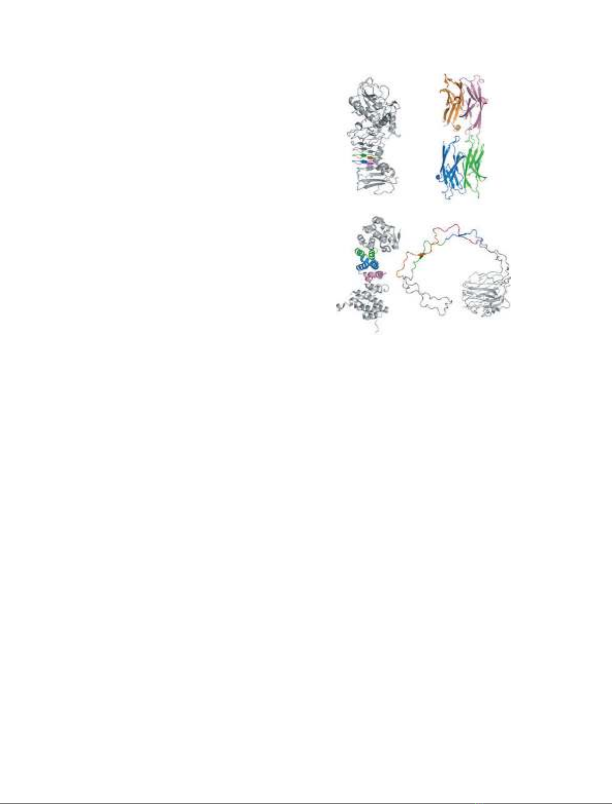
[31] (Fig. 1). Three of these four structures are formed
by bacterial proteins.
De novo designed proteins with perfect repeats
fold into stable 3D structures
In the PDB, majority (80%) of the proteins with per-
fect TRs are proteins designed de novo (Table 1). The
TR of a large proportion of these proteins fold into
the well-defined repetitive 3D structures such as colla-
gen triple helices, a-helical coiled coils, and a-helical
solenoids [2,17]. The fact that the designed perfect TRs
can form the stable 3D structures indicates that the
absence of such structures in natural proteins results
from evolution and not from problems with their fold-
ing propensities per se.
Prediction of intrinsically disordered regions in
SwissProt supports the tendency of TRs to be
unfolded
The ability of TRs to be structured or disordered was
further tested by using a larger dataset extracted from
SwissProt. The analysed dataset of TRs from the
Protein Repeat DataBase (http://bioinfo.montp.cnrs.fr/
?r=repeatDB) was filled in by the t-reks program
[21]. The TRs with P
sim
values ranging from 0.7 to 1
consist of 51 685 repeats found in 33 151 proteins,
which represent 9.1% of all proteins in the SwissProt
release of January 2009 (364 403 sequences). The level
of intrinsic disorder in these repeats and repeat-
containing proteins was evaluated by using several
computational tools.
Compositional profiling
Intrinsically disordered proteins (IDPs) and intrinsi-
cally disordered regions (IDRs) are known to be differ-
ent from structured globular proteins and domains
with regard to many attributes, including amino acid
composition, sequence complexity, hydrophobicity,
charge, flexibility, and type and rate of amino acid
substitutions over evolutionary time. For example,
IDPs ⁄IDRs are significantly depleted in a number of
so-called order-promoting residues, including bulky
hydrophobic (Ile, Leu, and Val) and aromatic (Trp,
Tyr, and Phe) residues, which would normally form
the hydrophobic core of a folded globular protein, and
also possess low contents of Cys and Asn residues. On
the other hand, IDPs ⁄IDRs were shown to be sub-
stantially enriched in so-called disorder-promoting
residues: Ala, Arg, Gly, Gln, Ser, Pro, Glu, and Lys
[32–36]. These biases in the amino acid composition of
1GO7 1D2P
2AJA 1JHN
Fig. 1. The 3D structures of proteins with almost perfect TRs.
Repeat regions are shown in colour.
J. Jorda et al. Structural state of perfect protein repeats
FEBS Journal 277 (2010) 2673–2682 ª2010 The Authors Journal compilation ª2010 FEBS 2675
IDPs and IDRs can be visualized using a normaliza-
tion procedure known as compositional profiling
[32,33,37]. In brief, compositional profiling is based on
the evaluation of the (C
s1
)C
s2
)⁄C
s2
values, where C
s1
is the content of a given residue in a set of interest
(regions and proteins with TRs), and C
s2
is the corre-
sponding value for the reference dataset (set of ordered
proteins or set of well-characterized IDPs). Negative
values of the profiling correspond to residues that are
depleted in a given dataset in comparison with a refer-
ence dataset, and the positive values correspond to res-
idues that are overrepresented in the set of interest.
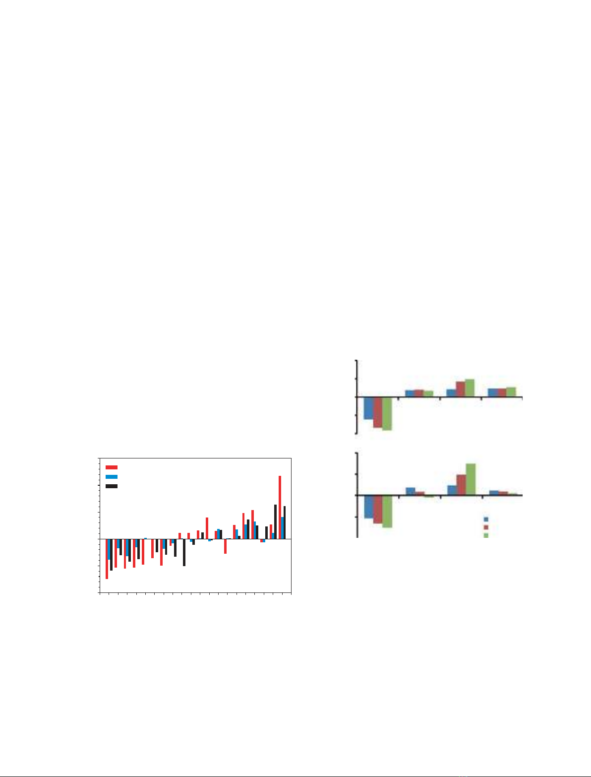
Figure 2 compares the amino acid compositions of
(a) all TRs analysed in this study, (b) proteins contain-
ing these TRs and (c) a dataset of IDPs with the com-
positions of ordered proteins. The datasets of IDPs
and fully structured proteins were taken from our pre-
vious analysis [38,39]. This shows that the composi-
tions of proteins containing TRs and of TRs
themselves are different from the compositions of
ordered proteins. They follow the trend for IDPs,
being generally depleted in major order-promoting res-
idues. This tendency for disorder is stronger for the
TRs, indicating that they contribute to this trend. At
the same time, the amino acid compositions of the
TRs have a bias when compared with the compositions
of ‘typical’ disordered proteins (Fig. 2). TRs have an
especially low occurrence of order-promoting Met and
the disorder-promoting charged residues Asp, Glu, and
Lys. On the other hand, TRs are highly enriched in
Cys and the disorder-promoting Pro, Gly, Ser, and
His.
To test the tendency of TRs to be disordered as a
function of their level of perfection, the TRs were sub-
divided into four subsets according to their P
sim
values
[0.7 < P
sim
£0.8 (32691 TRs), 0.8 < P
sim
£0.9 (8322
TRs), 0.9 < P
sim
£1.0 (1471 TRs), and homorepeats
with P
sim
= 1.0 (5259 TRs). Homorepeats were analy-
sed separately from the other TRs, because they signif-
icantly outnumber the other types of repeats, and
having them in the same group would obscure the
effect related to the other repeats. The amino acid
compositions of these subsets were compared with the
compositions of fully structured proteins. Figure 3 rep-
resents the results of compositional profiling for TRs
with different level of perfection. Both homorepeats
and the other TRs show the same trend. With the
increase in the perfection of the repeated segment, the
amount of order-promoting residues is gradually
reduced, whereas the relative contents of disorder-
promoting polar residues are gradually increased.
1.0
1.5
TRs
Entire sequences
Typical IDPs
–0.5
0.0
0.5
WFY I MLVNCTAG DRHQSKP
(CAA
Dataset–CAA
Struct)/CAA
Struct
–1.0 E
Fig. 2. Compositional profiling of TRs, entire sequences of proteins
containing these TRs, and a set of fully disordered proteins from
DisProt in comparison with the composition of fully structured pro-
teins from the PDB. CStruct
AA is the content of a given amino acid in
the set of structured proteins; CDataset
AA is the content of this amino
acid in the dataset of interest. Amino acids are arranged in order of
decreasing structure-promoting ability as suggested by the TopIDP
scale [37].
Nonpolar G Polar P
20
10
0
–10
–20
40
20
0
–20
–40
0.7–0.8
0.8–0.9
0.9–1
A
B
Chr
–C
AA
struct
AA Ctr –C
AA
struct
AA
Fig. 3. (A) Differences in amino acid compositions between TRs,
subdivided into groups with different levels of repeat perfection
and fully structured proteins. The homorepeats are analysed sepa-
rately (B), owing to their unusually high occurrence in comparison
to the other TRs. For this purpose, a dataset of perfect and cryptic
homorepeats was created and subdivided into three groups
depending on the P
sim
values. Ctr
AA and Chr
AA are the contents of a
given amino acid in the set of TRs (excluding homorepeats) and
only homorepeats, respectively. Amino acids are arranged in four
sets: order-promoting aromatic and aliphatic amino acids (Trp, Phe,
Tyr, Ile, Met, Leu, Val, and Ala) which are denoted as nonpolar;
order-neutral Gly, disorder-promoting polar residues (Asn, Cys, Thr,
Gln, Ser, Arg, Asp, His, Glu, and Lys) and disorder-promoting
nonpolar Pro.
Structural state of perfect protein repeats J. Jorda et al.
2676 FEBS Journal 277 (2010) 2673–2682 ª2010 The Authors Journal compilation ª2010 FEBS

The contents of Gly and Pro residues do not change
significantly.
Prediction of intrinsic disorder
As the compositional profiling showed that TRs and
repeat-containing proteins have a noticeable increase
in the number of disorder-promoting residues, we fur-
ther analysed the abundance of predicted intrinsic dis-
order in these sequences with several computational
tools, including the pondr
vlxt [34,40] and vsl2
[41,42] algorithms, as well as predictors such as iupred
[43,44], foldindex [45], and topidp [37]. The results of
this analysis are summarized in Table 2, which clearly
shows that both TRs and repeat-containing proteins
are highly disordered. Furthermore, TRs have higher
percentage of disordered residues than the entire
TR-containing sequences. Prediction of intrinsic disor-
der also confirmed an observation that the amounts of
disorder in both datasets increase with increases in the
repeat perfection (Table 2).
This observation is further illustrated by the distri-
butions of values representing the number of predicted
disorder residues divided by the number of residues in
the considered region (Fig. 4). These distributions are
generated for TR regions of different levels of perfec-
tion (Fig. 4A) and for the corresponding repeat-con-
taining proteins (Fig. 4B). Figure 4A shows that all
analysed TRs are highly disordered, irrespective of the
level of their perfection. At the same time, as the
perfection of TRs increases, the relative content of dis-
order also increases. For example, at least 70% of TRs
with 0.7 < P
sim
£0.8 are predicted to have disorder
ratios of more than 0.95. For TRs with 0.8 <
P
sim
£0.9, this percentage increases to 85%, for those
with 0.9 < P
sim
£1.0 it is 86%, and for perfect ho-
morepeats it reaches 97% (Fig. 4A). Figure 4B shows
that only 6% of the whole sequences of proteins con-
taining perfect repeats are well structured (disorder
ratio less than 0.2). The rest of these sequences have
widespread disorder ratios, ranging from 0.25 to 1.
Proteins containing the least perfect repeats
(0.7 < P
sim
£0.8), about 5%, are almost evenly distrib-
uted among the various disorder ratios. Thus, perfect
repeats preferentially occur in proteins that have disor-
der ratios of more than 0.2 and are poorly represented
in more structured proteins, whereas less perfect
repeats are equally probable in sequences with differ-
ent disorder ratios.
Intrinsic disorder of tandem repeats across
species and subcellular localizations
The pondr
vlxt predictor and TopIDP index were
used to establish variation of the disorder level among
TRs of viral, eukaryotic and prokaryotic proteins. The
tested dataset included TRs with P
sim
‡0.9 identified
in SwissProt. The homorepeats were excluded and
analysed separately from the other TRs, because their
predominant occurrence in eukaryotic proteins would
obscure the results. Prior to the analysis, the redun-
dancy of the dataset related to the existence of protein
sequences from different strains of the same species
(especially for bacteria and viruses) had been filtered
out by using the species name, consensus motif, and
number and location of repeats. As a result, the data-
set contained 245 repeats from prokaryotic proteins,
1059 repeats from eukaryotic proteins, and 70 repeats
Table 2. Analysis of intrinsic disorder distribution in TRs and TR-containing proteins.
P
sim
= 0.7–0.8 P
sim
= 0.8–0.9 P
sim
= 0.9–1 Homorepeats
TRs
Total no. 34 286 5519 1382 5259
Average length 25.5 41.0 59.1 13.8
Intrinsic disorder ratio (%): VSL2 80.4 88.6 88.9 98.4
Intrinsic disorder ratio (%): IUPRED 56.0 62.7 67.2 86.5
Intrinsic disorder ratio (%): FOLDINDEX 62.4 68.6 70.3 79.9
Intrinsic disorder ratio (%): TOPIDP 85.6 88.8 91.1 74.4
Sequences
a
Total no. 25 649 4915 1295 3663
Average length 643.4 752.0 840.2 790.4
Intrinsic disorder ratio (%): VSL2 49.3 58.6 57.0 61.6
Intrinsic disorder ratio (%): IUPRED 32.1 41.7 41.7 45.4
Intrinsic disorder ratio (%): FOLDINDEX 46.6 52.3 52.3 52.7
Intrinsic disorder ratio (%): TOPIDP 71.2 75.3 72.2 74.9
a
Whole proteins containing these TRs.
J. Jorda et al. Structural state of perfect protein repeats
FEBS Journal 277 (2010) 2673–2682 ª2010 The Authors Journal compilation ª2010 FEBS 2677


