
Purification and characterization of a membrane-bound enzyme
complex from the sulfate-reducing archaeon
Archaeoglobus fulgidus
related to heterodisulfide reductase from methanogenic archaea
Gerd J. Mander
1
, Evert C. Duin
1
, Dietmar Linder
2
, Karl O. Stetter
3
and Reiner Hedderich
1
1
Max-Planck-Institut fu
¨r terrestrische Mikrobiologie, Marburg, Germany;
2
Biochemisches Institut, Fachbereich Humanmedizin,
Justus-Liebig-Universita
¨t Giessen, Germany;
3
Lehrstuhl fu
¨r Mikrobiologie und Archaeenzentrum, Universita
¨t Regensburg, Germany
Heterodisulfide reductase (Hdr) is a unique disulfide reduc-
tase that plays a key role in the energy metabolism of
methanogenic archaea. The genome of the sulfate-reducing
archaeon Archaeoglobus fulgidus encodes several proteins of
unknown function with high sequence similarity to the
catalytic subunit of Hdr. Here we report on the purification
of a multisubunit membrane-bound enzyme complex from
A. fulgidus that contains a subunit related to the catalytic
subunit of Hdr. The purified enzyme is a heme/iron-sulfur
protein, as deduced by UV/Vis spectroscopy, EPR spec-
troscopy, and the primary structure. It is composed of four
different subunits encoded by a putative transcription unit
(AF499, AF501–AF503). A fifth protein (AF500) encoded
by this transcription unit could not be detected in the purified
enzyme preparation. Subunit AF502 is closely related to the
catalytic subunit HdrD of Hdr from Methanosarcina bark-
eri. AF501 encodes a membrane-integral cytochrome, and
AF500 encodes a second integral membrane protein. AF499
encodes an extracytoplasmic iron-sulfur protein, and AF503
encodes an extracytoplasmic c-type cytochrome with three
heme c-binding motifs. All of the subunits show high
sequence similarity to proteins encoded by the dsr locus of
Allochromatium vinosum and to subunits of the Hmc
complex from Desulfovibrio vulgaris. The heme groups of the
enzyme are rapidly reduced by reduced 2,3-dimethyl-1,4-
naphthoquinone (DMNH
2
), which indicates that the
enzyme functions as a menaquinol–acceptor oxidoreduc-
tase. The physiological electron acceptor has not yet been
identified. Redox titrations monitored by EPR spectroscopy
were carried out to characterize the iron-sulfur clusters of the
enzyme. In addition to EPR signals due to [4Fe-4S]
+
clus-
ters, signals of an unusual paramagnetic species with gvalues
of 2.031, 1.994, and 1.951 were obtained. The paramagnetic
species could be reduced in a one-electron transfer reaction,
but could not be further oxidized, and shows EPR properties
similar to those of a paramagnetic species recently identified
in Hdr. In Hdr this paramagnetic species is specifically
induced by the substrates of the enzyme and is thought to be
an intermediate of the catalytic cycle. Hence, Hdr and the
A. fulgidus enzyme not only share sequence similarity, but
may also have a similar active site and a similar catalytic
function.
Keywords:Archaeoglobus fulgidus; heterodisulfide reductase;
Hmc complex; iron-sulfur proteins; sulfate-reducing
bacteria.
Heterodisulfide reductase (Hdr) is a key enzyme in the
energy metabolism of methanogenic archaea. In the final
step of methanogenesis, the mixed disulfide of the metha-
nogenic thiol coenzymes coenzyme M and coenzyme B is
generated in a reaction catalyzed by methyl-coenzyme M
reductase [1]. This disulfide is reduced by a unique disulfide
reductase, designated heterodisulfide reductase (Hdr). Two
types of Hdr from phylogenetically distantly related meth-
anogens have been identified and characterized. Neither
type of enzyme belongs to the family of pyridine nucleotide
disulfide oxidoreductases [2].
Hdr from Methanothermobacter marburgensis is an
iron-sulfur flavoprotein composed of the subunits HdrA,
HdrB, and HdrC. The enzyme has been purified from the
soluble fraction, and none of its subunits are predicted to
form transmembrane helices. From sequence data, it has
been deduced that HdrA contains an FAD-binding motif
and four binding motifs for [4Fe-4S] clusters. HdrC
contains two additional binding motifs for [4Fe-4S]
clusters [2].
Hdr in the two closely related Methanosarcina species
M. barkeri and M. thermophila is tightly membrane-
bound [3–5]. The enzyme is composed of two subunits,
a membrane-bound b-type cytochrome (HdrE) and a
hydrophilic subunit (HdrD) containing two binding
motifs for [4Fe-4S] clusters. Subunit HdrD of the
M. barkeri enzyme is a homologue of a hypothetical
fusion protein of the M. marburgensis HdrCB subunits
Correspondence to R. Hedderich, Max-Planck-Institut fu
¨r terrestrische
Mikrobiologie, Karl-von-Frisch-Strabe, D-35043 Marburg/Germany.
Fax: + 49 6421 178299, Tel.: + 49 6421 178230,
E-mail: hedderic@mailer.uni-marburg.de
Abbreviations: Hme, Hdr-like menaquinol-oxidizing enzyme; Hdr,
heterodisulfide reductase; DMN, 2,3-dimethyl-1,4-naphthoquinone;
H-S-CoM, coenzyme M or 2-mercaptoethanesulfonate; H-S-CoB,
coenzyme B or 7-mercaptoheptanoylthreonine phosphate;
CoM-S-S-CoB, heterodisulfide of H-S-CoM and H-S-CoB; Hmc,
high-molecular-mass c-type cytochrome; Dsr, dissimilatory sulfite
reductase.
Enzyme: heterodisulfide reductase (EC 1.99.4.-).
(Received 10 October 2001, revised 12 February 2002, accepted 15
February 2002)
Eur. J. Biochem. 269, 1895–1904 (2002) ÓFEBS 2002 doi:10.1046/j.1432-1033.2002.02839.x

[4]. A homologue of the M. marburgensis HdrA subunit is
lacking in Hdr from Methanosarcina species. It has
therefore been suggested that the conserved subunits
HdrD and HdrCB must harbor the catalytic site for the
reduction of the disulfide substrate. The active site of Hdr
was recently shown to contain a [4Fe-4S] cluster that is
directly involved in mediating heterodisulfide reduction
[6,7]. This extra iron-sulfur cluster has been proposed to
be co-ordinated by cysteine residues of the highly
conserved sequence motif CX
31)38
CCX
33)34
CXXC found
in subunits HdrD and HdrB. The nonconserved subunits
HdrE and HdrA are thought to interact with the physio-
logical electron donor, which differs in the two types of
Hdr. The physiological electron donor of Hdr from
Methanosarcina species is thought to be the membrane-
soluble electron carrier methanophenazine [8]. Hdr from
M. marburgensis forms a functional complex with the
MvhAGD hydrogenase [9]. This complex catalyzes the
reduction of CoM-S-S-CoB by H
2
.
Hdr was originally thought to be unique to methanogenic
archaea. However, in recent years, genes encoding pro-
teins related to the catalytic subunit of Hdr have been identi-
fied in a broad range of prokaryotes unable to perform
methanogenesis [2]. No function has so far been assigned to
these Hdr-like proteins, and none has been purified and
characterized. Archaeoglobus fulgidus is one of the organ-
isms that encode the largest number of proteins related to
Hdr [10].
This extremely thermophilic sulfate-reducing archaeon
completely oxidizes organic substrates, such as lactate, to
CO
2
[11]. Acetate is oxidized to CO
2
by a modified acetyl-
CoA pathway using typical methanogenic coenzymes
[12,13]. Some of the reducing equivalents generated in the
oxidative branch of the pathway are transferred to the
deazaflavin coenzyme F
420
, which is reoxidized by the
F
420
H
2
–menaquinone oxidoreductase. F
420
H
2
–menaqui-
none oxidoreductase is an integral membrane protein that
shows high sequence similarity to energy-conserving
NADH–quinone oxidoreductases [10,14]. It is assumed to
function as a proton or sodium ion pump as well. In
addition, the membrane fraction of A. fulgidus catalyzes the
reduction of 2,3-dimethyl-1,4-naphthoquinone (DMN) by
L
-lactate, which indicates that lactate dehydrogenase direct-
ly channels the reducing equivalents generated in the
oxidation of lactate to pyruvate into the menaquinone pool
[12]. A. fulgidus has been shown to contain a modified
menaquinone as a membrane-soluble electron carrier [15]. It
is, however, not yet known how the reduced menaquinone
pool is electrically connected to the enzymes of sulfate
reduction, namely adenosine 5¢-phosphosulfate reductase
and sulfite reductase.
Here we report on the isolation and characterization of a
heme-containing membrane protein from A. fulgidus related
to Hdr from M. barkeri. A function of this enzyme as reduced
menaquinone–acceptor oxidoreductase is discussed.
MATERIALS AND METHODS
Materials
Redox dyes were obtained from Aldrich–Sigma. DMN was
from Sigma. Potassium trithionate was a gift from Peter
M. H. Kroneck (Universita
¨t Konstanz). All other chemicals
were from Merck. The chromatographic materials were
from Amersham Pharmacia Biotech.
Growth of the organism
A. fulgidus (VC16, DSMZ 304) was grown in a 300-L
fermenter at 83 °C on lactate/sulfate medium as described
previously [11]. Cells were harvested after shock cooling to
4°C with a continuous flow centrifuge (Z61; Padberg Lahr,
Germany) at 17 000 g; the pellet was frozen in liquid
nitrogen and stored at )80 °Cbeforeuse.
Enzyme purification
All purification steps were carried out under strictly anoxic
conditions under an atmosphere of N
2
/H
2
(95 : 5, v/v) at
18 °C. Cells were lysed by sonication and then centrifuged
at 6400 gfor 1 h. The supernatant was ultracentrifuged at
150 000 gfor 2 h. The pellet was resuspended in 50 m
M
Mops (pH 7.0) using a Teflon homogenizer. Protein was
solubilized from the membranes with 15 m
M
dodecyl-b-
D
-
maltoside [2 mg dodecyl-b-
D
-maltosideÆ(mg protein)
)1
]at
4°C for 12 h. The unsolubilized proteins and the mem-
branes were removed by ultracentrifugation as described
above. The supernatant was applied to a Q-Sepharose
HighLoad column (2.6 ·10 cm) equilibrated with 50 m
M
Mops/KOH (pH 7.0) containing 2 m
M
dodecyl-b-
D
-malto-
side (buffer A). Protein was eluted in a stepwise NaCl
gradient (80 mL each in buffer A): 0 m
M
, 300 m
M
,
400 m
M
,500m
M
,600m
M
,and1
M
. The majority of the
heme-containing protein(s) were eluted at 600 m
M
NaCl.
These fractions were applied to a Superdex 200 gel-filtration
column (2.6 ·60 cm) equilibrated with buffer A containing
100 m
M
NaCl. Protein was eluted using the same buffer.
Heme-containing protein(s) were eluted after 120 mL (peak
maximum). These fractions were applied to a Mono Q
anion-exchange column (HR 10/10) equilibrated with
buffer A. Protein was eluted using a linear NaCl gradient
(0–1
M
, 100 mL). Heme-containing protein(s) were eluted
at 600 m
M
NaCl. The enzyme was concentrated by
ultrafiltration (Molecular/Por ultrafiltration membranes;
100-kDa cut off; Spectrum, Houston, USA) and stored in
buffer A at 4 °C under N
2
. Protein was judged to be >95%
pure by SDS/PAGE.
UV/Vis spectroscopy
Spectra of samples in 1-mL quartz cuvettes in an anaerobic
chamber under N
2
/H
2
(95 : 5, v/v) were recorded using a
Zeiss Specord S10 diode array spectrophotometer connected
to a quartz photoconductor (Hellma Mu
¨llheim, Germany).
The oxidation or reduction of the heme groups of the
enzyme by DMN or DMNH
2
were followed spectropho-
tometrically. DMN or DMNH
2
was added to the enzyme
solution [1 mg proteinÆmL
)1
in 50 m
M
Mops/KOH (pH 7.0)]
to a final concentration of 150 l
M
, and spectra were
recorded every 5 s. DMNH
2
was prepared as described
previously [16].
Analytical methods
Non-heme iron was quantified colorimetrically with neo-
cuproin (2,9-dimethyl-1,10-phenanthroline) and ferrozine
1896 G. J. Mander et al.(Eur. J. Biochem. 269)ÓFEBS 2002

[3-(2-pyridyl)-5,6-bis-(4-phenylsulfonate)-1,2,4-triazine] as
described by Fish [17]. Acid-labile sulfur was analyzed as
methyleneblue[18].
The protein concentration was routinely determined by
the method of Bradford (Rotinanoquant; Roth Karlsruhe,
Germany) using BSA as standard.
Heme was extracted with acetone/HCl and the pyridine
hemochrome derivate was formed as described. Reduced
minus oxidized difference spectra were recorded at room
temperature [19]. The spectra obtained were compared with
the pyridine hemochrome spectra obtained with heme
extracted from hemoglobin.
EPR spectroscopy measurements
EPR spectra at X-band (9 GHz) were obtained with a
Bruker EMX spectrometer. All spectra were recorded with
a field modulation frequency of 100 kHz. The sample was
cooled by an Oxford Instruments ESR 900 flow cryostat
with an ITC4 temperature controller. Spin quantitations
were carried out under nonsaturating conditions using
10 m
M
copper perchlorate as the standard (10 m
M
CuSO
4
,
2
M
NaClO
4
,10m
M
HCl). When the EPR signals over-
lapped with other signals, e.g. radical signals from the redox
dyes, the signals were simulated, and the simulations were
double integrated to obtain the spin intensity. Temperature-
dependence studies were carried out under nonsaturating
conditions where possible. For all signals, the peak ampli-
tude was measured at different temperatures. These values
were used to obtain Curie plots describing the temperature
behavior of the respective signal. EPR signals were simu-
lated using noncommercial programs based on formulas
described previously [20].
Redox titrations
Redox titrations were carried out at 18 °Cinananaerobic
chamber under N
2
/H
2
(95 : 5, v/v). Potentials were adjusted
with small amounts of freshly prepared sodium dithionite
(20 m
M
stock solution) or freshly prepared potassium
ferricyanide (20 m
M
stock solution). All redox potentials
quoted here are relative to the standard hydrogen electrode.
In these titrations, a selection of the following mediators
(final concentration 20 l
M
) were added individually to the
enzyme solution: 1,2-naphthoquinone (E°¢ ¼+134 mV),
duroquinone (E¢¼+86 mV), 1,4-naphthoquinone
(E¢¼+69 mV), thionine (E¢¼+64 mV), methylene blue
(E¢¼+11 mV), indigodisulfonate (E¢¼)125 mV),
2-hydroxy-1,4-naphthoquinone (E¢¼)145 mV), anthra-
quinone-1,4-disulfonate (E¢¼)170 mV), phenosafranin
(E¢¼)252 mV), anthraquinone-2-sulfonate (E°¢¼
)255 mV), safranin O (E¢¼)289 mV), and neutral red
(E¢¼)325 mV). The final concentration of Hdr-like
menaquinol-oxidizing enzyme (Hme) was 7 l
M
in 50 m
M
Mops/KOH (pH 7.0) containing 2 m
M
dodecyl-b-
D
-malto-
side. After equilibration of the desired potential, a 0.3-mL
aliquot was transferred to a calibrated EPR tube and
immediately frozen in liquid nitrogen. The redox potential
wasmeasuredwithanAg/AgClredoxcombinationelec-
trode (Mettler Toledo Giessen, Germany). To obtain
potentials relative to the standard hydrogen electrode, a
value of 207 mV (corresponding to the potential of Ag/
AgCl at 25 °C) was added to the measured redox potentials.
Determination of amino-acid sequences
For determination of N-terminal amino-acid sequences,
polypeptides were separated by SDS/PAGE and blotted
on to poly(vinylidene difluoride) membranes (Applied
Biosystems) as described previously [4]. Sequences were
determined using an Applied Biosystems 4774 protein/
peptide sequencer and the protocol given by the manu-
facturer.
Amino-acid sequence analysis
For the prediction of transmembrane helices in proteins,
noncommercial programs were used (http://www.sbc.su.se/
miklos/DAS/; http://www.cbs.dtu.dk/services/TMHMM-
2.0/). For sequence comparisons, multiple sequence align-
ments were generated using the
FASTA
3server(http://
www.ebi.ac.uk/fasta3/).
RESULTS
Purification of a heme-containing enzyme complex
from the membrane fraction of
A. fulgidus
The genome of A. fulgidus encodes several membrane-
bound oxidoreductases that share sequence similarity with
subunits of Hdr from methanogenic archaea, in particular
with the membrane-bound enzyme from M. barkeri [4,10],
which is anchored in the cytoplasmic membrane via a b-
type cytochrome [3]. We used this knowledge to identify
and purify heme-containing membrane-bound enzymes
from A. fulgidus cells cultivated on lactate/sulfate medium
by following the characteristic absorption of heme proteins.
The membrane fraction was isolated, and proteins were
solubilized with the detergent dodecyl-b-
D
-maltoside. On
anion-exchange chromatography on Q-Sepharose, the
major heme-containing fraction was eluted at 600 m
M
NaCl. Approximately 70% of the heme present in solubi-
lized membranes was found in this fraction. A further
purification of the proteins in this heme-containing fraction
by gel filtration on Superdex 200 resulted again in only one
heme-containing fraction eluted after 120–150 mL. In the
final purification step, the sample was chromatographed on
a Mono Q anion-exchange column. The protein thus
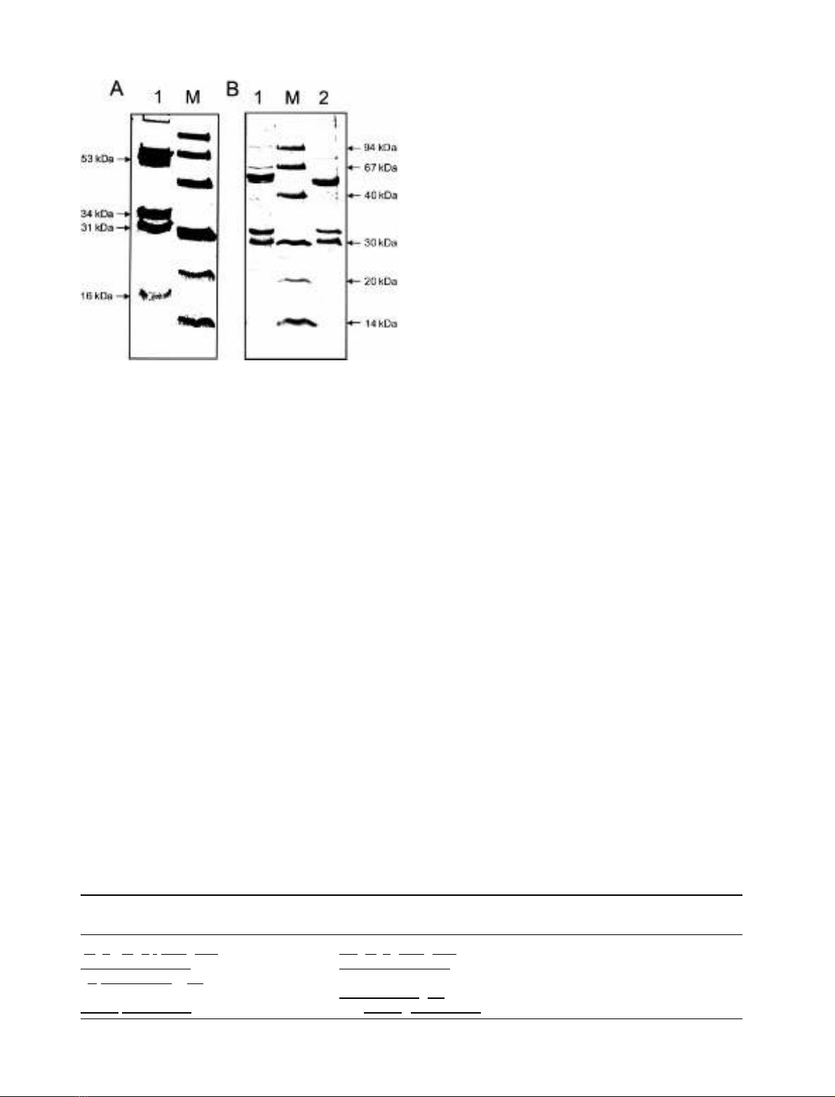
purified was subjected to SDS/PAGE (Fig. 1). Samples
were either boiled for 5 min in SDS buffer or incubated in
SDS buffer at room temperature for 1 h before electro-
phoresis. The samples incubated at room temperature
yielded four major polypeptide bands with apparent
molecular masses of 53, 34, 31, and 16 kDa after SDS/
PAGE (Fig. 1, lane A1). In the boiled sample, the 34-kDa
polypeptide was only detectable at lower intensities. This
may be due to protein aggregation, which is typical of
integral membrane proteins (Fig. 1, lane B2). From the
results of SDS/PAGE, it can be deduced that the 16-kDa
protein is only present in substoichiometric amounts. In
some preparations, this protein was completely absent
(Fig. 1B).
As will be described below, the enzyme complex purified
from A. fulgidus shows similarity to Hdr and has a
menaquinol-oxidizing activity. The enzyme was therefore
preliminarily designated Hdr-like menaquinol-oxidizing
enzyme complex, abbreviated as Hme complex.
ÓFEBS 2002 Hdr-like enzyme complex from A. fulgidus (Eur. J. Biochem. 269) 1897
Identification of the genes encoding the subunits
of the Hme complex and sequence analysis
The N-terminal sequences of the four polypeptides present
in the purified enzyme preparation were determined by
Edman degradation (Table 1). Using these sequences, the
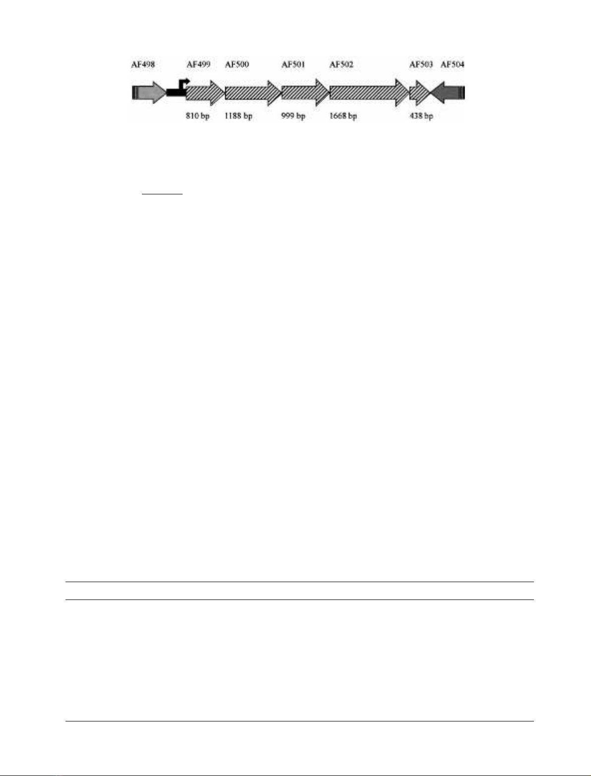
corresponding genes (AF499, AF501–503) were identified in
thegenomeofA. fulgidus [10]. The noncoding regions
between the different genes are short (less than 12 bp) or
nonexistent (the genes overlap). The sequence region
upstream of AF499 is AT-rich and contains typical archaeal
promoter elements. The sequence AAAGGTTAATATA
was found 64 bp upstream of the start codon of AF499; this
corresponds to the BRE element and the box A element of
archaeal promoters [21,22]. The AF499–AF503 gene cluster
can therefore be predicted to form a transcription unit
(Fig. 2). This transcription unit contains one gene (AF500)
for which no corresponding protein was found in the
purified enzyme preparation. The results of the sequence
analyses of the deduced proteins are given in Table 2.
The protein encoded by AF502 has a calculated mole-
cular mass of 64.4 kDa. The protein shows about 35%
sequence identity with the proposed catalytic subunit HdrD
from M. barkeri. The closest relative of the protein encoded
by AF502 (40% sequence identity) is the dissimilatory
sulfite reductase (Dsr)K protein from the sulfur-oxidizing
phototrophic bacterium Allochromatium vinosum. The DsrK
protein is encoded by the dsr locus, which also encodes the
subunits of the siroheme sulfite reductase [23]. Another
relative of the protein encoded by AF502 is the high-
molecular-mass c-type cytochrome (Hmc)F protein of
Desulfovibrio vulgaris (20% sequence identity) [24].
A characteristic of HdrD of M. barkeri is the presence of
two typical [4Fe-4S] cluster binding motifs in the N-terminal
part of the protein. HdrD contains 10 additional cysteine
residues found in two CX
31)38
CCX
33)34
CXXC sequence
motifs at the C-terminal part of the protein [4]. Multiple
sequence alignments of HdrD, AF502, DsrK, and HmcF
clearly identified the two typical CXXCXXCXXXCP
binding motifs for [4Fe-4S] clusters in the N-terminal part
of these proteins. AF502, DsrK, and HmcF also contain
one of the two CX
31)38
CCX
33)34
CXXC motifs present in
HdrD. Only in AF502 does an aspartate residue replace one
of the five cysteines present in this motif [25].
The AF501 protein has a calculated molecular mass of
38 kDa. The molecular mass of this protein was estimated
by SDS/PAGE to be 34 kDa. The protein shows the highest
sequence similarity (30% identity) to the DsrM protein
from A. vinosum, encoded by the dsr locus,andtotheb-type
cytochrome subunit of nitrate reductase from various
organisms, such as NarI of nitrate reductase of Escherichia
coli [26]. AF501 also has low sequence similarity to the
b-type cytochrome (HdrE) of Hdr. A topological analysis
suggests that AF501, like NarI, has five membrane-span-
ning helices. In the b-type cytochromes of nitrate reductases,
four histidine residues are conserved, two in helix b and two
Fig. 1. SDS/PAGE of the purified Hme complex. Proteins were sepa-
ratedina12%slabgel(8·7 cm) which was subsequently stained
with Coomassie Brilliant Blue R250. The polypeptide with an apparent
molecular mass of 16 kDa, identified as a c-type cytochrome by
N-terminal sequencing, was not found in all preparations. The pre-
paration shown in (A) still contains the c-type cytochrome, while the
preparation shown in (B) lacks this polypeptide. M, Low-molecular-
mass markers (Amersham Pharmacia Biotech). The molecular masses
of the marker proteins are given on the right side. Lane A1, 15 lgof
the A. fulgidus Hme complex denatured for 30 min at room temper-
ature in SDS sample buffer; lane B1, 10 lg Hme complex denatured
for 30 min at room temperature in SDS sample buffer; lane B2, 10 lg
Hme complex denatured for 5 min at 100 °C in SDS sample buffer.
The polypeptide with an apparent molecular mass of 34 kDa, identi-
fied as a b-type cytochrome-like protein by N-terminal sequencing,
shows a lower intensity in the boiled sample; it probably forms
aggregates that do not run into the gel (lane B2). This behavior is
typical of integral membrane proteins. The polypeptide with an
apparent molecular mass of 53 kDa appears as a double band in
unboiled samples (lanes A1 and B1).
Table 1. N-Terminal sequences of the polypeptides of the purified enzyme. N-Terminal sequences were either obtained by Edman degradation
(column 1) or derived from the genome sequence of A. fulgidus (column 2). The corresponding genes are given in column 3. Amino acids present in
both sequences are underlined, and amino acids that could not be determined with certainty in the Edman degradation are given in parentheses.
Sequence derived by Edman
degradation
Sequence derived from the A. fulgidus
genome sequence Identified ORF
(M)(E)RMRE(I)IEIKAKFP MEEMPERIEIKQKFP AF502
MIGVIFGVIVFYIAV MIGVIFGVIVFYIAV AF501
(K)TQFIESPEEV(V)EK MMSRRKFLLLTGAAAAGAILTPQISA
KTQFIESPEEVREK
AF499
MYNK-YVIPLILVFL MSEMYNKKYVIPLILVFL AF503
1898 G. J. Mander et al.(Eur. J. Biochem. 269)ÓFEBS 2002
in helix d. These histidine residues have been assigned as
b-heme axial ligands for two heme groups that are located
on different halves of the membrane bilayer [26]. AF501 not
only has the same topology as NarI, but also contains the
two histidine residues in helix b and two in helix d. AF501 is
therefore predicted to ligate two heme groups.
The AF499 protein has a calculated molecular mass of
30.5 kDa. Sequence analysis revealed that the protein
belongs to a group of iron-sulfur proteins with 16 conserved
cysteine residues predicted to co-ordinate four [4Fe-4S]
clusters. Members of this family include DsrO from
A. vinosum, HmcB from D. vulgaris, and HybA, DmsB,
and NrfC from E. coli. Some members, including AF499,
have an N-terminal Ôtwin-arginineÕsignal sequence that is
characteristic of cofactor-containing proteins translocated
into the periplasm via the Tat translocase [27]. As deduced
from the N-terminal sequence of AF499, the signal peptide
is not present in the mature enzyme (Table 1).
The AF503 protein has a calculated molecular mass of
16.7 kDa. The protein contains three CxxCH sequence
motifs characteristic of proteins that co-ordinate heme c.
The protein is therefore predicted to co-ordinate three
heme cmolecules. AF503 shows the highest sequence
similarity to a protein encoded by the dsr locus of
A. vinosum, the DsrJ protein. The mature form of the
AF503 protein contains an N-terminal hydrophobic stretch
predicted to form a transmembrane ahelix, which may
anchor the protein in the membrane. This stretch may
function as a signal peptide of the Sec pathway [28].
The AF500 protein, which was not detected in the
purified enzyme, has a calculated molecular mass of
43 kDa. This protein shows highest sequence identity to
the DsrP protein from A. vinosum. It shows low sequence
similarity to the HmcC protein of D. vulgaris. Topological
analysis suggests that AF500, like DsrP and HmcC, has 10
membrane-spanning helices. These three proteins are also
related to the DmsC protein of dimethylsulfoxide reductase
[29]. The latter protein contains only eight predicted
transmembrane helices.
Catalytic properties of the Hme complex
and characterization by UV/Vis spectroscopy
To determine whether the cytochrome present in the Hme
complex is reduced by menaquinone, in vitro assays were
performed using the more hydrophilic analogue of men-
aquinone, DMN. The enzyme purified under anoxic condi-
tions generally contained the heme groups in the reduced
state. Any enzyme molecules that contained oxidized heme
groups could be rapidly reduced by sodium dithionite.
Addition of DMN to the reduced enzyme resulted in rapid
oxidation of the heme present in the enzyme. The oxidized
heme groups could be rapidly reduced using DMNH
2
as
electron donor. The rates of heme reduction by DMNH
2
or
oxidation by DMN were too rapid to be resolved. Figure 3
shows the dithionite-reduced minus air-oxidized absorbance
difference spectrum of an enzyme preparation containing
only minor amounts of the 16-kDa c-type cytochrome. The
Fig. 2. Genomic organization of the genes encoding the subunits of the Hme complex from A. fulgidus.ThegenenamesannotatedbyTIGRaregiven
above the arrow representing the genes and their direction of transcription. The size in bp is given below each gene. Between the genes AF498 and
AF499 is a 385-bp-long noncoding region. The genes within the putative transcription unit from AF499 to AF503 have an intergenic region ranging
from 1 to 11 bp or even overlap (AF500 and AF501 overlap by 3 bp). The region 81–65 bp upstream of the start codon of AF499 was identified as
an archaeal promoter element by sequence analysis. The sequence AAAGGTTAATATA shows a high level of identity with the consensus sequence
()35 to )23, AAANNNTTATATA); the sequence of the so-called BRE (transcription factor B recognition element) is in italics; the sequence of the
so-called Box A is underlined. These elements have been identified as essential elements for archaeal transcription [21,22].
Table 2. Features of the subunits of the Hme complex from A. fulgidus.Data are either derived from the analysis of the sequence (calculated
molecular mass, predicted transmembrane helices, cofactor binding sites, sequence identities) or obtained experimentally (apparent molecular mass,
cofactor content).
Gene AF502 AF501 AF499 AF503 AF500
Apparent/calculated
molecular mass
53/64.4 kDa 34/38 kDa 31/30.5 kDa 16/16.7 kDa – /43 kDa
Transmembrane helices None 5 None 1 10
Cofactor binding sites 2[4Fe-4S],
4 highly conserved
cysteine residues
2 heme
groups
4[4Fe-4S] 3 heme c
(CX
2
CH)
–
Highest sequence
identity with
DsrK DsrM DsrO DsrJ DsrP
Further comments Related to the
catalytic subunit
of Hdr
Cytochrome,
integral membrane
protein
Extracytoplasmic
iron-sulfur protein
Extracytoplasmic
c-type cytochrome
Integral membrane
protein
ÓFEBS 2002 Hdr-like enzyme complex from A. fulgidus (Eur. J. Biochem. 269) 1899


