
RESEARC H Open Access
Arthrogenicity of type II collagen monoclonal
antibodies associated with complement
activation and antigen affinity
Thongchai Koobkokkruad
†
, Tatsuya Kadotani
†
, Pilaiwanwadee Hutamekalin
†
, Nobuaki Mizutani
†
and Shin Yoshino
*
Abstract
Background: The collagen antibody-induced arthritis (CAIA) model, which employs a cocktail of monoclonal
antibodies (mAbs) to type II collagen (CII), has been widely used for studying the pathogenesis of autoimmune
arthritis. In this model, not all mAbs to CII are capable of inducing arthritis because one of the initial events is the
formation of collagen-antibody immune complexes on the cartilage surface or in the synovium, and subsequent
activation of the complement by the complexes induces arthritis, suggesting that a combination of mAbs showing
strong ability to bind mouse CII and activate the complement may effectively induce arthritis in mice. In the
present study, we examined the relationship between the induction of arthritis by the combination of IgG2a (CII-6
and C2A-12), IgG2b (CII-3, C2B-14 and C2B-16) and IgM (CM-5) subclones of monoclonal antibodies (mAb) of anti-
bovine or chicken CII and the ability of mAbs to activate complement and bind mouse CII.
Methods: DBA/1J mice were injected with several combinations of mAbs followed by lipopolysaccharide.
Furthermore, the ability of mAbs to activate the complement and bind mouse CII was examined by ELISA.
Results: First, DBA/1J mice were injected with the combined 4 mAbs (CII-3, CII-6, C2B-14, and CM-5) followed by
lipopolysaccharide, resulting in moderate arthritis. Excluding one of the mAbs, i.e., using only CII-3, CII-6, and C2B-
14, induced greater inflammation of the joints. Next, adding C2A-12 but not C2B-16 to these 3 mAbs produced
more severe arthritis. A combination of five clones, consisting of all 5 mAbs, was less effective. Histologically, mice
given the newly developed 4-clone cocktail had marked proliferation of synovial tissues, massive infiltration by
inflammatory cells, and severe destruction of cartilage and bone. Furthermore, 4 of the 6 clones (CII-3, CII-6, C2B-14,
and C2A-12) showed not only a strong cross-reaction with mouse CII but also marked activation of the
complement in vitro.
Conclusion: The combination of 4 mAbs showing strong abilities to activate the complement and bind mouse CII
effectively induced arthritis in DBA/1J mice. This in vitro system may be useful for the selection of mAbs associated
with the development of arthritis.
Background
Rheumatoid arthritis (RA) is an autoimmune disease
characterized by chronic inflammation of the joints and
the subsequent destruction of cartilage and bone asso-
ciated with elevated levels of autoantibodies to type II
collagen (CII) in both cartilage and synovium [1,2]. The
most commonly used animal model for RA is collagen-
induced arthritis (CIA), showing chronic inflammation
of multiple joints, induced by immunizing rodents with
CII [3-5]. In patients with RA [6] and the CIA model
[7-9], increased levels of complement C3a in serum
have been described [10-14], suggesting that the activa-
tion of complement-producing pathways through anti-
gen-antibody immune complexes regulates arthritis.
Arthritis similar to that in the CIA model can be
induced in naïve mice by transferring serum containing
autoantibodies to CII from arthritic mice [15]. Further-
more, the collagen antibody-induced arthritis (CAIA)
model, which employs a cocktail of monoclonal antibo-
dies (mAbs) to CII, has been widely used for studying
* Correspondence: yoshino@kobepharma-u.ac.jp
†Contributed equally
Department of Pharmacology, Kobe Pharmaceutical University, 4-9-1
Motoyamakita-machi, Higashinada-ku, Kobe-shi, Hyogo-ken, Japan
Koobkokkruad et al.Journal of Inflammation 2011, 8:31
http://www.journal-inflammation.com/content/8/1/31
© 2011 Koobkokkruad et al; licensee BioMed Central Ltd. This is an Open Access article distributed under the terms of the Creative
Commons Attribution License (http://creativecommons.org/licenses/by/2.0), which permits unrestricted use, distribution, and
reproduction in any medium, provided the original work is properly cited.

the pathogenesis of autoimmune arthritis and evaluating
therapeutics [16-18]. It is an exceedingly valuable tool
because consistent and severe arthritis can be induced
within days instead of the 4 weeks required to induce
CIA in mice [19]. On the other hand, not all mAbs to
CII are capable of inducing arthritis because the initial
event in this model is the formation of collagen-anti-
body immune complexes on the cartilage surface or in
the synovium, and subsequent activation of the comple-
ment by the complexes may induce arthritis, suggesting
that a combination of mAbs showing strong ability to
bind mouse CII and activate the complement may effec-
tively induce arthritis in mice; however, the relationship
between the development of arthritis and the ability of
mAbs to activate complement and bind mouse CII has
not fully been examined.
We have previously developed IgG2a (CII-6) and
IgG2b (CII-3) subtypes of anti-CII mAbs from spleen
cells of DBA/1J mice immunized with bovine CII (Huta-
mekalin et al., 2009). In the present study, we developed
IgG2a (C2A-12), IgG2b (C2B-14 and C2B-16), and IgM
(CM-5) subtypes of anti-CII mAbs from DBA/1J mice
immunized with chicken CII. Therefore, we examine
whether arthritis is induced by i.p. injection with several
combinations of anti-CII mAbs followed by lipopolysac-
charide (LPS), shown to exacerbate arthritis in both CIA
[20] and CAIA models [16,17]. Furthermore, to examine
the relationship between the development of arthritis
and the ability of mAbs to activate the complement and
bind mouse CII, we measured cross-reactions with
mouse CII and activation of the complement in vitro.
Materials and methods
Animals
Male DBA/1J mice (8 weeks of age) were bred in the
animal breeding unit of Kobe Pharmaceutical University,
Kobe, Japan. The mice were housed in a specific patho-
gen-free environment and fed standard rodent chow and
water ad libitum. All procedures were performed with
the approval of the Institutional Animal Care and Use
Committee.
mAbs to CII
In this study, we developed IgG2a (C2A-12), IgG2b
(C2B-14 and C2B-16) and IgM (CM-5) subtypes of
anti-CII mAbs from spleen cells of DBA/1J mice
immunized with chicken CII (Sigma-Aldrich Fine Che-
micals, MI, USA) emulsified with CFA (Difco Labora-
tories, Detroit, MI, USA) as described previously
[16,18]. Briefly, mice were given a booster injection of
0.1 mg chicken CII dissolved in 100 μl JG buffer on
days 11-13. Three days after the injection, spleen cells
(1 × 10
8
) were obtained and fused with NS-1 myeloma
cells (2 × 10
7
) using PEG1500 (Roche Diagnostics
GmbH, Mannheim, Germany) according to the manu-
facturer’sinstructions.
Hybridoma cells producing antibodies against chicken
CII were screened by ELISA using plates coated with
chicken CII (10 μg/ml in JG buffer). The wells were
blocked with 1% casein (Sigma-Aldrich) dissolved in
PBS at room temperature for 1 h. Fifty microliters of
culture medium mixed with an equal volume of PBS
containing 1% Tween 20 (Sigma-Aldrich) was reacted at
37°C for 1 h. mAbs bound to collagen were detected by
phosphatase-labeled anti-mouse IgG (Fc) (Sigma-
Aldrich). Color was developed by adding 100 μlof3
mM p-nitrophenylphosphate (Bio-Rad, Richmond, CA,
USA), and absorbance was measured at 405 nm using
an IMMUNO-MINI NJ-2300 (Thermo Fisher Scientific,
Roskilde, Denmark).
The selected hybridoma cells were cloned by limited
dilution and cultured in a serum-free CM-B medium
(Sanko Junyoku Co. Ltd., Tokyu, Japan) in nunc™96-
microwell plates (Thermo Fisher Scientific). mAbs were
purified by HiTrap IgG Protein A or HiTrap IgM (GE
Healthcare, Uppsala, Sweden) affinity chromatography,
and concentrated by Vivaspin-20 (Sartorius Stedim Bio-
tech Gmbh, Goettingen, Germany) to 10 mg/ml in PBS
based on an OD280 of IgG mAb at 1 mg/ml of 1.42.
Induction of arthritis
The 3-or 4-clone cocktail was prepared by mixing an
equal volume of 10 mg/mL, and mice were given 0.6 or
0.8 mL of the cocktail (6 or 8 mg/mouse) by i.p. injec-
tion on day 0, respectively, followed by an i.p. injection
of LPS (50 μg/mouse) on day 3.
The mice were observed daily after the injection of
mAbs for the development of arthritis until day 10. The
severity of arthritis was scored as: 0 = normal; 1 = mild
erythema or swelling of wrist or ankle or erythema and
swelling of any severity for 1 digit; 2 = more than three
inflamed digits or moderate erythema and swelling of
the ankle or wrist; 3 = severe erythema and swelling
inflammation of wrist or ankle; 4 = complete erythema
and swelling of the wrist and ankle including all digits.
Histopathology and immunohistochemistry assessment of
arthritis
Front paw joints were dissected on day 10, fixed in 10%
neutral-buffered formalin, decalcified in decalcifying
solution (Wako, Osaka, Japan), and embedded in paraf-
fin. The front ankle joints were sectioned at 4 μmand
stained with hematoxylin and eosin (H&E) by the stan-
dard technique.
For immunohistochemical staining, the sections were
deparaffinizedandhydratedthroughxyleneanda
graded alcohol series. The sections were depleted of
endogenous peroxidase by incubating in 3% H
2
O
2
in
Koobkokkruad et al.Journal of Inflammation 2011, 8:31
http://www.journal-inflammation.com/content/8/1/31
Page 2 of 7
distilled water for 30 min. After blocking non-specific
binding with diluted normal rabbit or goat serum in
PBS for 20 min, the sections were incubated for 1 h at
room temperature with a primary antibody against IL-
1beta (SC-1251, goat IgG; Santa Cruz Biotechnology,
Santa Cruz, CA) or TNF-alpha (HP8001, rabbit IgG;
Hycult Biotechnology BV, Uden, Netherlands). The sec-
tions for IL-1beta and TNF-alpha were developed using
a VECTASTAIN Elite ABC goat kit and rabbit IgG kit,
respectively, and a DAB substrate kit for peroxydase
(Vector Laboratories, South San Francisco, CA). Coun-
terstaining was performed with hematoxylin. As a nega-
tive control, goat or rabbit IgG was used.
Activation of C3 in vitro by mAbs
The activation of C3 in vitro by mAbs (CII-6, C2A-12,
CII-3, C2B-14, C2B-16, and CM-5) was examined by
ELISA with modification of the system developed by
Banda et al. [12]. Dilutions (100-800 μg/ml) of mAbs
were detected using plates coated with chicken CII (25
μg/ml) and adding complement (Rockland Immuno-
chemicals, PA). Horseradish peroxidase-conjugated goat
IgG anti-mouse C3 antibody (MP Biomedical, OH,
USA) was added and the color reaction was examined
by adding TMB substrate (BD Pharmingen, MA, USA)
at 450 nm using a microplate reader. Values for the
activation of C3 by mAbs were expressed as a percen-
tage of the CII-3 value (800 μg/ml).
Cross-reaction of mAbs with mouse or chicken CII
The cross-reaction of mAbs (CII-6, C2A-12, CII-3, C2B-
14, C2B-16, and CM-5) with mouse or chicken CII (1
μg/ml) was determined by ELISA with affinity for col-
lagen. Dilutions (0.001-1000 μg/ml) of mAbs were
detected using plates coated with mouse or chicken CII
and adding phosphate-labeled anti-mouse IgG (Fc) or
IgM (Sigma-Aldrich). The plates were developed with p-
nitro phenyl phosphatase and read at 405 nm using a
microplate reader. Values for the cross-reaction of
mAbs with mouse or chicken CII were expressed as a
percentage of the CII-3 value (1000 μg/ml).
Results
Time course of changes in the arthritis score induced by
arthritogenic mAbs
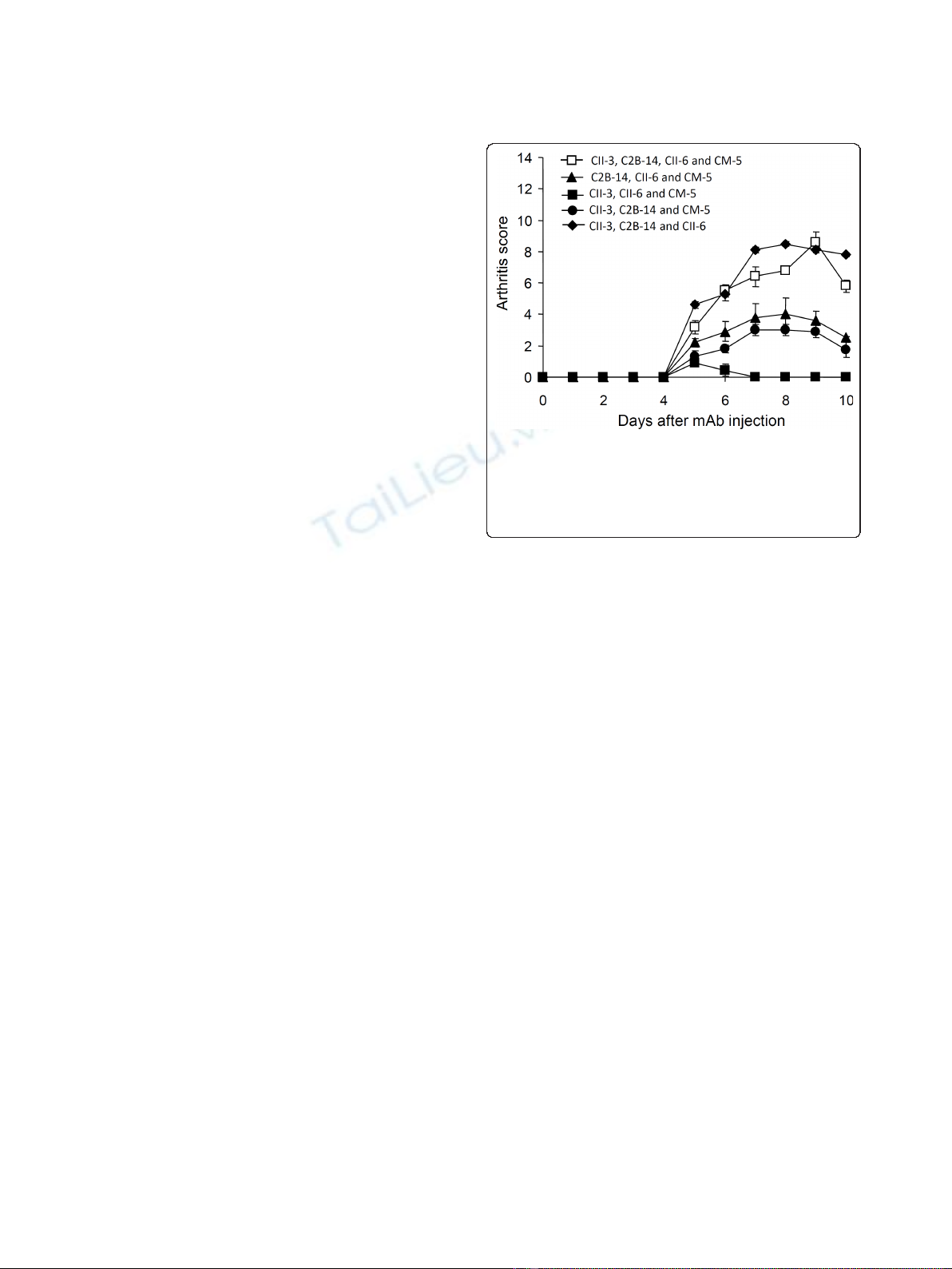
First, we investigated whether arthritis is induced by
combinations of CII-3, CII-6, C2B-14, and CM-5 in
DBA/1J mice (Figure 1). The 4 mAbs combined caused
arthritis, the severity of which was 6.8 ± 0.2 on day 8.
Furthermore, a cocktail of 3 mAbs (CII-3, CII-6, and
C2B-14) induced greater inflammation of the joints than
any other combination (arthritic score: 8.5 ± 0.2 on day
8). On the other hand, the combination of CII-3, CII-6,
and CM-5 (without C2B-14) caused no arthritis.
Consequently, the combination of CII-3, CII-6, and
C2B-14 was used in subsequent experiments.
Effect of an extra mAb on the arthritogenicity of the 3-
clone cocktail
We subsequently added C2A-12 and/or C2B-16 to the 3-
clone cocktail (CII-3, CII-6, and C2B-14) to test the arthri-
togenicity (Figure 2A). The results showed that adding
C2A-12 (arthritic score: 10.3 ± 1.0 on day 8) but not C2B-
16 (5.0 ± 1.5) to CII-3, CII-6, and C2B-14 was effective in
producing more severe arthritis; however, the combination
of all 5 mAbs was less effective (arthritic score: 9.2 ± 1.2
on day 8). Furthermore, the severity of the arthritis
induced by the combination of CII-3, CII-6, C2B-14, and
C2A-12 was dependent on the dose (Figure 2B).
Figure 1 shows the importance of C2B-14, without
which CII-3, CII-6, and CM-5 showed no arthritogeni-
city. Thereafter, we examined the effect of excluding
C2B-14 from the new cocktail. CII-3, CII-6, and C2A-12
(without C2B-14) caused no arthritis (Figure 3).
Histological examination of the arthritis induced by the
new 4-clone cocktail
Histopathological examination of joints in DBA/1J mice
was performed on day 10 after injection of the 4-clone
cocktail. Figure 4A and 4C show the naïve front paw
and ankle joints as a control, respectively. Mice given
the cocktail developed severe arthritis (Figure 4B), and
showed marked proliferation of synovial tissues, massive
infiltration by inflammatory cells, and severe destruction
of cartilage and bone in the ankle joints (Figure 4D).
Figure 1 Time course of changes in the arthritic score after the
administration of arthritogenic mAbs. DBA/1J mice received i.p.
injections of 4 clones (CII-3, C2B-14, CII-6 and CM-5), 3 clones (C2B-
14, CII-6 and CM-5), 3 clones (CII-3, CII-6 and CM-5), 3 clones (CII-3,
C2B-14 and CM-5), and 3 clones (CII-3, C2B-14 and CII-6) on day 0
followed by LPS. Each value is the mean ± SEM for five animals.
Koobkokkruad et al.Journal of Inflammation 2011, 8:31
http://www.journal-inflammation.com/content/8/1/31
Page 3 of 7
Figure 4E and 4G show the staining of TNF-alpha and
IL-1beta, respectively, in normal joints. Cells expressing
TNF-alpha and IL-1beta were detected in inflammatory
regions in the treated mice (Figure 4F and 4H).
Activation of complement and cross-reaction with mouse
or chicken CII in vitro
Figure 5 shows the activation of complement by the
mAbsasapercentageoftheCII-3valueat800μg/mL.
C2B-14andC2A-12showedstrong effects compared
with the other clones. For example, values for C2B-14
and C2A-12 were 123 and 142% at 400 μg/mL. The
levels for C2B-16 (73%) and CII-6 (70%) were similar to
that for CII-3 (60%) at 400 μg/mL. On the other hand,
complement activation by CM-5 (26%) was less than
that by CII-3 at 400 μg/mL. The order of the mAbs in
terms of the activation of complement was C2A-12 =
C2B-14 > CII-3 = C2B-16 = CII-6 > CM-5.
Figure 6A and 6B show cross-reaction with mouse and
chicken CII, respectively, as a percentage of the CII-3
value at 1000 μg/mL.C2B-14andCII-3boundexten-
sively to mouse CII: 103 and 90% at 1 μg/mL, respec-
tively. Furthermore, CII-6 and C2A-12 showed rates of
67 and 48% at 1 μg/mL, respectively; however, C2B-16
and CM-5 did not show binding activity at 1 μg/mL. On
the other hand, for chicken CII, CII-6, C2B-16, and CM-
5 did not show binding activity at 1 μg/mL, although
C2B-14, C2A-12 and CII-3 showed 101, 51 and 24%,
respectively. In terms of the cross-reaction of the mAbs
with mouse and chicken CII, the order was CII-3 = C2B-
14 > CII-6 > C2A-12 > C2B-16 = CM-5, and C2B-14 >
C2A-12 > CII-3 > CII-6 = C2B-16 = CM-5, respectively.
Discussion
The present study demonstrated that a combination of
CII-6, CII-3, C2A-12, and C2B-14 induced severe arthri-
tis in DBA/1J mice. Importantly, these 4 anti-CII mAbs
showed both marked cross-reactions with mouse CII
and the activation of complement, indicating that the
initial event in this model is the formation of collagen-
antibody immune complexes on the cartilage surface or
in the synovium, and subsequent activation of comple-
ment by the complexes may induce arthritis.
Figure 2 Effect of an extra monoclonal antibody on the arthritogenicity of the 3-clone cocktail (CII-3, C2B-14, and CII-6).A:DBA/1J
mice were given i.p. injections of a cocktail of CII-3, C2B-14, and CII-6, the cocktail plus C2A-12, the cocktail plus C2B-16 and the cocktail plus
C2A-12 and C2B-16 on day 0 followed by an injection of LPS on day 3. B: DBA/1J mice received a new 4-clone cocktail (CII-3, C2B-14, CII-6, and
C2A-12, total 2, 4 and 8 mg/mouse) on day 0 followed by LPS. Each value is the mean ± SEM for five animals.
Figure 3 Effect of excluding C2B-14 on the arthritogenicity of
the cocktail (CII-3, C2B-14, CII-6, and C2A-12). DBA/1J mice
received an injection of 4-clones (CII-3, CII-6, C2B-14, and C2A-12) or
3-clones (CII-3, CII-6, and C2A-12) on day 0 followed by an injection
of LPS. Each value is the mean ± SEM for five animals.
Koobkokkruad et al.Journal of Inflammation 2011, 8:31
http://www.journal-inflammation.com/content/8/1/31
Page 4 of 7
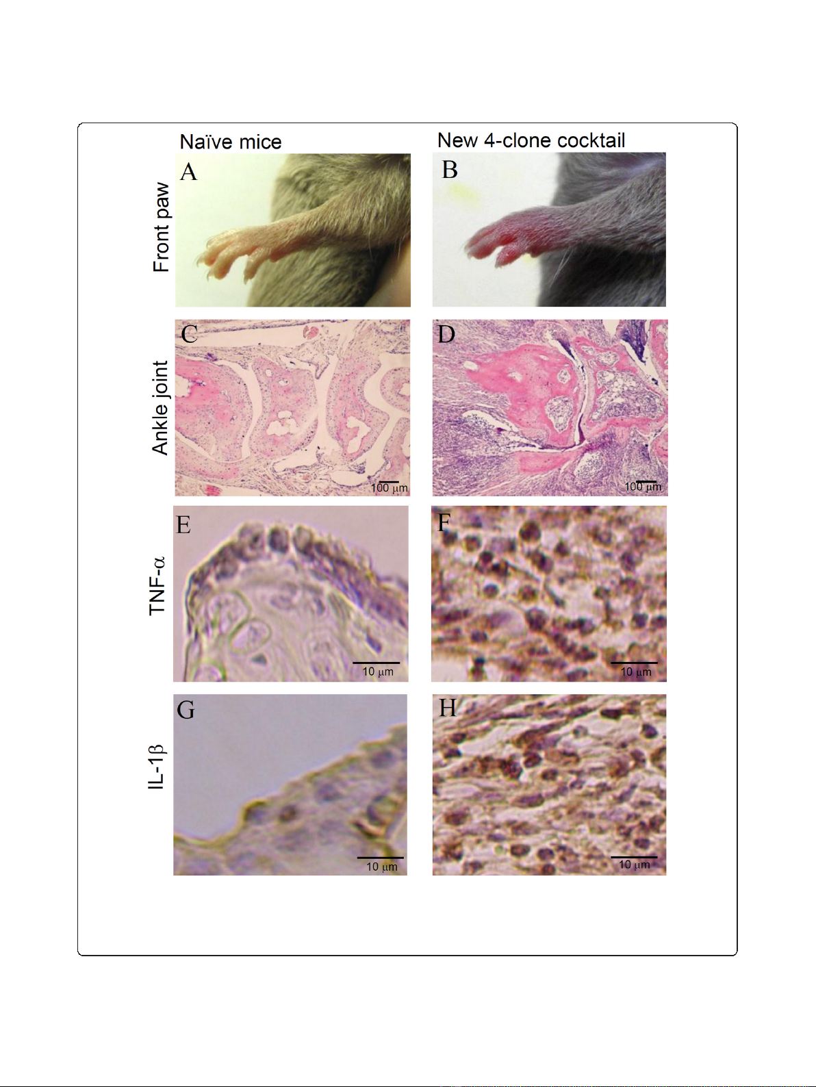
Figure 4 Histological changes induced by the new 4-clone cocktail (CII-3, CII-6, C2B-14, and C2A-12). DBA/1J mice were injected with the
new 4-clone cocktail on day 0 followed by LPS. On day 10, the front paws were amputated for histological examination. The tissues were
stained with H&E and for immunohistochemistry (TNF-alpha and IL-1beta). Results shown are representative histological pictures of five mice
ankle joints in each group. A: normal paw, B: arthritis, C: normal ankle joint, D: arthritic ankle joint, E: normal TNF- alpha, F: arthritic TNF- alpha, G:
normal IL-1 beta, H: arthritic IL-1 beta.
Koobkokkruad et al.Journal of Inflammation 2011, 8:31
http://www.journal-inflammation.com/content/8/1/31
Page 5 of 7


![Báo cáo seminar chuyên ngành Công nghệ hóa học và thực phẩm [Mới nhất]](https://cdn.tailieu.vn/images/document/thumbnail/2025/20250711/hienkelvinzoi@gmail.com/135x160/47051752458701.jpg)


