
BioMed Central
Page 1 of 6
(page number not for citation purposes)
AIDS Research and Therapy
Open Access
Methodology
Assessment of HIV-1 entry inhibitors by MLV/HIV-1 pseudotyped
vectors
Sandra Siegert1,2,5, Sonja Thaler1,3, Ralf Wagner4 and Barbara S Schnierle*1,2
Address: 1Georg-Speyer-Haus, Institute for Biomedical Research, Paul-Ehrlich-Strasse 42-44, D-60596 Frankfurt am Main, Germany, 2Paul-Ehrlich
Institute, Abt. 2/01, Paul-Ehrlich Strasse 51-59, D-63225 Langen, Germany, 3Department of Medicine III, Johannes Gutenberg University, 55101
Mainz, Germany, 4Institute of Medical Microbiology and Hygiene, University of Regensburg, Franz-Josef-Strauss Allee 11, 93053 Regensburg,
Germany and 5Friedrich Miescher Institute, Maulbeerstrasse 66, CH-4058 Basel, Switzerland
Email: Sandra Siegert - sandra.siegert@fmi.ch; Sonja Thaler - thaler@3-med.klinik.uni-mainz.de; Ralf Wagner - ralf.wagner@klinik.uni-
regensburg.de; Barbara S Schnierle* - schba@pei.de
* Corresponding author
Abstract
Background: Murine leukemia virus (MLV) vector particles can be pseudotyped with a truncated
variant of the human immunodeficiency virus type 1 (HIV-1) envelope protein (Env) and selectively
target gene transfer to human cells expressing both CD4 and an appropriate co-receptor. Vector
transduction mimics the HIV-1 entry process and is therefore a safe tool to study HIV-1 entry.
Results: Using FLY cells, which express the MLV gag and pol genes, we generated stable producer
cell lines that express the HIV-1 envelope gene and a retroviral vector genome encoding the green
fluorescent protein (GFP). The BH10 or 89.6 P HIV-1 Env was expressed from a bicistronic vector
which allowed the rapid selection of stable cell lines. A codon-usage-optimized synthetic env gene
permitted high, Rev-independent Env expression. Vectors generated by these producer cells
displayed different sensitivity to entry inhibitors.
Conclusion: These data illustrate that MLV/HIV-1 vectors are a valuable screening system for
entry inhibitors or neutralizing antisera generated by vaccines.
Background
The acquired immunodeficiency syndrome (AIDS) was
first described about 20 years ago. Since then almost 20
million people have died from human immunodeficiency
virus (HIV-1) infection and 42 million are infected with.
New drugs and an effective vaccine are urgently needed. In
particular, new drugs that block the HIV type 1 (HIV-1)
entry into host cell have clear advantages over the cur-
rently used drugs. They should abrogate the establishment
of a productive infection and consequently could dimin-
ish the chances of HIV-1 developing resistance. Further-
more, a vaccine that prevents AIDS should elicit broadly
cross-reactive neutralizing antibodies to prevent infection.
A safe and simple assay for measuring neutralizing activi-
ties against different HIV-1 strains is critical for the devel-
opment of such a vaccine or entry inhibiting drugs.
We previously generated a retroviral vector which specifi-
cally transfers genes into human CD4+ cells [1,2]. This
vector was derived by pseudotyping murine leukemia
virus (MLV) capsid particles with a variant of the HIV-1
envelope protein (Env) containing the surface glycopro-
tein gp120-SU and a carboxyl-terminally truncated trans-
membrane (TM) protein with only 7 cytoplasmic amino
Published: 12 September 2005
AIDS Research and Therapy 2005, 2:7 doi:10.1186/1742-6405-2-7
Received: 25 July 2005
Accepted: 12 September 2005
This article is available from: http://www.aidsrestherapy.com/content/2/1/7
© 2005 Siegert et al; licensee BioMed Central Ltd.
This is an Open Access article distributed under the terms of the Creative Commons Attribution License (http://creativecommons.org/licenses/by/2.0),
which permits unrestricted use, distribution, and reproduction in any medium, provided the original work is properly cited.

AIDS Research and Therapy 2005, 2:7 http://www.aidsrestherapy.com/content/2/1/7
Page 2 of 6
(page number not for citation purposes)
acids. HIV-1 Env facilitates vector attachment to target
cells and membrane fusion, which is initiated by the inter-
action of HIV-1 Env with the CD4 receptor molecule on
the surface of the target cell. CD4 binding induces a con-
formational change in the envelope glycoprotein and
allows the binding of a co-receptor of the chemokine
receptor family [3]. The co-receptor usage is virus strain
dependent: R5 viruses, which infect monocytes and mac-
rophages, use CCR5 and X4 viruses, which infect T cell
lines, use CXCR4. X4R5 strains can use CXCR4 as well as
CCR5 for entry. The transfer of a marker gene by MLV/
HIV-1 vectors is therefore a safe and simple method to
assay entry mediated by HIV-1 Env and can be used to
evaluate HIV-1 entry inhibitors, such as small molecules
or neutralizing antibodies in sera of vaccinated animals or
patients.
Here, we optimized production of the MLV/HIV-1 vector
to allow analysis of different HIV-1 Envs and demonstrate
that the responsiveness to viral entry inhibitors was
dependent on the HIV-1 strain the Env was derived from.
This illustrates that MLV/HIV-1 pseudotyped vectors are
useful tools for analyzing HIV-1 entry.
Results and discussion
Generation of a stable producer cell line encoding the 89.6
P HIV-1 Env
We and others have previously reported that MLV capsids
can be pseudotyped with cytoplasmatically truncated var-
iants of the HIV-1 or HIV-2 envelope glycoproteins pos-
sessing only 7 cytoplasmic amino acids. These MLV/HIV
pseudotyped vectors have the HIV host range [4,1,5]. We
used a X4 HIV-1 Env variant (BH10) for pseudotyping,
which restricted vector entry to CD4 and CXCR4 receptor-
positive cells [2]. HIV-1 Env was expressed from an expres-
sion construct that also encoded Rev, which is required to
transport the rev responsive element (RRE)-containing
env mRNA from the nucleus to the cytoplasm. In the
present study we evaluated a codon-usage-optimized HIV-
1 env gene that encodes the truncated Env variant of the
X4R5 89.6 P HIV-1 isolate and lacks rev sequences. West-
ern blot analysis of transfected 293T cells showed that the
change from lentiviral to mammalian codon usage
allowed high HIV-1 Env protein expression in the absence
of Rev (Figure 1). The 89.6 P Env showed different migra-
tion in polyacrylamide gels from the BH10 isolate, which
might be caused by different glycosylation patterns of the
protein and reflects strain-specific differences in Env.
We previously constructed a MLV/HIV-1 producer cell line
based on FLY cells [6], which expresses the HIV-1 Env of
the X4 HIV-1 BH10 strain and a retroviral vector encoding
the green fluorescent protein (GFP) [2]. These cells are fur-
ther referred to as FLY-HIV-87-GFP cells. HIV-1 Env pro-
tein expression is driven by the strong human elongation
factor 1α promoter and stable clones were selected via the
puromycin resistance gene (pac) encoded on the bicis-
tronic messenger RNA.
We cloned a codon-usage-optimized 89.6 P HIV-1 Env
into this vector and stable cell clones were rapidly isolated
by puromycin selection. Protein expression of HIV-1 Env
was ensured by expansion of the cells in the continued
presence of puromycin. A single clone was further trans-
duced with a GFP-encoding retroviral vector to obtain the
producer cell line FLY-syn-GFP.
Characterization of vectors particles derived from FLY-
HIV-87-GFP or FLY-syn-GFP
The characterization of the two producer cell lines was
started by determining the amount of infectious retroviral
vector particles released from the producer cells. Infec-
tious particles can be easily detected by the transfer of the
gfp gene. NIH3T3 CD4/CXCR4 cells that express the CD4
and CXCR4 receptors were transduced with supernatants
derived from FLY-HIV-87-GFP or FLY-syn-GFP cells and
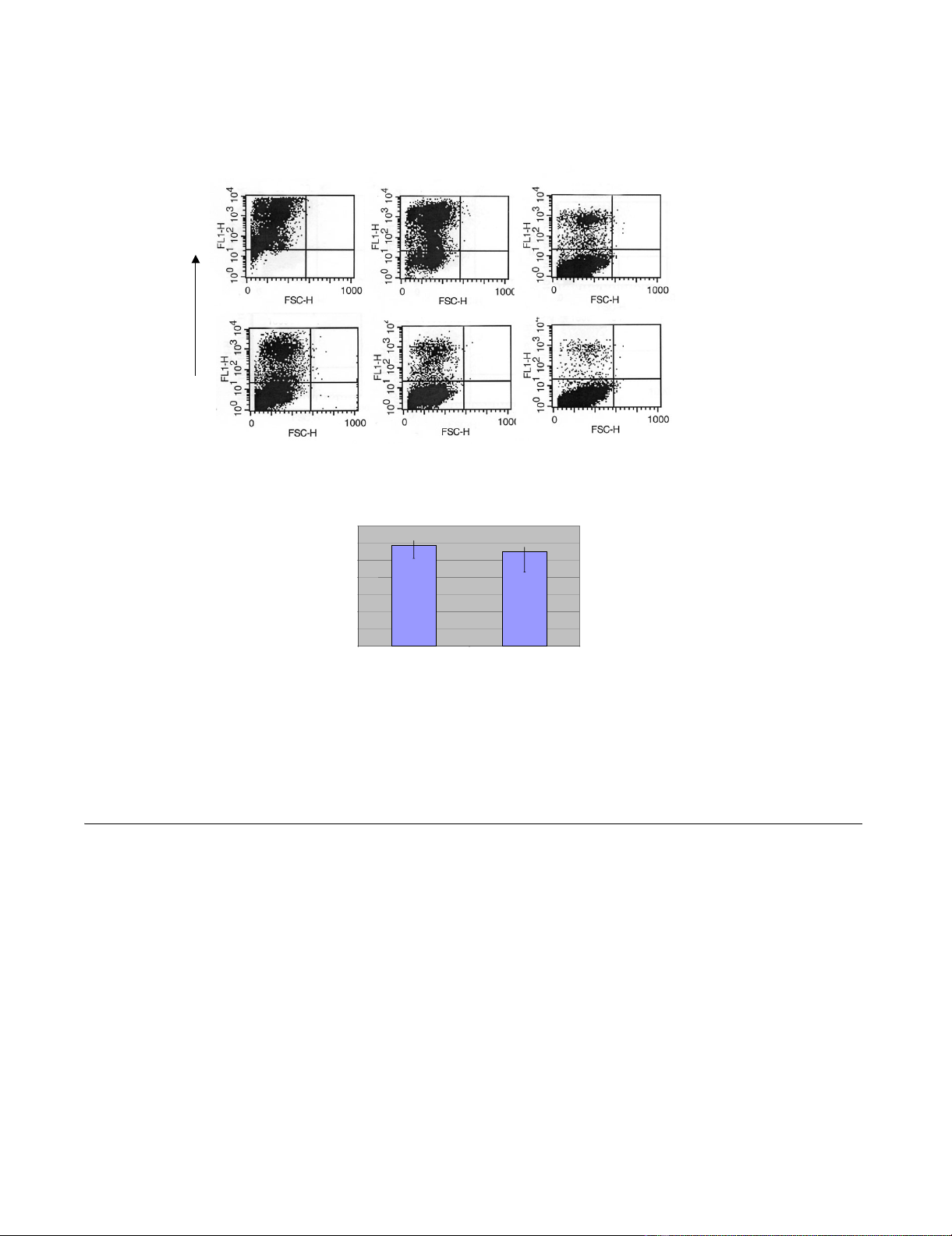
analyzed after two days by flow cytometry. Figure 2A gives
a typical FACS analysis of NIH3T3-CD4/X4 cells trans-
duced with serially diluted vector supernatants. Titers are
given in Figure 2B in infectious units per ml and represent
the average values of five experiments. The titers produced
by FLY-HIV-87-GFP cells were reproducibly higher than
those obtained from FLY-syn-GFP cells. However, the
X4R5 phenotype of the 89.6 P HIV-1 Env was retained by
the pseudotypes. Only vector particles derived from FLY-
syn-GFP cells were able to transduce NIH3T3 cells express-
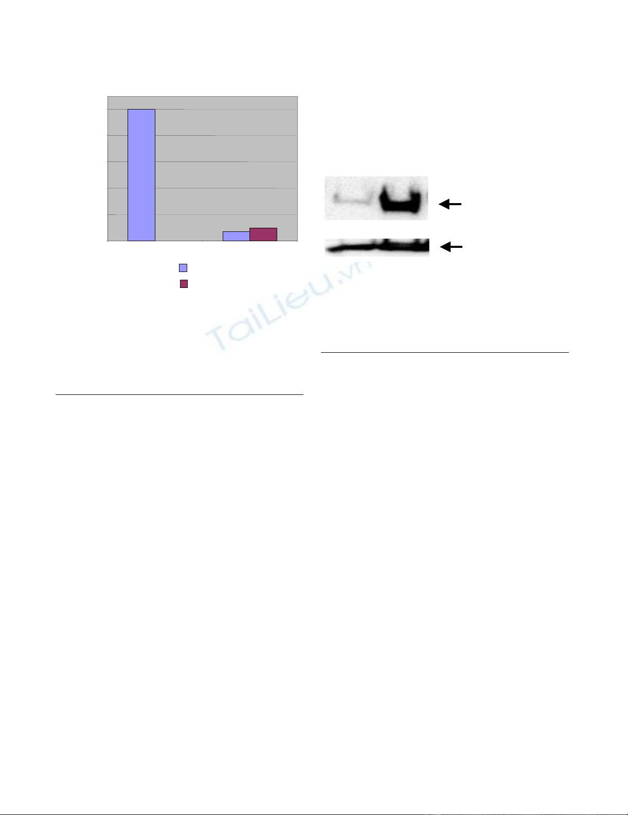
ing CD4 and CCR5 (Figure 3).
The changed codon usage of the 89.6 P Env resulted in
high expression in FLY cells; however, vector titers were
always lower than those of vectors containing BH10 Env.
Expression of the 89.6 P HIV-1 EnvFigure 1
Expression of the 89.6 P HIV-1 Env. 293T cells were trans-
fected with 3 µg plasmid DNA and cell lysates were analyzed
after two days for HIV-1 Env expression by Western blot
analysis. The two forms of Env are indicated as gp140 (C-ter-
minally truncated precursor) and gp120 (SU).
∆CT Env precursor
gp120
89.6PBH10
AIDS Research and Therapy 2005, 2:7 http://www.aidsrestherapy.com/content/2/1/7
Page 3 of 6
(page number not for citation purposes)
To analyze the amount of Env incorporated into particles,
the supernatants of the producer cells were collected and
centrifuged through a 30% sucrose cushion. The Western
blot analysis of the pellets revealed that high levels of the
89.6 P Env was incorporated into vector particles (Figure
4). Equal loading was verified by detection of the MLV
p30 Gag protein. These data imply that the amount of Env
in viral vector particles does not correlate with their titer.
Instead, it seems likely that the fusion ability of Env,
which displays strain-specific differences, affects vector
infectivity.
Evaluation of HIV-1 entry inhibitors
Attachment and entry of HIV-1 into CD4 cells involve a
series of conformational changes in Env which allow co-
receptor binding and finally fusion of viral and cell mem-
branes. AMD-3100 is a small molecule inhibitor of gp120
attachment to the CXCR4 receptor, and T-20 is a synthetic
peptide corresponding to a helical region of HIV-1 gp41
that blocks fusion of the cellular and the viral membrane.
While AMD-3100 is only active against X4 and X4R5 HIV-
1 strains, T20 inhibits fusion of most HIV-1 strains.
As shown in Figure 5A, the sensitivity of the BH10 and
89.6 P Env-containing vectors to AMD-3100 was slightly
different. BH10 was less sensitive, indicating a higher
Titer of MLV/HIV-1 pseudotyped vector particles released from producer cellsFigure 2
Titer of MLV/HIV-1 pseudotyped vector particles released from producer cells. A: NIH3T3-CD4/X4 cells (1 × 10⁵) were incu-
bated with 1, 0.1 or 0.01 ml supernatant from producer cells in a total volume of 1 ml. For titer determination, the number of
GFP+ cells (upper left) was determined. B: Titers were calculated by measuring the percentage of GFP-positive cells after
transduction. Values are the average of 5 experiments.
FLY-HIV-87-GFP FLY-syn-GFP
Infectious units/ml
1,E+00
1,E+01
1,E+02
1,E+03
1,E+04
1,E+05
1,E+06
1,E+07 7,6E+05 3,1E+05
FLY-syn-GFP
FLY-HIV-87-GFP
1 ml 0.1 ml 0.01 ml
GFP
A
B
AIDS Research and Therapy 2005, 2:7 http://www.aidsrestherapy.com/content/2/1/7
Page 4 of 6
(page number not for citation purposes)
affinity for CXCR4. However, the responsiveness to T20
was not significantly different between both Env proteins
(Figure 5B). The inhibitor concentrations used in these
studies were higher than those previously published
because C-terminally truncated Envs have a fast fusion
kinetic and are thus less sensitive to entry inhibitors [7]. It
has been shown that the cytoplasmic tail slows the folding
of HIV-1 Env from a late prebundle configuration into the
six-helix bundle, and thereby also slows down the fusion
process [8]. Inhibition of HIV-1 Env mediated transduc-
tion was specific, since amphotropic MLV could not be
inhibited by AMD3100 or T20 (data not shown).
We present here two producer cell lines that release MLV/
HIV-1 pseudotyped retroviral vectors particles. Transfer of
the gfp gene can be used as an indication of an infection
process mediated by the HIV-1 envelope glycoprotein.
This assay is robust and simple to perform and GFP
expression can be rapidly monitored by flow cytometry
without further staining of the target cells. Expression of
the HIV-1 Env from a bicistronic vector allowed fast
establishment of stable producer cell lines. Optimization
of HIV-1 Env codon usage led to high expression without
the need for Rev co-expression and will, in combination
with the bicistronic vector, facilitate the easy exchange of
Env sequences. The system can also be applied to transient
vector production, and synthetic genes will permit fast
testing of diverse HIV-1 Envs, including those from drug
resistant strains.
Conclusion
MLV/HIV-1 vectors are a valuable screening system for
entry inhibitors or neutralizing antisera generated by
vaccines.
Methods
Plasmids
The truncated variant of the envelope glycoprotein HIV-1
Env Tr712 [1] was derived from the plasmid pLßAc/env-
Tr712-neo as a 3.1 kb SalI/XhoI fragment and was cloned
into the XhoI site of the bicistronic vector pEF-IRES-P [9].
The sequence of the 89.6 P HIV-1 Env isolate (aa 1 – 712)
was chemically synthesized and the codons were modi-
fied to high GC content without changing the coding
sequence. The Env signal peptide sequence was exchanged
with that of the CD5 receptor. Env was excised as an
EcoRI/XhoI fragment, blunt-ended and cloned into the
XhoI site of the vector pEF-IRES-P, resulting in the clone
pEF-IRES-P-89.6 P.
Cells
NIH 3T3 derivatives [10], 293T (ATCC #CRL-11268) and
FLY [11] cells were grown in Dulbecco's modified Eagle's
medium (GIBCO BRL, Eggenstein) supplemented with
10% fetal bovine serum, 1% penicillin/streptomycin and
Co-receptor usage of MLV/HIV-1 vectorsFigure 3
Co-receptor usage of MLV/HIV-1 vectors. NIH3T3 cells
expressing the CD4 receptor and either CXCR4 or CCR5
were transduced with vector particles derived from FLY cell
lines, and the titers were calculated by measuring the per-
centage of GFP-positive cells. The X4R5 89.6 P Env in vector
particles derived from FLY-syn-GFP cells allowed the trans-
duction of both target cell lines.
0,0E+00
2,0E+05
4,0E+05
6,0E+05
8,0E+05
1,0E+06
FLY-HIV-87-GFP FLY-syn-GFP
CD4, CXCR4
CD4, CCR5
Infectious units/ml
Incorporation of HIV-1 Env into vector particlesFigure 4
Incorporation of HIV-1 Env into vector particles. Incorpora-
tion of Env into MLV/HIV-1 particles was analyzed by West-
ern blot analysis of particles concentrated from FLY cell
supernatants by ultracentrifugation. Equal loading was con-
firmed after stripping the blot and incubating with an anti-
MLV-Gag (p30) antibody.
FLY-HIV-87-GFP
FLY-syn-GFP
gp120
p30 MLV Gag
AIDS Research and Therapy 2005, 2:7 http://www.aidsrestherapy.com/content/2/1/7
Page 5 of 6
(page number not for citation purposes)
1% L-glutamine. FLY cells are based on human HT1080
cells and express the MLV Gag/Pol gene product [11]. Sta-
bly transfected FLY-HIV-87-GFP or FLY-syn-GFP cells were
grown in the medium described above supplemented
with 2.5 µg/ml puromycin (Sigma, Deisenhofen).
Stable transfection of cells
FLY cells (106) were seeded in a 10 cm tissue culture plate.
The following day, cells were transfected with 10 µg of the
bicistronic construct pEF-IRES-P-89.6 P. Transfection was
performed with SuperFect™ Transfection Reagent (Qia-
gen, Hilden). Forty-eight hours after transfection, 2.5 µg/
ml puromycin was added to the medium. After the selec-
tion and isolation of single cell clones, cells were tested for
HIV-1 envelope glycoprotein expression by Western blot
analysis and the best expressing clone was transduced
with VSV-G pseudotyped retroviral vectors encoding GFP
(pMX-EGFP) [12].
Retroviral transduction and titer determination
Serial dilutions of vector supernatants from packaging
cells were passed through 0.45-µm filters and incubated
with 1 × 10⁵ NIH 3T3-CD4/CXCR4 or NIH 3T3-CD4/
CCR5 cells for 6 h and longer incubation (e.g. over night)
did not change the titer. Supernatants were mixed with
different amounts of T20 (generous gift of Prof. von Laer,
Georg-Speyer-Haus, Frankfurt) or AMD-3100 (NIH AIDS
Research and Reference Reagent Program) for inhibition
assays. The numbers of GFP-expressing cells were detected
by FACS analysis 48 – 72 hours after transduction. The tit-
ers are given in infectious units per ml (IU/ml) and were
determined by calculating the percentage of GFP-positive
cells. GFP expression was monitored by a shift to green
fluorescence (FL-1). FACS analysis was performed with a
FACScan (Becton Dickinson, Heidelberg) using the Cel-
lquest software.
Preparation of viral proteins and immunoblots
For the analysis of incorporation of Env proteins into the
vector particles, supernatant of producer cells was filtered
through a 0.45-µm filter (Greiner, Frickenhausen, Ger-
many) and centrifuged through a 30% sucrose cushion for
90 min at 26,000 rpm in an SW28 rotor. The pellet was
resuspended in 50 µl SDS loading buffer [13] and 25 µl
Inhibition of MLV/HIV-1 mediated gene transfer by HIV-1 entry inhibitorsFigure 5
Inhibition of MLV/HIV-1 mediated gene transfer by HIV-1 entry inhibitors. Indicated amounts of either (A) AMD-3100 or (B)
T20 were added to vector supernatants during the transduction of NIH3T3 cells expressing CD4 and CXCR4. Transfer of the
gfp gene was measured and titers are given as percentage of those obtained from untreated supernatants. Inhibition of MLV/
HIV-1 vectors was done 2–3 times and had the same outcome. Inhibition of transduction of vectors derived from FLY-HIV-87-
GFP cells by AMD3100 was done only once.
AMD3100 concentration [nM]
Infectious titer in % of untreated
supernatants
FLY-HIV-87-GFP
FLY-syn-GFP
T20 concentration [µ
µµ
µM]
0
20
40
60
80
100
0 0.25 0.5 0.75 1 1.25 1.5 1.75 2 2.25 2.5
0
20
40
60
80
100
120
0 50 100 150 200 250 300
AB


![Báo cáo seminar chuyên ngành Công nghệ hóa học và thực phẩm [Mới nhất]](https://cdn.tailieu.vn/images/document/thumbnail/2025/20250711/hienkelvinzoi@gmail.com/135x160/47051752458701.jpg)


