
BioMed Central
Page 1 of 12
(page number not for citation purposes)
Theoretical Biology and Medical
Modelling
Open Access
Research
Binding site of ABC transporter homology models confirmed by
ABCB1 crystal structure
Aina W Ravna*, Ingebrigt Sylte and Georg Sager
Address: Department of Medical Pharmacology and Toxicology, Institute of Medical Biology, Faculty of Health Sciences, University of Tromsø,
N-9037 Tromsø, Norway
Email: Aina W Ravna* - Aina.W.Ravna@uit.no; Ingebrigt Sylte - ingebrigt.sylte@uit.no; Georg Sager - georg.sager@uit.no
* Corresponding author
Abstract
The human ATP-binding cassette (ABC) transporters ABCB1, ABCC4 and ABCC5 are involved in
resistance to chemotherapeutic agents. Here we present molecular models of ABCB1, ABCC4 and
ABCC5 by homology based on a wide open inward-facing conformation of Escherichia coli MsbA,
which were constructed in order to elucidate differences in the electrostatic and molecular
features of their drug recognition conformations. As a quality assurance of the methodology, the
ABCB1 model was compared to an ABCB1 X-ray crystal structure, and with published cross-
linking and site directed mutagenesis data of ABCB1. Amino acids Ile306 (TMH5), Ile340 (TMH6),
Phe343 (TMH6), Phe728 (TMH7), and Val982 (TMH12), form a putative substrate recognition site
in the ABCB1 model, which is confirmed by both the ABCB1 X-ray crystal structure and the site-
directed mutagenesis studies. The ABCB1, ABCC4 and ABCC5 models display distinct differences
in the electrostatic properties of their drug recognition sites.
Introduction
The human ATP-binding cassette (ABC) transporters
ABCB1, ABCC4 and ABCC5 belong to the ABC super-
family, a subgroup of Primary active transporters [1]. The
transporters in the ABC superfamily are structurally
related membrane proteins that have a common intracel-
lular motif that exhibits ATPase activity. This motif cleaves
ATP's terminal phosphate to energize the transport of
molecules from regions of low concentration to regions of
high concentration [1-3]. Since ABC genes are highly con-
served between species, it is likely that most of these genes
have been present since the beginning of eukaryotic evo-
lution [4].
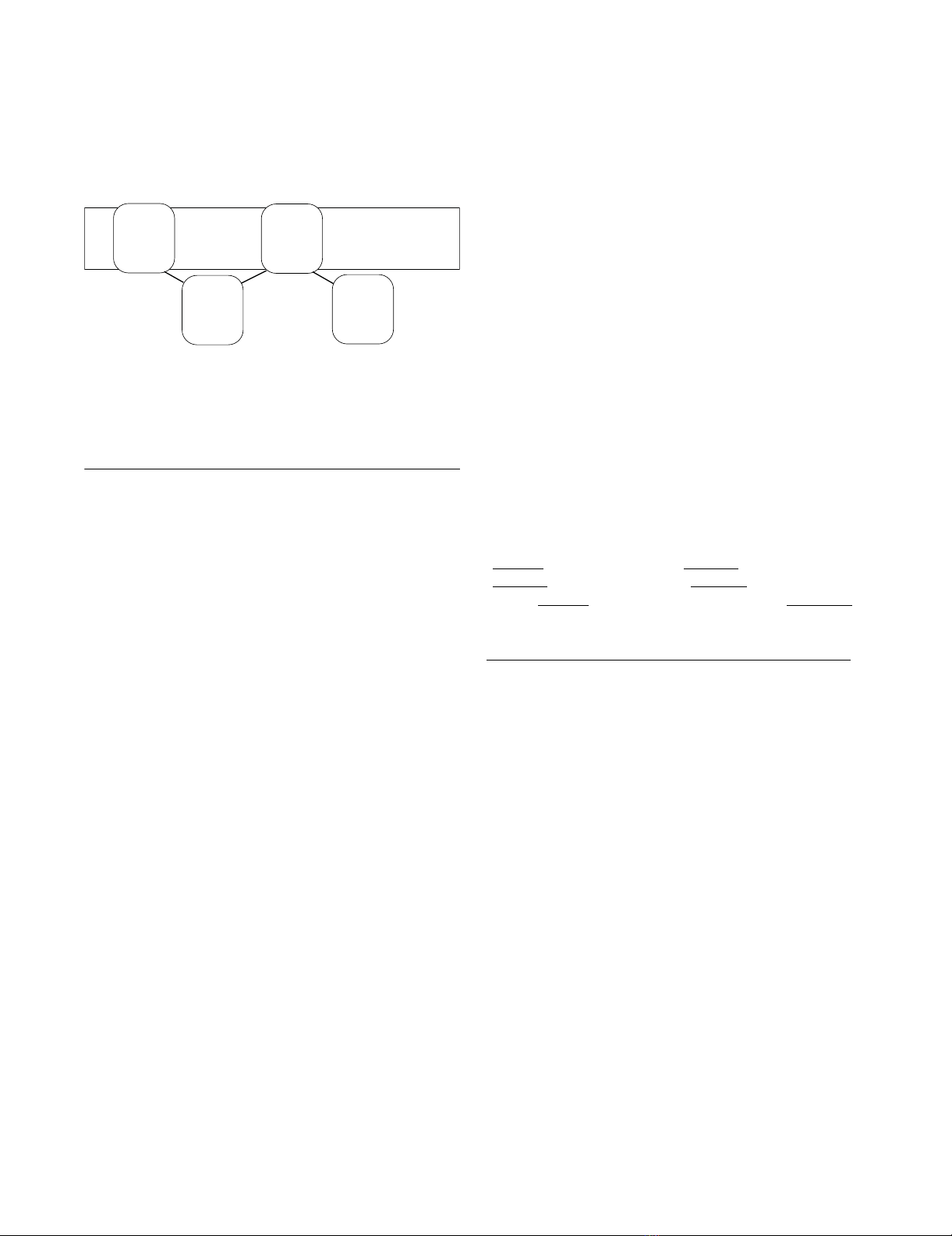
The overall topology of ABCB1, ABCC4 and ABCC5 is
divided into transmembrane domain 1 (TMD1) - nucle-
otide-binding domain 1 (NBD1) - TMD2 - NBD2 (Figure
1). The Walker A, or phosphate binding loop (P-loop),
and Walker B motifs, are localized in the NBDs, while the
TMDs contribute to the substrate translocation events
(recognition, translocation and release). ABCB1, ABCC4
and ABCC5 are exporters, pumping substrates out of the
cell.
Transporters have drug recognition sites that make them
specific for particular substrates, and drugs may interact
with these recognition sites and either inhibit the trans-
porter or act as substrates. Experimental studies have
shown that ABCB1 transports cationic amphiphilic and
lipophilic substrates [5-8], while ABCC4 and ABCC5
transport organic anions [9]. Both ABCC4 and ABCC5
transport cAMP and cGMP, however, with differences in
their kinetic parameters; ABCC4 with a preference for
cAMP and ABCC5 with a preference for cGMP [9,10].
Published: 4 September 2009
Theoretical Biology and Medical Modelling 2009, 6:20 doi:10.1186/1742-4682-6-20
Received: 4 June 2009
Accepted: 4 September 2009
This article is available from: http://www.tbiomed.com/content/6/1/20
© 2009 Ravna et al; licensee BioMed Central Ltd.
This is an Open Access article distributed under the terms of the Creative Commons Attribution License (http://creativecommons.org/licenses/by/2.0),
which permits unrestricted use, distribution, and reproduction in any medium, provided the original work is properly cited.
Theoretical Biology and Medical Modelling 2009, 6:20 http://www.tbiomed.com/content/6/1/20
Page 2 of 12
(page number not for citation purposes)
When chemotherapeutic agents are expelled from cancer
cells as substrates of ABCB1, ABCC4 or ABCC5, the result
is multidrug resistance. In order to overcome multidrug
resistance, development of inhibitors of drug efflux trans-
porters has been sought for use as supplement to drug
therapy [11]. However, clinical trials of potential anti-
MDR agents have been disappointing due to adverse
effects in vivo of agents being very effective in vitro. Even if
there is a long time since Victor Ling described MDR, (i.e.
ABCB1) [12], very little is known about subtype selective
recognition and binding of ABC proteins. Structural
insight into their mode of ligand interaction and func-
tional mechanisms will be an important contribution to
pinpoint potential drug targets and to design putative
inhibitors. Recent papers report a considerable difference
in substrate specificity of ABCC4 and ABCC5 [9], includ-
ing various chemotherapeutic agents [13], and with
potential impact on reversal of MDR [14]. Elucidating the
molecular aspects of ligand interactions with ABCB1,
ABCC4 or ABCC5 may therefore aid in the design of ther-
apeutic agents that can help to overcome multidrug resist-
ance.
We have previously constructed molecular models of
ABCB1 [15], ABCC4 [16] and ABCC5 [17] based on the
Staphylococcus aureus ABC transporter Sav1866, which has
been crystallized in an outward-facing ATP-bound state
[18]. In this study, we present molecular models of
ABCB1, ABCC4 and ABCC5 based on a wide open
inward-facing conformation of Escherichia coli MsbA [19].
Since the molecular modelling was carried out before the
X-ray crystal structure of the Mus musculus ABCB1 in a
drug-bound conformation was published [20], we got a
unique opportunity to test our methodology, molecular
modelling by homology, and the quality of the ABCB1
model, when the crystal structure was published. Since we
wanted to elucidate differences in the electrostatic and
molecular features of the drug recognition conformation
of these transporters, the wide open conformation of the
MsbA template [19] was of particular interest. The electro-
static potential surfaces (EPS) of the models were calcu-
lated, and the models were compared to the X-ray crystal
structure of the Mus musculus ABCB1 [20], and with pub-
lished cross-linking and site directed mutagenesis data on
ABCB1 [21-35].
Computational methods
Software
Version 3.4-9b of the Internal Coordinate Mechanics
(ICM) program [36] was used for homology modelling,
model refinements and electrostatic calculations. The
AMBER program package version 8.0 [37] was used for
molecular mechanics energy minimization.
Alignment
A multiple sequence alignment of (SWISS-PROT acces-
sion numbers are given in brackets) human ABCB1
(P08183), human ABCC4 (O15439), human ABCC5
(O15440), human ABCC11 (Q9BX80), Escherichia coli
MsbA (P60752) and Vibrio cholerae MsbA (Q9KQW9),
obtained using T-COFFEE [38], Version 4.71 available at
the Le Centre national de la recherche scientifique website
http://www.igs.cnrs-mrs.fr/Tcoffee/tcoffee_cgi/index.cgi,
was used as a basis for the homology modelling module
of ICM program [36]. ABCC11 was included in the align-
ment because it is closely related to ABCC5 phylogeneti-
cally [15], and its inclusion may strengthen the alignment.
The alignment was adjusted for sporadic gaps in the TMH
segments, and for secondary structure predictions defin-
ing the boundaries of the TMHs using the PredictProtein
server for sequence analysis and structure prediction [39],
and SWISS-PROT [40].
The alignment of human ABCB1 and Escherichia coli MsbA
was compared to previously published alignments of
human ABCB1 and Escherichia coli MsbA [19,41], and it
was observed that in our alignment, the ABCB1 sequence
was shifted 2 positions to the left relative to the E. coli
MsbA sequence in the alignment of TMH2, and 1 position
the left relative to the E. coli MsbA sequence in the align-
ment of TMH6, as compared to the previously published
alignments of human ABCB1 and Escherichia coli MsbA
[19,41]. Thus, 3 alignments were used to construct 3
ABCB1 models, 1 model with our original alignment, 1
model with TMH2 adjusted to correspond to the previ-
ously published alignments of human ABCB1 and
Escherichia coli MsbA [19,41], and 1 model with both
TMH2 and TMH6 adjusted, thus using the same align-
Overall domain topology of ABCB1, ABCC4 and ABCC5Figure 1
Overall domain topology of ABCB1, ABCC4 and
ABCC5.
Extracellular side
Cell membrane
Cytoplasm
TMD1 TMD2
ABC1 ABC2

Theoretical Biology and Medical Modelling 2009, 6:20 http://www.tbiomed.com/content/6/1/20
Page 3 of 12
(page number not for citation purposes)
ment as the previously published alignments of human
ABCB1 and Escherichia coli MsbA [19,41]. The alignment
of Escherichia coli MsbA, human ABCB1, human ABCC4
and human ABCC5 used for the homology modelling
procedure, with TMH2 adjusted to correspond to the pre-
viously published alignments of human ABCB1 and
Escherichia coli MsbA [19,41], is shown in Figure 2. For
illustrative purposes, only the sequences of the template
and the 3 target proteins ABCB1, ABCC4 and ABCC5 are
shown.
Homology modelling
A full atom version of the open inward facing Escherichia
coli MsbA X-ray crystal structure (PDB code: 3B5W[19])
was kindly provided by Geoffery Chang and used as a
template in the construction of the homology models of
ABCB1, ABCC4 and ABCC5. The ICM program constructs
the molecular model by homology from core sections
defined by the average of Cα atom positions in conserved
regions. Loops were searched for within several thousand
structures in the PDB databank [42] and matched in
regard to sequence similarity and sterical interactions with
the surroundings of the model, and the best-fitting loop
was selected based on calculating the maps around the
loops and scoring of their relative energies. The segment
connecting NBD1 and TMD2 was also included in the
loop search procedure.
Calculations
The ABCB1, ABCC4 and ABCC5 models were refined by
globally optimizing side-chain positions and annealing of
the backbone using the RefineModel macro of ICM. The
macro was comprised of (1) a side-chain conformational
sampling using 'Montecarlo fast' [43], (2) 5 iterative
annealings of the backbone with tethers (harmonic
restraints pulling an atom in the model to a static point in
space represented by a corresponding atom in the tem-
plate), and (3) a second side-chain conformational sam-
pling using 'Montecarlo fast'. 'Montecarlo fast' samples
conformational space of a molecule with the ICM global
optimization procedure, and its iterations consist of a ran-
dom move followed by a local energy minimization, and
calculation of the complete energy. The iteration is
accepted or rejected based on energy and temperature.
The refined ABCB1, ABCC4 and ABCC5 models were
energy minimized using the AMBER 8.0 program package
[37]. Two energy minimizations were performed for each
model, (1) with restrained backbone by 500 cycles of the
steepest descent minimization followed by 500 steps of
conjugate gradient minimization, and (2) with no
restraints by 1000 cycles of the steepest descent minimiza-
tion followed by 1500 steps of conjugate gradient mini-
mization. The leaprc.ff03 force field [37], and a 10 Å cut-
off radius for non-bonded interactions and a dielectric
multiplicative constant of 1.0 for the electrostatic interac-
tions, were used in the molecular mechanics calculations.
The EPS of the ABCB1, ABCC4 and ABCC5 models were
calculated with the ICM program, with a potential scale
from -10 to +10 kcal/mol.
Model validation
To check the stereochemical qualities of the ABCB1,
ABCC4 and ABCC5 models, the SAVES Metaserver for
analyzing and validating protein structures http://nih
server.mbi.ucla.edu/SAVES/ was used. Programs run were
Procheck [44], What_check [45], and Errat [46], and the
pdb file of the open inward facing Escherichia coli MsbA
template [19] was also checked for comparison with the
models.
For further validation, the ABCB1, ABCC4 and ABCC5
models were compared with the X-ray crystal structure of
the Mus musculus ABCB1 [20] and cross-linking and site
directed mutagenesis data published on ABCB1 [21-35].
Results
The 3 ABCB1 models, constructed based on 3 different
alignments, where compared with cross-linking data and
subsequently also the X-ray crystal structure of the Mus
musculus ABCB1 [20], and it was revealed that when
TMH2 was aligned as the previously published align-
ments of human ABCB1 and Escherichia coli MsbA
[19,41], amino acids in TMH2/TMH11 (Val133/Gly939
and Cys127/Ala935) where oriented towards each other
in accordance with both cross-linking data and the X-ray
crystal structure of the Mus musculus ABCB1 [20]. How-
ever, when TMH6 was aligned as the previously published
alignments of human ABCB1 and Escherichia coli MsbA
[19,41], ligand binding amino acids (Ile340 and Phe343)
pointed away from the drug binding site, while when
aligned as proposed from our T-COFFEE [38] alignment,
it was in accordance both with cross-linking data and the
X-ray crystal structure of the Mus musculus ABCB1 [20].
Thus, the ABCB1 model which was most in accordance
with cross-linking data and the X-ray crystal structure of
the Mus musculus ABCB1 [20] was based on the alignment
where TMH2 was adjusted according to the previously
published alignments of human ABCB1 and Escherichia
coli MsbA [19,41], while TMH6 was kept exactly as in our
T-COFFEE [38] alignment. The alignment of Escherichia
coli MsbA, human ABCB1 (TMH2 adjusted), human
ABCC4 and human ABCC5 used for the homology mod-
elling procedure is shown in Figure 2. For illustrative pur-
poses, only the sequences of the template and the 3 target
proteins ABCB1, ABCC4 and ABCC5 are shown.
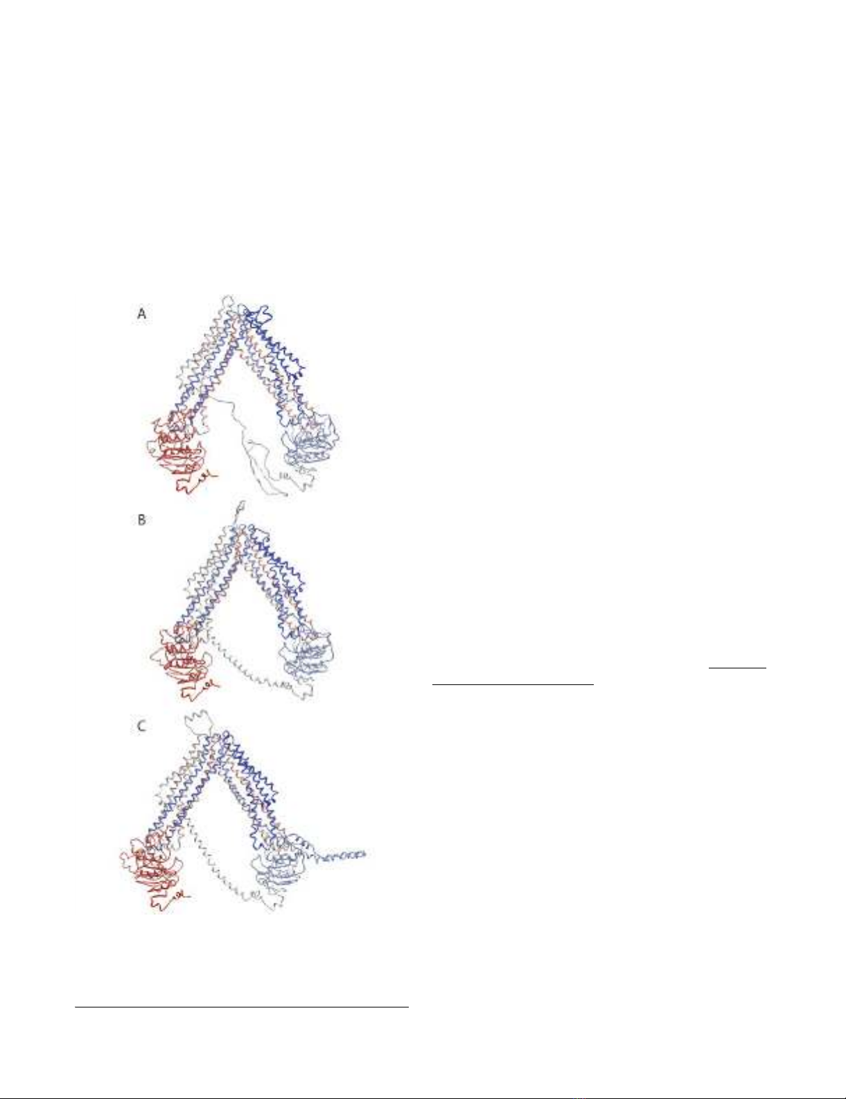
The energy minimized ABCB1, ABCC4 and ABCC5 mod-
els are shown in Figures 3A-C. Each transporter was in an

Theoretical Biology and Medical Modelling 2009, 6:20 http://www.tbiomed.com/content/6/1/20
Page 4 of 12
(page number not for citation purposes)
Alignment of Escherichia coli MsbA, human ABCB1, human ABCC4 and human ABCC5 used as input alignment for the ICM homology modelling moduleFigure 2
Alignment of Escherichia coli MsbA, human ABCB1, human ABCC4 and human ABCC5 used as input align-
ment for the ICM homology modelling module. TMHs, Walker A motifs and Walker B motifs are indicated as boxes.
15
92
191
292
386
486
572
THM1
THM2
THM3 THM4
THM5 THM6
WalkerA
WalkerB
THM7 THM8
THM9 THM10
THM11 THM12
WalkerA
WalkerB
Theoretical Biology and Medical Modelling 2009, 6:20 http://www.tbiomed.com/content/6/1/20
Page 5 of 12
(page number not for citation purposes)
open V-shaped inward conformation with their NBD1
and NBD2 ~50 Å apart. Both Walker A motifs of each
model consisted of a coiled loop and a short α-helix (P-
loop), and the ATP-binding half sites faced each other.
The Walker B motifs were in β-sheet conformation and
localized in the NBD's hydrophobic cores, which were
constituted of 5 parallel β-sheets. The amino acids local-
ized on the surface of each NBD were mainly charged. In
the "arms" of the V-shaped structure, NBD1 was associ-
ated with TMHs 1, 2, 3 and 6 (TMD1), and TMHs 10 and
11 (TMD2), while NBD2 was associated with TMHs 4 and
5 (TMD1), and TMHs 7, 8, 9 and 12 (TMD2). Thus, the
TMDs were twisted relative to the NBDs, such that TMH4
and TMH5 were crossed over ("cross-over motif" [19])
and associated with TMD2, and TMH10 and TMH11 were
crossed over and associated with TMD1. All TMHs con-
tributed to substrate translocation pore, which was closed
towards the extracellular side.
The loop connecting NBD1 and TMD2 of each transporter
was abundant with charged amino acids. The loop con-
necting NBD1 and TMD2 of ABCB1 was in extended con-
formation forming a β-sheet between amino acids
sections Lys645-Glu652 and Lys665-Ser671, while the
loops connecting the subunits of ABCC4 and ABCC5 were
α-helical. ABCB5 featured an insertion loop (as compared
with the amino acid sequences of Escherichia coli MsbA)
from Ile479 to His548 in NBD1, and as displayed in Fig-
ures 3C and 4C, this loop was pointing away from NBD1
parallel to the membrane. However, modelling loops of
lengths as that of the connection between NBD1 and
TMD2 is relatively inaccurate and consequently the mod-
elled loop structures must be regarded as uncertain.
Figures 4A-I show the EPS of the substrate recognition
area of each of the ABC models. The EPS of the substrate
recognition area in the TMDs of ABCB1 was neutral with
negative and weakly positive areas, while the EPS of the
ABCC5 substrate recognition area was generally positive.
The substrate recognition area of ABCC4 was generally
positive with negative area "spots".
The results from the stereochemical validations retrieved
from the SAVES Metaserver http://nih
server.mbi.ucla.edu/SAVES/ are shown in Table 1. Overall
factors from the Errat option at ~90 indicate that the mod-
els were of high quality.
Site directed mutagenesis studies on ABCB1 have indi-
cated that Ile306 (TMH5) [27,35], Ile340 (TMH6) [33],
Phe343 (TMH6) [21,27], Phe728 (TMH7) [27], and
Val982 (TMH12) [33,35] may participate in ligand bind-
ing. As shown in Figure 5A, these residues may form a sub-
strate recognition site in the ABCB1 model. The
involvement of these residues in ligand binding is con-
firmed in the X-ray crystal structure of the Mus musculus
ABCB1 [20] (Figure 5B). Table 2 shows the corresponding
residues in ABCC4 and ABCC5. Measured Cα-Cα dis-
tances in the human ABCB1 model, in the X-ray crystal
structure of the Mus musculus ABCB1 [20] and experimen-
tal distance ranges from cross-linking studies and are
listed in Table 3.
Discussion
Visualization of the molecular structures of human ABC
transporters in 3D models contributes to the comprehen-
Backbone Cα-traces of ABCB1 model (A), ABCC4 model (B) and ABCC5 model (C) viewed in the membrane plane, cytoplasm downwardsFigure 3
Backbone Cα-traces of ABCB1 model (A), ABCC4
model (B) and ABCC5 model (C) viewed in the
membrane plane, cytoplasm downwards. Colour cod-
ing: blue via white to red from N-terminal to C-terminal.


