
RESEARC H Open Access
Higher levels of Zidovudine resistant HIV in the
colon compared to blood and other
gastrointestinal compartments in HIV infection
Guido van Marle
1*
, Deirdre L Church
1,2,3,4
, Kali D Nunweiler
1
, Kris Cannon
1
, Mark A Wainberg
5,6
, M John Gill
1,3
Abstract
Background: The gut-associated lymphoid tissue (GALT) is the largest lymphoid organ infected by human
immunodeficiency virus type 1 (HIV-1). It serves as a viral reservoir and host-pathogen interface in infection. This
study examined whether different parts of the gut and peripheral blood lymphocytes (PBL) contain different drug-
resistant HIV-1 variants.
Methods: Gut biopsies (esophagus, stomach, duodenum and colon) and PBL were obtained from 8 HIV-1 infected
preHAART (highly active antiretroviral therapy) patients at three visits over 18 months. Patients received AZT, ddI or
combinations of AZT/ddI. HIV-1 Reverse transcriptase (RT)-coding sequences were amplified from viral DNA
obtained from gut tissues and PBL, using nested PCR. The PCR fragments were cloned and sequenced. The
resulting sequences were subjected to phylogenetic analyses, and antiretroviral drug mutations were identified.
Results: Phylogenetic and drug mutation analyses revealed differential distribution of drug resistant mutations in
the gut within patients. The level of drug-resistance conferred by the RT sequences was significantly different
between different gut tissues and PBL, and varied with antiretroviral therapy. The sequences conferring the highest
level of drug-resistance to AZT were found in the colon.
Conclusion: This study confirms that different drug-resistant HIV-1 variants are present in different gut tissues, and
it is the first report to document that particular gut tissues may select for drug resistant HIV-1 variants.
Introduction
Science has been confronted with the problem of drug-
resistance virtually since the introduction of the first
antiretroviral drugs to treat infection by human immu-
nodeficiency virus type 1 (HIV-1) (reviewed in [1]).
The first approach to antiretroviral therapy (ART)
used single nucleoside reverse transcriptase inhibitors
(NRTIs) which were found to select for drug-resistant
variants very quickly [1,2]. The development of many
new NRTI, non-nucleoside RT inhibitors (NNRTI),
andproteaseinhibitors(PI)offeredadditionaltreat-
ment options in cases of drug-resistance. It also
offered the possibility of combination therapies (i.e.
highly active antiretroviral therapy (HAART)) able to
suppress HIV replication and reduce the likelihood of
developing drug-resistance [1-3].
The ability of HIV-1 to rapidly develop drug-resistance
is linked to its highly divergent nature as a result of the
error-prone reverse transcription step in its life cycle [4].
Due to the high mutation rate, HIV-1 exists in the infected
individual as a collection of many different viral variants,
also known as a quasi-species [5]. The extent of quasi-
species diversity during infection is amongst others
affected by factors such as viral fitness, availability of cells
for infection, selective pressure from antiretroviral therapy,
duration of infection, and host immune responses [5-8].
Studies of patients on antiretroviral therapy revealed
that viral sequences continued to evolve in genes not
targeted by the drugs, despite successful suppressive
therapy [9-11]. This phenomenon can be explained by
continued viral replication in other tissues and/or cell
compartments due to inefficient action or penetration of
the antiretroviral drugs (ARVs) in these compartments.
* Correspondence: vanmarle@ucalgary.ca
1
Department of Microbiology and Infectious Diseases, University of Calgary,
Calgary, Alberta, Canada
Full list of author information is available at the end of the article
van Marle et al.Retrovirology 2010, 7:74
http://www.retrovirology.com/content/7/1/74
© 2010 van Marle et al; licensee BioMed Central Ltd. This is an Open Access article distributed under the terms of the Creative
Commons Attribution License (http://creativecommons.org/licenses/by/2.0), which permits unrestricted use, distribution, and
reproduction in any medium, provided the original work is properly cited.

These inefficiently targeted compartments are referred
to as sanctuary sites (reviewed in [12-14]). The central
nervous system (CNS) is well known as a sanctuary site,
because certain antiretroviral drugs do not easily cross
the blood-brain barrier [13]. Recent studies postulated
the gut may also be an important sanctuary site, where
HIV-1 can persist despite successful antiviral therapy
[15,16]. This is consistent with observations in the SIV
model [17]. The gut-associated lymphoid tissue (GALT)
is known as a major site for viral replication, CD4
+
T-cell depletion, and immune dysfunction [18-22]. How-
ever, relatively little is known about the distribution of
HIV-1 antiretroviral drug-resistance across different
parts of the gastrointestinal (GI) tract. We recently
showed that HIV-1 quasi-species varied within different
parts of the GI tract of pre-HAART patients, indicating
that HIV-1 replication in the gut is compartmentalized
[23]. Now, we have extended these observations to show
that variability exists in the distribution of drug-resistant
variants in different gut tissues and peripheral blood
lymphocytes of these pre-HAART patients. The number
of drug-resistant HIV variants differed in the colon
compared to blood and other gut tissues, depending on
the antiretroviral therapy received. This suggests that
antiretroviral drug-resistance is highly variable in the
different gut compartments.
Results
Diversity of the HIV-1 RT-coding region in different gut
tissues
The samples were obtained from a preHAART cohort
study of HIV-1 seropositive men who have sex with
men (MSM) [24,25]. The 8 patients in the current
report were used in an earlier study of HIV-1 diversity
in the gut [23]. For the current study, gut and peripheral
blood lymphocyte (PBL) samples from these 8 patients
from 3 subsequent visits over 18 months were used
(Table 1). All patients were on mono- or dual therapies
of primarily AZT (azidothymidine, zidovudine) and ddI
(dideoxyinosine). One patient (#42) died during the
study and only samples from the first visit were avail-
able. This patient was still included in our analyses as a
patient with end stage disease and suspected drug resis-
tance. In addition, five patients (#1, #3, #7, #8, #19)
were still alive at the time of this study (2007) and
received HAART. For patients #3, #7, #8, #19, year 2007
PBL samples (indicated as Visit 2007) were collected,
and drug-resistance mutations were assessed to get
insight in the drug resistance mutations 15 years after
the original visit. HIV-1 viral sequences were most con-
sistently amplified from DNA from most PBL and
biopsy tissues, using our nested PCR protocol. There-
fore, our analyses focused on these viral DNA derived
sequences. For some patient visits RT-coding sequences
from only two gut-tissues and PBL could be obtained, in
particular for visit 1 (Additional File 1). In total, around
1000 RT-coding sequences were obtained and analyzed.
The mean total (d), and nonsynonymous (d
N
)pair-
wise distances between patients were calculated for the
RT-coding sequences obtained from PBL, esophagus,
stomach, duodenum, and colon for all patients at all
visits (Figure 1). Although we were not able to obtain
sequences from the duodenum and colon for visit 1 for
a number of patients, the overall interpatient distances
(d) of RT coding sequences tended to decrease
(p< 0.05) at the last visit for sequences derived from
the PBL, esophagus, stomach, and the duodenum
(Figure 1). This suggested evolution towards a more
conserved RT coding region between patients in these
tissues in this particular sample of patients. In contrast,
the overall interpatient distance of the RT-coding
sequences in the colon increased over time (p<0.05)
(Figure 1). A decrease in the d
N
values (i.e. codon/
amino acid changing substitutions) (p< 0.05) towards
the last visit between the patients was observed for the
RT-coding region for both PBL and duodenum, while in
the esophagus and stomach the d
N
decreased but fluctu-
ated over time. In the colon the d
N
values increased
(Figure 1), suggesting greater interpatient diversity in
RT coding sequences in the colon in this group of
patients.
Compartmentalization of antiretroviral drug-resistance in
the gut
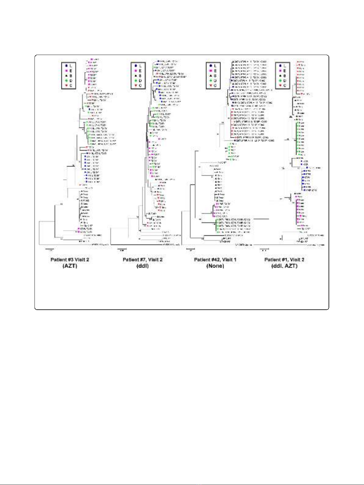
The RT sequences were subsequently subjected to phy-
logenetic analyses. Neighbour-joining trees of all RT
sequences and bootstrap analysis revealed clustering of
some sequences by tissue and patient (bootstrap values
>70).Aswepreviouslyreportedsuchclusteringwas
not consistent for most of the sequences [23] (data not
shown). Bootstrap analyses of RT sequences by indivi-
dual patient and visit, revealed more consistent cluster-
ing on the basis of tissue (bootstrap values > 70),
although this also varied by patient (Figure 2). The
representative neighbour-joining trees for the RT
sequences for patient #3 (Visit 2), #42 (Visit 1), and #1
(Visit 2) revealed clustering of sequences (bootstrap
values > 70) on the basis of tissue (Figure 2). Similar
trees were obtained for the other patients and visits
(data not shown). Further analysis of the clustering pat-
tern using the Slatkin-Maddison test [26-28] revealed
that there was no consistent significant compartmentali-
zation of the RT sequences. However, many sequences
grouped together by tissue in the phylogenetic trees (for
example patient #7, Visit 2, Figure 2).
We analyzed the presence of mutations associated
with NRTI resistance using the Stanford HIV-1 Drug-
Resistance Database [29]. As shown in Figure 2, distinct
van Marle et al.Retrovirology 2010, 7:74
http://www.retrovirology.com/content/7/1/74
Page 2 of 13
drug-resistance mutations were found in each tissue
compartment for patient #3 (Visit 2), consistent with
grouping together of the nucleic acid sequences. For
patient #1 (visit 2), we observed grouping of sequences
by tissue, but very few drug-resistance mutations, prob-
ably due to the greater efficiency of therapy with two
NRTIs (AZT and ddI). For patient #42 (Visit 1), various
drug resistance mutations were found in the stomach,
colon, and PBL, which was consistent with the sus-
pected viral failure due to antiviral drug resistance. Dif-
ferent or no mutations were present in the duodenum,
stomach and esophagus of this patient suggesting that
antiretroviral drug resistance can differ significantly
among tissues. Although the clustering of RT sequences
ofPatient#7(Visit2)wasnotindicativeofcompart-
mentalization according to the Slatkin-Maddison cri-
teria, the different tissue compartments could still be
separated and grouped based on drug-resistance muta-
tions. This observation was consistent for all patients
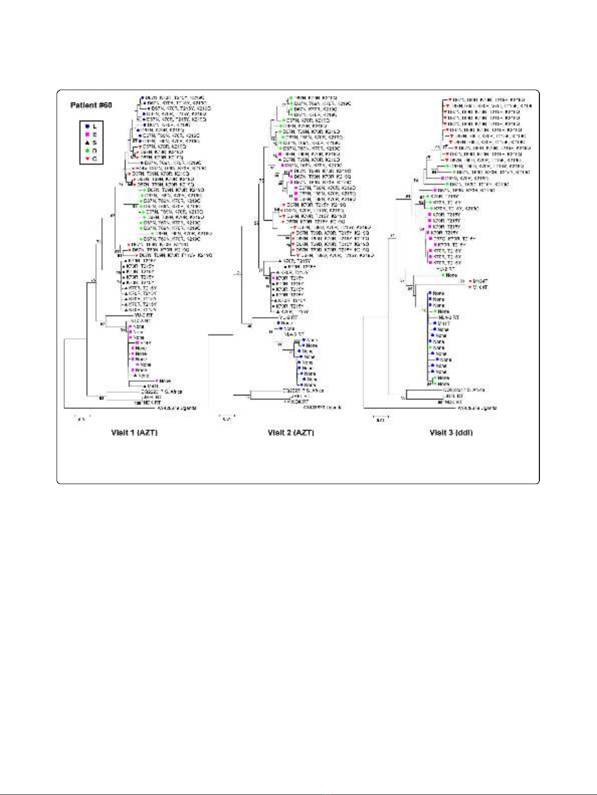
and visits, as illustrated for patient #60 in Figure 3.
Repeating the phylogenetic analyses after removing the
drug resistance conferring sites from the sequences
Table 1 Patient Information
Patient Date
HIV
+
Date
Death
1
Prior
Therapy
2
Visit 1 Visit 2 Visit 3
Date VL
3
CD4
Count
4
Therapy Date VL
3
CD4
Count
4
Therapy Date VL
3
CD4
Count
4
Therapy
#1 Jun. 1
1986
–AZT Apr. 7
1993
2.7 264 ddI, AZT Jan. 19
1994
2.4 210 ddI, AZT Oct. 26
1994
3.5 190 AZT
#2 Jan. 1
1989
Oct. 16
1994
ddI Apr. 7
1993
6.4 187 ddI Jan. 26
1994
5.6 40 D4T Sept. 14
1994
5.8 18 None
#3* Nov. 1
1989
–ddI Apr. 6
1993
4.3 144 ddI Jan. 26
1994
5.3 162 AZT Sept. 13
1994
5.6 77 AZT
#7* Jun. 1
1987
–AZT Apr. 21
1993
4.5 270 ddI Jan. 26
1994
4.9 234 ddI Mar. 9
1995
4.5 146 AZT
#8* Oct. 1
1988
–AZT Apr. 21
1993
3.3 475 ddI Jan. 12
1994
3.5 338 ddI Nov. 16
1994
4.4 325 ddI
#19* Jan. 1
1991
–ddI Jun. 2
1993
3.9 77 ddI Jan. 26
1994
5.3 22 AZT Nov. 14
1994
4.7 42 AZT
#60 Dec. 1
1988
Apr. 3
1998
AZT Sept. 14
1994
4.5 48 AZT Jun. 14
1995
4.9 51 AZT Feb. 28
1996
5.3 21 ddI
#42 Nov. 1
1989
Oct. 21
1993
AZT, ddC Oct. 6
1993
5.6 9 None
1
Patient #42 passed away during the study and only visit 1 samples were available.
2
Antiretroviral therapy in the 6 months preceding visit 11.
3
VL - log plasma viral load in log
10
(copies)/mL.
4
CD4
+
cell counts in cells/μL.
*Patients for which a 2007 PBL sample was analyzed.
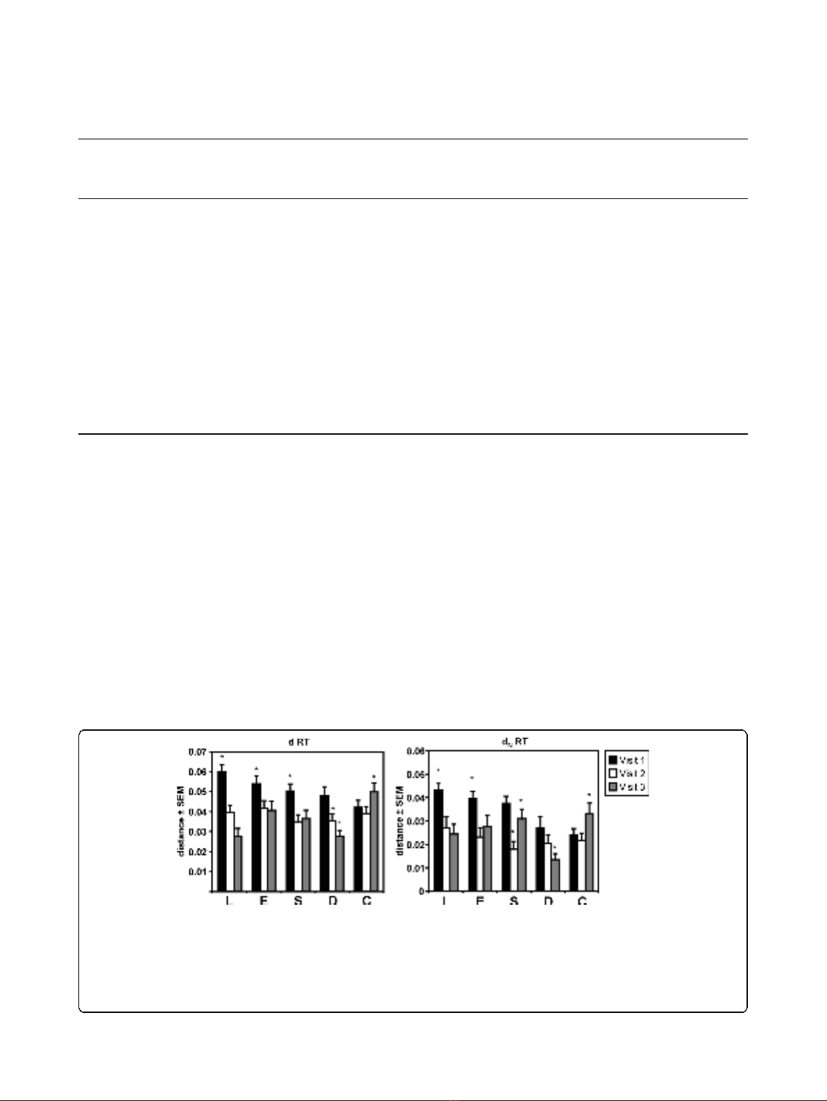
Figure 1 Viral interpatient diversity of the RT-coding region in the gut tissues (esophagus (E), stomach (S), duodenum (D), colon (C))
and PBL (L) of HIV-1 infected patients at different visits. Viral RT-coding sequences tended to a more conserved sequence among patients
in the esophagus, stomach, duodenum and PBL, as reflected by the lower mean total distance (d) between patients, while the sequences in the
colon became more diverse over time. Similarly, the decrease in mean total non-synonomous distance (d
N
, i.e. amino acid changing mutations)
for PBL and duodenum suggested evolution towards more conserved RT protein sequences over time among patients, while the increased d
N
reflected the RT protein sequence becoming more diverse over time in the colon among patients. These observations indicated that the RT-
coding region evolved differently in the different gut tissues and PBL in this group of patients. (*p< 0.05, Dunnett C post-hoc analysis)
van Marle et al.Retrovirology 2010, 7:74
http://www.retrovirology.com/content/7/1/74
Page 3 of 13
resulted in the same tree topologies (data not shown),
indicating that the drug resistance conferring sites were
not solely responsible for the observed clustering of RT
sequences by tissue.
These observations strongly suggest a differential dis-
tribution of antiretroviral drug-resistance in the different
gut tissues, with drug-resistance mutations differing
from those observed in the blood. These observations
were further corroborated by sorting drug-mutations by
tissue compartment (summarized in Table 2 and Addi-
tional File 1), indicating drug-resistance mutations dif-
fered significantly between tissues within each patient
(p< 0.05 Chi-square test), and varied over time (p<
0.05, Chi-square test). Furthermore, the different tissues
also differed significantly in distribution of drug-resis-
tance mutations in the viral quasi-species (p< 0.05 Chi-
square test). Our analysis revealed no evidence for a
preferential presence of any specific drug-resistance
mutations for any individual tissue compartment.
Table 2 also shows the drug-resistance mutation profile
of the PBL samples for two surviving patients currently
on HAART, collected in 2007, 15 years after the original
study. Again, drug-resistance mutations differed from the
original historical samples (p< 0.05 Chi-square test), and
similar results were obtained for the other two surviving
patients (Additional File 1). Thisanalysisconfirmsthat
the changes observed in the viral DNA samples were the
result of changes in the viral population close to the time
of sampling and not the result of picking up viral DNA
sequences that were archived over many years.
Different drug-resistance levels in different parts of the gut
The development of drug-resistance is dependent on the
drugs used in therapy. We analyzed the percentage of
Figure 2 Representative bootstrap Neighbor-Joining trees of RT-coding sequences obtained from gut tissues (esophagus (E), stomach
(S), duodenum (D), colon (C)) and PBL (L) (indicated by different shapes and shading). RT sequences grouped by individual gut tissue and
PBL to varying degrees in the different patients. Upon closer examination of the drug-resistance mutations indicated at each branch, grouping
of resistance mutations by gut tissue and PBL was observed. Differences in drug-resistance mutations were found in the different tissues and
PBL. Similar differences were observed for RT sequences recovered from the tissues of other patients, indicating difference in distribution of
drug-resistance in the gut. (Bootstrap values > 70 are indicated.)
van Marle et al.Retrovirology 2010, 7:74
http://www.retrovirology.com/content/7/1/74
Page 4 of 13
drug-resistant sequences and the average drug-resistance
score for all sequences recovered from each tissue, tak-
ing into account the antiretroviral therapy received prior
to the time samples were collected (Figure 4). The Stan-
ford database was used to determine the drug-resistance
score for each RT sequence. RT sequences with a drug-
resistance score ≥30 were designated drug-resistant.
Following AZT or ddI treatment, different numbers of
respectively AZT- or ddI-resistant RT-coding sequences
were found in the GI tissues (esophagus, stomach, duo-
denum and colon) and PBL (i.e. blood) (p< 0.05, Figure
4A and 4B). Thus, the distribution of drug-resistant
sequences is diverse, and antiretroviral therapy selects
for different numbers of drug-resistant HIV-1 variants
in each tissue.
Next, we analyzed the average drug-resistance score of
all drug-resistant RT sequences (i.e drug-resistance
score ≥30) among the different tissues following AZT
or ddI treatment (Figure 4C and 4D). This analysis
revealed that RT sequences with the highest drug-resis-
tance score for AZT were recovered from the colon
(Figure 4C, p< 0.05). No significant differences in ddI
resistance scores were observed following ddI treatment,
although they tended to be higher in the stomach (Fig-
ure 4D). Together with our other observations, these
results suggested that antiretroviral therapies (AZT and
ddI) affected each gut tissue compartment differently,
and that AZT preferentially selected for more AZT
resistant HIV-1 variants in the colon.
Discussion
The presence of HIV-1 antiretroviral drug-resistance in
different tissues, such as the CNS, has been well docu-
ment [30-33]. However, little is known about HIV-1
antiretroviral drug-resistance in different tissues of
the gut, despite its importance as a reservoir for viral
replicationandahostpathogeninterphaseinHIV/
AIDS [18-22]. To provide insight into the potential
Figure 3 Bootstrap Neighbor-Joining trees of the RT-coding sequences of patient #60 at visits 1, 2 and 3. Differences in drug-resistance
mutations (indicated at the tree branches) and grouping of RT-coding sequences was observed. However, at all visits differences were observed
in the drug-resistance mutations between the various tissues, consistent with differential distribution of drug-resistance in the gut. Similar results
were obtained for the RT-coding sequence of the other patients. (Bootstrap values > 70 are indicated.)
van Marle et al.Retrovirology 2010, 7:74
http://www.retrovirology.com/content/7/1/74
Page 5 of 13


![Vaccine và ứng dụng: Bài tiểu luận [chuẩn SEO]](https://cdn.tailieu.vn/images/document/thumbnail/2016/20160519/3008140018/135x160/652005293.jpg)


%20--%3e%3cdefs%3e%3cstyle%3e%20.st0%20{%20fill:%20%23fff;%20}%20.st1%20{%20fill:%20%237800fa;%20}%20%3c/style%3e%3c/defs%3e%3cpath%20class='st1'%20d='M117.78,12.18H43.11c2.9,3.47,4.65,7.94,4.65,12.82,0,5.6-2.3,10.66-6.01,14.29h76.02l7.22-13.56-7.22-13.56Z'/%3e%3cg%3e%3cpath%20class='st0'%20d='M53.58,26.17h-.59v-1.46h.59v-4.96h2.83c1.78,0,2.67.94,2.67,2.82v5.76c0,1.87-.89,2.81-2.67,2.81h-2.83v-4.96ZM55.36,21.37v3.34h1.1v1.46h-1.1v3.34h1.01c.61,0,.91-.37.91-1.1v-5.93c0-.74-.3-1.1-.91-1.1h-1.01Z'/%3e%3cpath%20class='st0'%20d='M65.99,31.14h-1.8l-.31-2.07h-2.19l-.31,2.07h-1.64l1.82-11.39h2.62l1.82,11.39ZM65.28,18.04c-.25.46-.51.77-.75.94-.21.15-.47.22-.79.22-.26,0-.57-.07-.92-.22l-.38-.15c-.14-.05-.26-.07-.37-.07-.3,0-.53.18-.71.54l-.91-.68c.25-.46.51-.77.75-.94.21-.14.48-.21.79-.21.26,0,.57.07.92.21l.38.15c.14.05.26.07.37.07.3,0,.53-.18.71-.54l.91.68ZM61.91,27.52h1.73l-.87-5.76-.87,5.76Z'/%3e%3cpath%20class='st0'%20d='M74.53,26.89v1.52c0,1.91-.89,2.86-2.67,2.86s-2.67-.95-2.67-2.86v-5.93c0-1.91.89-2.86,2.67-2.86s2.67.95,2.67,2.86v1.11h-1.69v-1.22c0-.75-.31-1.12-.93-1.12s-.93.37-.93,1.12v6.15c0,.74.31,1.11.93,1.11s.93-.37.93-1.11v-1.63h1.69Z'/%3e%3cpath%20class='st0'%20d='M81.4,31.14h-1.8l-.31-2.07h-2.19l-.31,2.07h-1.64l1.82-11.39h2.62l1.82,11.39ZM75.9,19.2l1.52-1.91h1.71l1.51,1.91h-1.61l-.76-.95-.75.95h-1.61ZM77.32,27.52h1.73l-.87-5.76-.87,5.76ZM83.1,15.99l-1.76,1.91h-1.26l1.17-1.91h1.86Z'/%3e%3cpath%20class='st0'%20d='M84.86,19.75c1.78,0,2.67.94,2.67,2.82v1.48c0,1.87-.89,2.81-2.67,2.81h-.85v4.28h-1.79v-11.39h2.64ZM84.01,21.37v3.86h.85c.58,0,.87-.36.87-1.08v-1.71c0-.71-.29-1.07-.87-1.07h-.85Z'/%3e%3cpath%20class='st0'%20d='M93.51,19.75c1.78,0,2.67.94,2.67,2.82v1.48c0,1.87-.89,2.81-2.67,2.81h-.85v4.28h-1.79v-11.39h2.64ZM92.66,21.37v3.86h.85c.58,0,.87-.36.87-1.08v-1.71c0-.71-.29-1.07-.87-1.07h-.85Z'/%3e%3cpath%20class='st0'%20d='M98.8,31.14h-1.79v-11.39h1.79v4.88h2.03v-4.88h1.83v11.39h-1.83v-4.88h-2.03v4.88Z'/%3e%3cpath%20class='st0'%20d='M105.36,24.55h2.46v1.62h-2.46v3.34h3.09v1.63h-4.88v-11.39h4.88v1.63h-3.09v3.18ZM108.17,17.29l-1.76,1.91h-1.26l1.17-1.91h1.86Z'/%3e%3cpath%20class='st0'%20d='M112.2,19.75c1.78,0,2.67.94,2.67,2.82v1.48c0,1.87-.89,2.81-2.67,2.81h-.85v4.28h-1.79v-11.39h2.64ZM111.35,21.37v3.86h.85c.58,0,.87-.36.87-1.08v-1.71c0-.71-.29-1.07-.87-1.07h-.85Z'/%3e%3c/g%3e%3ccircle%20class='st1'%20cx='25'%20cy='25'%20r='20'/%3e%3cpath%20class='st0'%20d='M32.78,19.27c2.92,0,4.43,2.55,5.28,5.33l.71,2.17c.14.38-.33.75-.71.75h-5.61c.19-.33.24-.71.09-1.08l-.75-2.45c-.43-1.32-.99-2.64-1.79-3.77.75-.57,1.65-.94,2.78-.94h0ZM25,18.38c3.25,0,4.9,2.78,5.89,5.89l.76,2.45c.14.42-.33.8-.8.8h-11.69c-.42,0-.94-.38-.8-.8l.75-2.45c.99-3.11,2.64-5.89,5.89-5.89h0ZM25,11.35c1.74,0,3.11,1.37,3.11,3.11s-1.37,3.11-3.11,3.11-3.11-1.41-3.11-3.11,1.41-3.11,3.11-3.11h0ZM17.27,19.27c1.08,0,1.98.38,2.73.94-.8,1.13-1.37,2.45-1.74,3.77l-.8,2.45c-.14.38-.05.75.09,1.08h-5.56c-.42,0-.9-.38-.75-.75l.71-2.17c.9-2.78,2.41-5.33,5.33-5.33h0ZM17.27,12.91c1.51,0,2.78,1.27,2.78,2.83s-1.27,2.83-2.78,2.83-2.83-1.27-2.83-2.83,1.27-2.83,2.83-2.83h0ZM32.78,12.91c1.56,0,2.78,1.27,2.78,2.83s-1.23,2.83-2.78,2.83-2.83-1.27-2.83-2.83,1.27-2.83,2.83-2.83h0ZM27.07,28.56v.09c0,.57-.24,1.08-.61,1.46h0v.05c-.38.33-.9.57-1.46.57s-1.08-.24-1.46-.61h0c-.38-.38-.61-.9-.61-1.46v-.09h1.41v.09c0,.19.05.38.19.47v.05c.09.09.28.19.47.19s.38-.09.47-.19v-.05c.14-.09.24-.28.24-.47t-.05-.09h1.41ZM30.99,28.56v.09c0,1.65-.66,3.16-1.74,4.24-1.08,1.08-2.59,1.79-4.24,1.79s-3.16-.71-4.24-1.79l-.05-.05c-1.04-1.08-1.7-2.55-1.7-4.2v-.09h1.41v.09c0,1.27.47,2.4,1.27,3.25h.05c.85.85,1.98,1.37,3.25,1.37s2.4-.52,3.25-1.37c.85-.8,1.37-1.98,1.37-3.25v-.09h1.37ZM34.99,28.56v.09c0,2.78-1.13,5.28-2.92,7.07-1.79,1.79-4.29,2.92-7.07,2.92s-5.23-1.13-7.07-2.92c-1.79-1.79-2.92-4.29-2.92-7.07v-.09h1.41v.09c0,2.4.94,4.53,2.5,6.08,1.56,1.56,3.72,2.5,6.08,2.5s4.52-.94,6.08-2.5c1.56-1.56,2.5-3.68,2.5-6.08v-.09h1.41Z'/%3e%3c/svg%3e)