
Ligand-induced heterodimerization between the ligand binding
domains of the
Drosophila
ecdysteroid receptor and ultraspiracle
Markus Lezzi
1
, Thomas Bergman
1,
*, Vincent C. Henrich
2
, Martin Vo¨ gtli
1,†
, Christina Fro¨mel
1
,
Marco Grebe
3
, Sabina Przibilla
3
and Margarethe Spindler-Barth
3
1
Institute for Cell Biology, ETH-Ho
¨nggerberg, Zu
¨rich, Switzerland;
2
Department of Biology, University of North Carolina,
Greensboro, NC, USA;
3
Abteilung fu
¨r allgemeine Zoologie und Endokrinologie, Universita
¨t, D-89069 Ulm, Germany
The insect ecdysteroid receptor consists of a heterodimer
between EcR and the RXR-orthologue, USP. We addressed
the question of whether this heterodimer, like all other RXR
heterodimers, may be formed in the absence of ligand and
whether ligand promotes dimerization. We found that
C-terminal protein fragments that comprised the ligand
binding, but not the DNA binding domain of EcR and USP
andwhichwereequippedwiththeactivationorDNA
binding region of GAL4, respectively, exhibit a weak ability
to interact spontaneously with each other. Moreover, the
heterodimer formation is greatly enhanced upon adminis-
tration of active ecdysteroids in a dose-dependent manner.
This was shown in vivo by a yeast two-hybrid system and
in vitro by a modified electromobility shift assay. Further-
more, the EcR fragment expressed in yeast was functional
and bound radioactively labelled ecdysteroid specifically.
Ligand binding was greatly enhanced by the presence of a
USP ligand binding domain. Therefore, ecdysteroids are
capable of inducing heterodimer formation between EcR
and USP, even when the binding of these receptor proteins to
cognate DNA response elements does not occur. This
capability may be a regulated aspect of ecdysteroid action
during insect development.
Keywords:Drosophila melanogaster;yeast;two-hybrid;
ecdysone receptor; dimerization; ultraspiracle.
Ecdysteroids are widespread steroid hormones found in
invertebrates [1] and plants [2,3] that regulate a variety of
developmental, physiological, and reproductive processes
[1,3]. Among insects, these hormones regulate the expression
of genes through a highly orchestrated and coordinated
transcriptional network [4–6]. The widespread and diverse
effects of ecdysteroids on transcriptional regulation have
served as a powerful model for investigating the diverse
mechanisms by which steroid hormones, acting via nuclear
receptors, exert their effects on a variety of life processes [4,7].
The ecdysone receptor (EcR) [8], responsible for medi-
ating these responses, occupies a special position among
nuclear hormone receptors because it shows a unique
combination of characteristics [9]. Unlike the vertebrate
steroid receptors [10–12], EcR heterodimerizes with the
insect RXR orthologue, ultraspiracle (USP) [13–15]. Nev-
ertheless, while other nuclear receptors that dimerize with
RXR normally are bound to DNA response elements
already in their nonliganded state [11,16], this apparently is
not true for the EcR/USP heterodimer (see however, [17]).
Immunostaining has shown that the polytene chromosomes
of a Chironomid or Sciarid are devoid of EcR/USP signals
when prepared from developmental stages associated with
low ecdysteroid titers [18,19]. A short in vitro incubation of
the tissues with 20-hydroxyecdysone, however, is followed
by the appearance of immunostaining signals at known
ecdysteroid-responsive gene loci [18,19]. The affinity of
EcR/USP dimers for ecdysone response elements (EcREs)
clearly increases in the presence of the ecdysteroid muri-
sterone A as demonstrated by electromobility shift assays
(EMSAs) [20–23].
Neither immunostaining assay nor EMSA studies can
distinguish between three possibilities concerning how
ecdysteroids influence EcR/USP binding to DNA: (a)
ecdysteroids promote dimerizing of EcR and USP, and
enhanced DNA affinity arises as a spontaneous conse-
quence of this partnering, as indicated for the glucocorticoid
receptor [24]; (b) ecdysteroids induce binding of EcR to its
cognate EcRE half-site, analagous to the effect noted for the
thyroid hormone receptor [17]; or (c) EcR and USP
dimerize spontaneously and ecdysteroids promote the
binding of the dimer to the DNA response element [20].
Correspondence to M. Lezzi, Institute for Cell Biology,
ETH-Ho
¨nggerberg, CH-8093 Zu
¨rich, Switzerland.
Fax: + 41 1 633 10 69, Tel.: + 41 1 371 97 39,
E-mail: lezzi@cell.biol.ethz.ch
Abbreviations: EcR, ecdysone receptor protein; USP, ultraspiracle
protein; RXR, 9-cis retinoic acid receptor; EcRE, ecdysone receptor
protein receptor response element; EMSA, electromobility shift assay;
ST-EMSA, supershift-type electromobility shift assay; LBD, ligand
binding domain; GBD, DNA binding domain of GAL4; GAD,
activation domain of GAL4; DBD, DNA binding domain of EcR.
Definition: in the present work the terms monomer,dimer’,
homodimer,andheterodimerare being used although the protein
complexes in question may in fact constitute multimers. Our nomen-
clature follows the common use and describes the state of interaction
of the respective nuclear receptor in a protein complex with respect to
another nuclear receptor.
*Present address: Affibody AB, Bromma, Sweden.
Present address:BankVontobelAG,Zurich,Switzerland.
(Received 16 January 2002, revised 3 May 2002,
accepted 16 May 2002)
Eur. J. Biochem. 269, 3237–3245 (2002) FEBS 2002 doi:10.1046/j.1432-1033.2002.03001.x
The primary purpose of this work is to address the
possibility that EcR and USP are capable of dimerization
in the absence of a bipartite EcRE and to monitor the
potential influence of ecdysteroid on dimerization. These
studies were carried out by expressing the ligand-binding
domain (LBD) of EcR and USP on two-hybrid vectors,
and examining their ability to dimerize in the absence
and presence of ecdysteroids. The experiments presented
demonstrate that the EcR and USP LBD are capable of
dimerization in the absence of a bipartite EcRE, and that
this protein–protein interaction is dramatically enhanced
by the presence of ecdysteroids.
MATERIALS AND METHODS
Plasmids
All plasmids used in the present work were purchased from
Clontech (Palo Alto). For a description of the plasmids, see
the supplier’s protocol and the references therein. Test
plasmid pCL1 encodes for full-length wild-type GAL4 and
served to monitor nonspecific effects on the reporter
enzyme, whereas test plasmids pTD1-1 and pVA3-1 were
used to test the general conditions for two-hybrid formation
and reporter gene activation. The other plasmids used are
mentioned below.
Plasmids coding for fusion proteins with various
C-terminal fragments of EcR
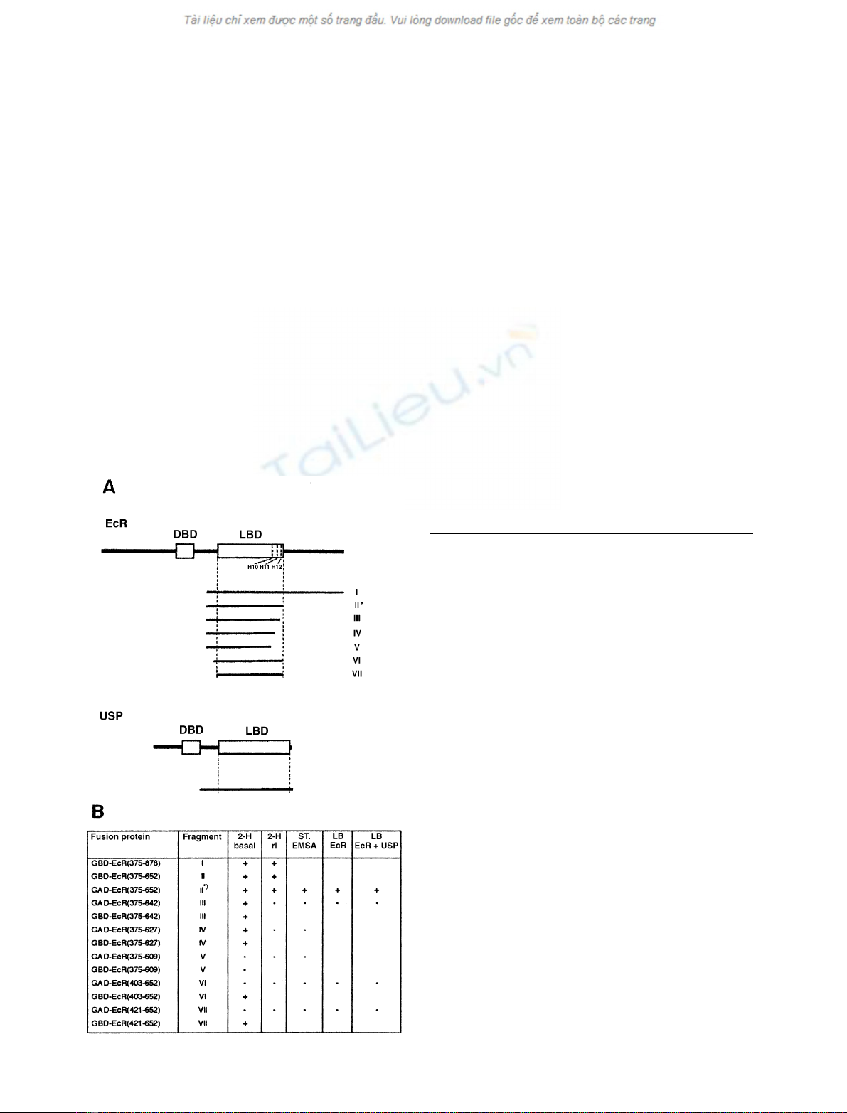
Schematic representations of the fragments are shown in
Fig. 1A.
Construct encoding fragment I
A 1.5-kb cDNA fragment of the Drosophila EcR cDNA
sequence [8] was produced by PCR using the forward
primer 5¢-CGACATATGGGCCAAGACTTTGTTAAG
AAGG-3¢and the reverse primer 5¢-TCCCCCGGGTCTA
GACTATGCAGTCGTCGAGTGCTC-3¢.Thereby,an
NdeI site was introduced at the 5¢end, and XbaIandSmaI
sites were introduced at the 3¢end. The fragment was
digested with NdeI/SmaI and cloned in-frame into pAS2
giving rise to clone pAS2-EcR(375–878), coding for fusion
protein GBD–EcR(375–878) which consists of the GAL4
DNA binding domain fused to the N-terminal end of a
fragment comprising a portion of hinge region, LBD, and
the entire C-terminal domain (also called F domain) of EcR.
A 679-nucleotide portion of the EcR fragment between
AatII (nucleotide 2441) and NarI (nucleotide 3120) was
Fig. 1. Heterodimerization and ligand binding abilities of the EcR
fragments investigated in the present studies as fusion proteins with
GAL4 DNA binding domain (GBD) or GAL4 activation domain (GAD).
(A) Delineation of EcR and USP fragments tested. EcR stands for the
B1 isoform [46] of the Drosophila melanogaster ecdysone receptor [8]. It
spans amino acids 1–878. DBD: DNA binding domain (amino acids
264–329). LBD: ligand binding domain (amino acids 417–651) as
defined by [37]. Helices 10, 11 and 12 (H10, H11, H12) start at amino
acids 610, 628, and 644, respectively [37]. Fragments are numbered
I–VII and correspond to those listed in (B). The dash in fragment I
indicates the location of the spontaneous mutation L442H. The
asterisk designates the fragment which was used most frequently in the
present studies. USP designates Ultraspiracle as characterized by [14].
Delineation of its DBD and LBD as proposed by [34]. (B) Results
obtained with EcR fragments described in (A). Explanation of column
heads and signs in columns: 2-H basal, spontaneous heterodimeriza-
tion with USP fragment in vivo as determined by the two-hybrid assay
and indicated by the appearance of basal (noninduced) b-galactosidase
activity; + or – signs mean this activity to be significantly or unsig-
nificantly higher than background, respectively; 2-H rI,relative
induction of heterodimerization by muristerone A (10
)6
to 5 ·10
)5
M
)
as assayed by the two-hybrid assay (rI: induced level divided by basal
level of b-galactosidase activity): + or – signs mean rI to be signifi-
cantly or unsignificantly higher than 1, respectively; ST-EMSA,
heterodimerization induced in vitro by 10
)5
M
muristerone A and
assayed by ST-EMSA; + or – signs mean supershifted band could or
could not be detected, respectively; LB EcR, ligand binding to EcR
fragment alone; + or – signs mean specific [
3
H] ponasterone A binding
to be significantly or nonsignificantly higher than background,
respectively. LB EcR ± USP: ligand binding to EcR fragment in
presence of USP fragment; GBD- and GAD-EcR fusion proteins were
combined with GAD- or GBD-USP(172–508), respectively; meaning
of + and – signs as in LB EcR column.
3238 M. Lezzi et al. (Eur. J. Biochem. 269)FEBS 2002

exchanged with the corresponding fragment of the original
cDNA clone [8].
Constructs encoding fragment II
Clone pAS2-EcR(375–652) encoding the fusion protein
GBD–EcR(375–652) between the GAL4 DNA binding
domain and a fragment comprising a large portion of hinge
plus the entire LBD of EcR was constructed in an
analogous manner to pAS2-EcR(375–878). The differences
concern the reverse primer 5¢-CGTCCCGGGTCTAGACT
AAACGTCCCAGATCTCCTCG-3¢, the length of PCR
fragment (800 nucleotides), and the exchanged fragment
(566 nucleotides from AatII at nucleotide 2441 to BglII at
nucleotide 3008). The related clone, pAS2-1-EcR(375–652),
was constructed by cutting out the NdeI–SmaIfragment
from pAS2-EcR(375–652) and recloning it into the NdeI–
SmaI site of the pAS2-1 vector. pAS2-EcR(375–652) and
pAS2-1-EcR(375–652) will not be distinguished in the
following. Clone pACT2-EcR(375–652) encodes a fusion
protein, GAD–EcR(375–652), consisting of the same EcR
fragment as pAS2-1-EcR(375–652) but combined with
GAL4 activation rather than DNA binding domain. It
was constructed by use of the forward PCR primer:
5¢-CATGCCATGGGCCAAGACTTTGTTAAGAAGG-3¢
and the reverse primer employed for constructing pAS2-
1-EcR(375–652). The NcoI–SmaI fragment encompassing
the EcR cDNA sequence nucleotide 2191–3024 was cloned
into the NcoI–SmaI site of vector pACT2.
Constructs encoding fragment III
Clone pAS2-1-EcR(375–642) encodes the same EcR-con-
taining fusion proteins as pAS2-1-EcR(375–652) except that
the C-terminal ahelix 12 of the LBD in EcR is missing. It
was produced by inserting an NcoI–EcoRI restriction
fragment of a PCR product into the pAS2-1 cloning site.
For producing the PCR fragment, the same forward primer
was used as for pACT2-EcR(375–652). The reverse primer
was 5¢-CGGAATTCTCACAGTTTGCGGTTTTTGAG
CTTTAG-3¢which generated a stop codon at nucleotide
position 2995. Clone pACT2-EcR(375–642) is analogous to
pAS2-1-EcR(375–642) and was produced by exchanging the
NcoI–EcoRI restriction fragment of pACT2-EcR(375–652)
by that of pAS2-1-EcR(375–642).
Constructs encoding fragments IV and V
The fusion proteins encoded by clones pACT2-1-EcR(375–
627) and pAS2-1-EcR(375–627) or by pACT2-1-EcR
(375–609) and pAS2-1-EcR(375–609) lack helices 11–12 or
10–12 of EcR LBD, respectively. They were produced in an
analogous manner as pAS2-1- and pACT2-EcR(375–642)
by using, however, as reverse primers 5¢-CGGAATTCTC
ACTGGTTGCCCAGCGTACGCAG-3¢and 5¢-CGGAA
TTCTCAGACGAGGCTCATTGAGTCGCC-3¢,which
introduced stop codons at nucleotide positions 2943 and
2896, respectively.
Constructs encoding fragments VI and VII
Clones pACT2-1-EcR(403–652) and pAS2-1-EcR(403–652)
encode fusion proteins that contain a smaller piece of the
EcR hinge region than clones pACT2-1-EcR(375–652) and
pAS2-1-EcR(375–652). They were produced as described
above by introducing into the respective vectors the NcoI/
EcoRI-cut PCR fragment by use of the forward and reverse
primers 5¢-CATGCCATGGAAATAT TGGCCAAGTGT
CAAGC-3¢and 5¢-CGGAATTCTCAAACGTCCCAGA
TCTCCTCGAG-3¢, respectively.
Clones pACT2-1-EcR(421–652) and pAS2-1-EcR
(421–652) lack the entire hinge region of EcR. They were
produced as described above using the forward primer
5¢-CATGCCATGGAGTTGGCCGTTATATACAAGTT
AATTTG-3¢.
Plasmids coding for fusion proteins with the C-terminal
fragment of USP
Clone pGAD424-USP(172–508) encodes a fusion protein
consisting of the activation domain of GAL4 and the
C-terminal part of USP including a portion of the hinge
region and the LBD. It was produced by cloning an EcoRI/
StuI-cutPCRfragmentintotheEcoRI/SmaI-cut pGAD424
vector. To generate the PCR fragment, the following
primers were used on the original USP cDNA clone of
Oro and coworkers [14] as a template: 5¢-AGGAATTCGA
AGCGGTCCAGGAGGAG-3¢and 5¢-AAGGCCTTCTA
GACTACTCCAGTTTCATCGCCAGGCC-3¢.
Although the pGAD424 construct yielded less fusion
protein than the corresponding pACT2 construct (see
below), the same relative effects were obtained when
comparing different EcR fragments or ligands in the two-
hybrid assay.
pAS2-1-USP(172–508) was produced by cutting
pGAD424-USP(172–508) with EcoRI and SalIandthe
resulting fragment was recloned in to the respective sites of
pAS2-1. From this plasmid, the clone pACT2-USP(172–
508) was constructed by producing a NcoI–SalIfragment
that was blunted at its SalI side (filling-in reaction) and then
cloned into the NcoI–SmaIsiteofpACT2.
PCR reaction, sequence verification
For PCR amplification, standard PCR conditions were
employed. All PCR fragments and the resulting inserts were
verified by commercial sequencing (Microsynth and GEN-
terprise; Gachnang, Switzerland and Mainz, Germany,
respectively).
Yeast strains
All yeast strains (Saccharomyces cerevisiae) were purchased
from Clontech. For routine two-hybrid work, strain Y187
was used; this strain harbors the reporter gene lacZ under the
control of a GAL1UAS-GAL1TATA element. In prelimi-
nary two-hybrid studies or experiments with cell-toxic
fusion proteins, the low expressing strains Y153 and Y157
were also employed, which carry the same reporter gene. For
ST-EMSA and ligand binding tests, strain Y190 was
employed, which is favorable for fusion protein expression.
Although these four strains differ in their overall fusion
protein expression and/or strength of reporter gene activity
(lacZ), comparative experiments showed that the relative
effects of different EcR/USP fragment combinations or
ligand types are not influenced by the yeast strain used.
FEBS 2002 Heterodimerization of EcR ligand binding domain (Eur. J. Biochem. 269) 3239

Preparation of yeast extracts for two-hybrid studies
Yeast cells were grown in YPD (1% yeast extract, 2%
Bacto-peptone, 2% glucose) at 30 C,andthentransformed
or cotransformed by the plasmids mentioned above. The
lithium acetate procedure was used, following the manu-
facturer’s protocol (Clontech). Yeast cells were plated on
synthetic dextrose (SD) minimal medium [0.67% yeast
nitrogen base (DIFCO) and 2% glucose] lacking leucine
and tryptophan to select for cells bearing the plasmids.
Colonies were grown at 30 C for 3 days and three colonies
were then selected for subsequent liquid culture. With
colonies that did not grow well, a colour reaction was
performed on filter lifts to monitor lacZ activity, according
to the manufacturer’s instructions, as a means to select
colonies for two-hybrid experiments and as an auxiliary
method to evaluate two-hybrid experiments. The selected
colonies were inoculated in 3 mL of an SD liquid medium,
and the culture was grown overnight with shaking. When
the D
600
of the culture reached 1.0, two 100-lL aliquots
were removed and transferred to a second tube with 1.9 mL
of SD liquid medium. One of the tubes was supplemented
with ligand (for type and final concentration, see Results)
and the other was supplemented with ethanol solvent of the
same final concentration, normally 0.25%. Triplicate sam-
ples from each liquid culture were processed according to
manufacturer’s instructions (freeze-thawing) and measured
for lacZ activity by assessing the colour change of Gal-ONp
as measured by absorbance spectrophotometry (D
420
). The
mean reading was then used to calculate the lacZ activity in
Miller Units, according to the manufacture’s protocol
(Clontech).
Ligands
Muristerone A (Sigma, Invitrogen or Alexis Biochemicals),
ponasterone A (Sigma), poststerone (a kind gift of
R. Lafont
4, ENS, Paris), 20-hydroxyecdysone (Sigma), and
the nonsteroidal ecdysone agonist RH 5992 (a kind gift of
Rohm and Haas company, Spring House, PA, USA)
5were
prepared as ethanol solutions (10 mgÆmL
)1
)andthen
diluted to the final concentration indicated in the text for
use in the two-hybrid experiments (for use in biochemical
analyses, see below).
Preparation of yeast extracts for biochemical analyses
For supershift-type EMSA and ligand binding studies,
growth and transformation of yeast cells were carried out as
for two-hybrid studies. Single colonies less than 4 days old
of yeast transformants expressing GAD–EcR(375–652) or
GBD–USP(172–508) were picked and cultured with sha-
king (150–200 r.p.m) at 30 C overnight in 5 mL of SD
medium. The overnight cultures were diluted in 50 mL
YPD and grown under the same conditions until
D
600
¼0.6–0.8. The cells were then prepared on ice. Cells
were harvested by centrifugation (1500 g,5min,4C) in
prechilled tubes. Pellets were washed with 50 mL ice-cold
wash buffer (20 m
M
Hepes, 20 m
M
NaCl, 20% glycerol,
1m
M
EDTA, 1 m
M
2-mercaptoethanol, pH 7.9), trans-
ferred into plastic tubes, and frozen in liquid nitrogen. The
frozen pellets were disrupted for 2 min at 2000 r.p.m.
6using
a Mikro-Dismembrator S (B. Braun Biotech International;
Melsungen, Germany). After thawing, the homogenates
were diluted with binding buffer [wash buffer with 1 m
M
phenylmethanesulfonyl fluoride, pH 7.9] supplemented
with a mixture of protease inhibitors (aprotinin, leupeptin,
pepstatin, benzamidine, antipapain, and cymostatin; final
concentration: 2 lgÆmL
)1
each) just before use. After a
short ultrasonication the samples were centrifuged
(100 000 g,1h,4C). Phenylmethanesulfonyl fluoride
(final concentration: 1 m
M
) was added to the supernatants.
The extracts were frozen in aliquots at )80 C until tested.
Supershift-type electrophoretic mobility shift
assays (ST-EMSAs)
A double-stranded probe of the GAL4 binding motif was
preparedandlabelledwith[a-
32
P]dCTP, as described
previously [25]. The reaction mix contained binding buffer
[20 m
M
Hepes, pH 7.4, 100 m
M
KCl, 5% (v/v) glycerol,
2m
M
dithiothreitol, 0.1% NP-40] and yeast cell extracts
with the EcR or USP fusion proteins, 1 lg of the nonspecific
competitor poly(dIdC), 10 fmol labeled oligonucleotide,
and muristerone A at a final concentration of 10
)5
M
or as
indicated in the Results. The reaction mix was incubated at
room temperature for 30 min and separated at 10 VÆcm
)1
on a 5% nondenaturing polyacrylamide gel in Tris/borate/
EDTA (45 m
M
Tris, 45 m
M
boric acid, 0.5 m
M
EDTA
pH 8.0) for 2 h. The gel was analyzed by a
PHOSPHORIMAGER
system (Molecular Dynamics, Sunnyvale, CA, USA).
Ligand-binding assays
Yeast cell extracts were diluted with binding buffer and
supplemented with protease inhibitors immediately before
use. Ligand-binding was determined with [
3
H]ponasterone
A (specific activity 7.9 TBqÆmmol
)1
; a kind gift from.
H. Kayser, Novartis, Switzerland) using a filter assay, as
described previously [26]. Yeast extracts expressing
C-terminal EcR or USP fusion proteins were incubated
with 4–5 ·10
)9
M
[
3
H]ponasterone A for 1 h at room
temperature, either separately or after mixing. For each
sample, the nonspecific binding, determined by addition of
10
)4
M
nonlabelled 20-hydroxyecdysone, was subtracted.
The purity of the [
3
H]ponasterone A was checked routinely
by HPLC analysis before use.
RESULTS
Spontaneous heterodimerization
in vivo
The results listed in Table 1 (see also Figs 1 and 2) indicate
that EcR and USP fragments lacking their own DNA
binding domain can form heterodimers in vivo. Yeast cells
cotransfected with plasmids expressing these fragment types
in the form of fusion proteins with GAL4 activation and
DNA binding domains, respectively, exhibited b-galacto-
sidase activity above background. Neither empty vector
pairs nor combinations of empty vector with a matching
vector coding for a fusion protein were able to bring about
b-galactosidase activity above background levels (not
shown), whether or not muristerone A was included as an
inducer. This indicates that heterodimerization between the
EcR–LBD and the USP–LBD containing fusion proteins is
not the result of an interaction between the GAL4
3240 M. Lezzi et al. (Eur. J. Biochem. 269)FEBS 2002
activation and DNA binding domains. Coexpression of
GAD-/GBD-fusion protein pairs containing only EcR or
USP fragments did not lead to induced b-galactosidase
activity (Table 1), even when muristerone A or the juvenile
hormone analogue methoprene (10
)5
M
)wasaddedtothe
culture (Table 1; T. Bergman, unpublished observation
7,
respectively). This suggests, first, that these fragments do not
homodimerize and that the reported homodimerization of
full length EcR and USP [22,27] is coordinated and
established by the respective multimeric binding motifs in
DNA. Second, together with experiments in which the
GBD-fusion proteins were expressed alone (results not
shown), the EcR/EcR and USP/USP combinations indicate
that the (putative) AF-2 functions within the LBD of EcR
and USP are inactive in yeast cells, unlike in insect cells [28],
presumably because the essential coactivators are missing
[29]. As expected for a true mutual protein–protein inter-
action, reciprocal exchange of the GAL4 DNA binding and
activation domain had no impact on the reporter gene
regulation. However, the growth rate was always drastically
reduced when the yeast cells were transformed with
GBD–EcR fusion peptides encoding constructs. The phe-
nomenon of cell-toxicityhas been encountered previously
with C-terminal progesterone receptor fragments fused to a
ubiquitin peptide [30]. It thus cannot be attributed to the
GAL4 moiety.
The EcR and USP fragments normally employed for our
studies comprised the whole ligand binding domains plus
the carboxy-terminal portion of the hinge region. In the
routine experiments, the F domain of EcR that lies on the
C-terminal side of the LBD was removed as its presence did
not affect heterodimerization appreciably (Fig. 1). Trunca-
ting helix 12 alone or helices 11 and 12 did not abolish
heterodimerization (Fig. 1). It was only after the additional
deletion of helix 10 that the EcR–LBD fragment became
incapable of interacting with USP–LBD. At the N-terminal
end, a gradual shortening of the EcR fragment resulted in a
peculiar dichotomy: GBD fusion proteins tolerated a
removal of the whole hinge region while GAD fusion
proteins failed to heterodimerize when their hinge portion
was further reduced by only 28 amino-acid residues (Fig. 1).
We interpret this negative effect as a steric hindrance of the
EcR–LBD functions through GAL4-AD when the inter-
vening region was too small or missing.
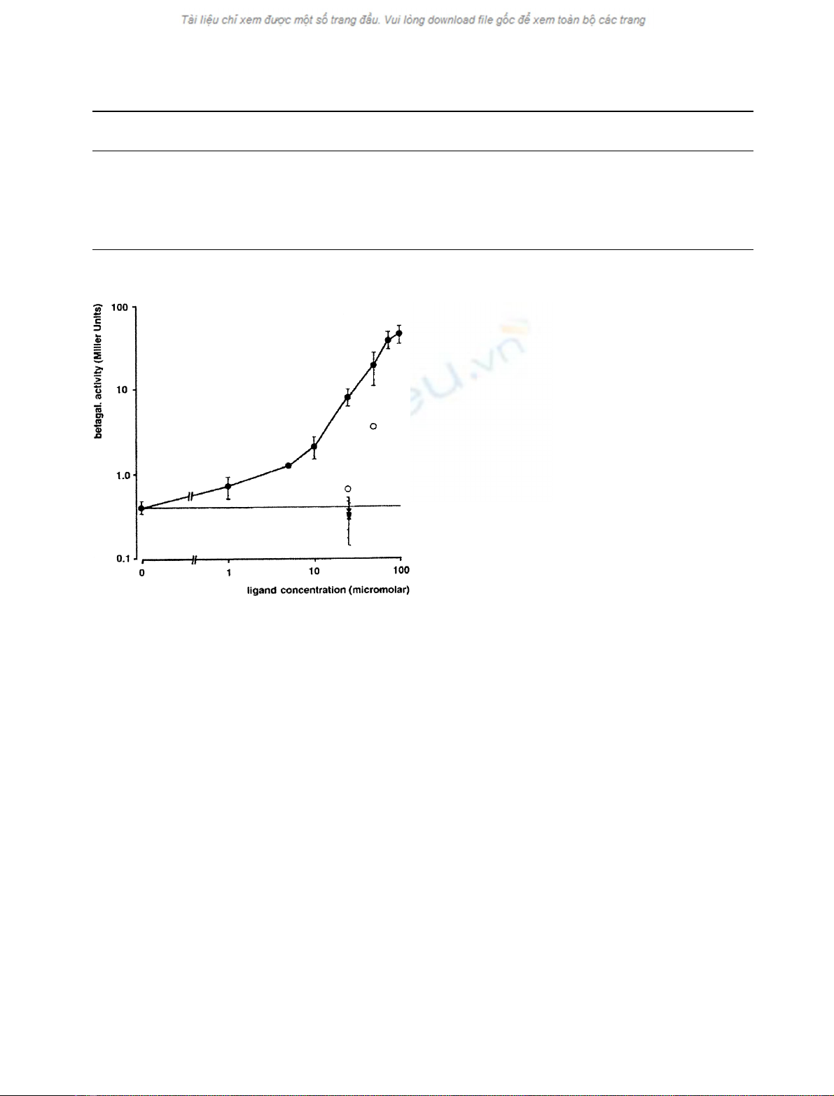
Ligand-induced heterodimerization
in vivo
Presence of muristerone A in the cultivation medium caused a
further increase in b-galactosidase activity of cotransfected
yeast cells (Table 1, Figs 1 and 2). As this effect was observed
neither with a plasmid (pCL1) constitutively expressing
GAL4 nor with a test two-hybrid pair (pTD1-1 and pAV3-
1), it was concluded that the ligand promotes interaction
between the EcR- and USP-LBDs rather than affecting
reporter enzyme or fusion protein stabilities. The induction by
ligand fully depends on the presence of helix 11 and 12 but not
theEcRFdomain.TheN-terminalportionofthehingeregion
is dispensible for ligand-induced heterodimerization. The
question of whether the same holds true for the C-terminal
region could not be assessed because of the technical problems
mentioned above: fusion proteins with EcR fragments
deprived of their hinge were either toxic to the yeast cells or
did not heterodimerize, possibly because of steric hindrance.
The effect of muristerone A on heterodimerization
between EcR–LBD and USP–LBD containing fusion
Table 1. Controls to two-hybrid experiments. Betagalactosidase activity is measured as averaged Miller Units± 95% confidence limit (number of
experiments).
12 ND, not determined.
DNA binding
construct
Activation
construct
Basal
activity
a
Induced betagalactosidase
activity (10
)6
M
)
a
Induced betagalactosidase
activity (1–5 ·10
)5
M
)
a
pCL1 pCL1
b
1244.00 ± 480.00 (5) 1021.00 ± 353.00 (3) 1508.00 (2)
pTD1-1 pVA3-1 55.10 ± 3.00 (8) 54.90 ± 2.40 (6) 59.20 ± 37.00 (3)
pAS2-1-USP(172-508) pACT2-EcR(375-652) 0.41 ± 0.07 (29) 0.71 ± 0.21 (8) 7.90 ± 1.80 (28)
pAS2-1-USP(172-508) pACT2-USP(172-508) 0.02 ± 0.02 (3) 0.06 ± 0.00 (1) 0.02 ± 0.00 (3)
pAS2-1-EcR(375-652) pACT2-USP(172-508) 0.68 ± 0.10 (12) 1.04 ± 0.19 (13) 1.77 ± 1.37 (4)
pAS2-1-EcR(375-652) pACT2-EcR(375-652) 0.03 ± 0.01 (5) 0.03 ± 0.01 (4) ND
a
Muristerone A concentration in yeast culture medium.
b
DNA binding and activation function in one protein.
Fig. 2. Dose–response curve. Effect of increasing concentrations of
muristerone A (d, solid line) on two–hybrid interaction between GBD-
USP(172–508) and GAD-EcR(375–652). The effectof 20-hydro-
xyecdysone (.), poststerone (j), RH5992 (m), and of ponasterone A
(s) is also shown at a concentration of 2.5 or 5 ·10
)5
M
. Ligand
concentration refers to concentration of ecdysteroids or agonist in the
yeast culture medium. Where error bars (spanning 95% confidence
limits) are given, the points represent averages of at least three
experiments, each.
FEBS 2002 Heterodimerization of EcR ligand binding domain (Eur. J. Biochem. 269) 3241


