
BioMed Central
Page 1 of 9
(page number not for citation purposes)
Virology Journal
Open Access
Methodology
Molecular approaches to the analysis of deformed wing virus
replication and pathogenesis in the honey bee, Apis mellifera
Humberto F Boncristiani Jr1, Gennaro Di Prisco2, Jeffery S Pettis1,
Michele Hamilton and Yan Ping Chen*1
Address: 1USDA-ARS Bee Research Laboratory, Beltsville, MD 20705, USA and 2Dipartimento di Entomologia e Zoologia Agraria "Filippo Silvestri"
- Via Università n. °100, 80055 Portici, Napoli, Italy
Email: Humberto F Boncristiani - humberto.boncristianai@ars.usda.gov; Gennaro Di Prisco - gennaro.diprisco@unina.it;
Jeffery S Pettis - jeff.pettis@ars.usda.gov; Michele Hamilton - Michele.hamilton@ars.usda.gov; Yan Ping Chen* - judy.chen@ars.usda.gov
* Corresponding author
Abstract
Background: For years, the understanding of the pathogenetic mechanisms that underlie honey
bee viral diseases has been severely hindered because of the lack of a cell culture system for virus
propagation. As a result, it is very imperative to develop new methods that would permit the in
vitro pathogenesis study of honey bee viruses. The identification of virus replication is an important
step towards the understanding of the pathogenesis process of viruses in their respective hosts. In
the present study, we developed a strand-specific RT-PCR-based method for analysis of Deformed
Wing Virus (DWV) replication in honey bees and in honey bee parasitic mites, Varroa Destructor.
Results: The results shows that the method developed in our study allows reliable identification
of the virus replication and solves the problem of falsely-primed cDNA amplifications that
commonly exists in the current system. Using TaqMan real-time quantitative RT-PCR incorporated
with biotinylated primers and magnetic beads purification step, we characterized the replication
and tissue tropism of DWV infection in honey bees. We provide evidence for DWV replication in
the tissues of wings, head, thorax, legs, hemolymph, and gut of honey bees and also in Varroa mites.
Conclusion: The strategy reported in the present study forms a model system for studying bee
virus replication, pathogenesis and immunity. This study should be a significant contribution to the
goal of achieving a better understanding of virus pathogenesis in honey bees and to the design of
appropriate control measures for bee populations at risk to virus infections.
Background
The viruses pose a serious threat to the health and well-
being of the honey bee, Apis mellifera, the most economi-
cally valuable pollinator of agricultural and horticultural
crops worldwide. In the U.S. alone, the honey bee has an
annual market value exceeding 14.6 billion dollars pro-
ducing honey and other hive products [1]. So far, honey
bees have been reported to be attacked by at least 18
viruses, most of which are single-strand positive sense
RNA viruses [2,3]. Recently, honey bees have drawn sig-
nificant attention to the scientific community and bee-
keeping industry due to the serious disease called Colony
Collapse Disorder (CCD), a malady that has killed bil-
lions of bees since 2006 across the U.S. and around the
Published: 11 December 2009
Virology Journal 2009, 6:221 doi:10.1186/1743-422X-6-221
Received: 24 August 2009
Accepted: 11 December 2009
This article is available from: http://www.virologyj.com/content/6/1/221
© 2009 Boncristiani et al; licensee BioMed Central Ltd.
This is an Open Access article distributed under the terms of the Creative Commons Attribution License (http://creativecommons.org/licenses/by/2.0),
which permits unrestricted use, distribution, and reproduction in any medium, provided the original work is properly cited.
Virology Journal 2009, 6:221 http://www.virologyj.com/content/6/1/221
Page 2 of 9
(page number not for citation purposes)
world [4-6]. A study using a metagenomic approach
found that Israeli Acute Paralysis Virus (IAPV), a species
that was originally identified in honey bees in Israel
showed that IAPV was detected in 25 of 30 (83%) CCD-
affected honey bee colonies but in only one of 21 healthy
colonies (Cox-Foster et al., 2007). The observed tight cor-
reclation between the IAPV and CCD affected colonies in
the U.S. has raised serious concerns about risks of virus
infections in honey bees. Although significant progress
has been made in honey bee virus research in the last few
decades (Reviewed in Chen and Siede, 2007 [7], investiga-
tion into virus replication and pathogenicity has been
severely hindered because of the lack of a cell culture sys-
tem for virus propagation. Therefore, observations of virus
cytopathic effect (CPE) in cultured cells, a standard
method used for unraveling the mechanisms of viral rep-
lication and the specific host responses to viral infections,
are not possible. As a result, it is to develop new methods
that would permit the study of virus replication in vitro.
Recent advances in molecular technology have greatly
expanded our ability to detect and elucidate the molecular
events associated with virus infections and pathogenesis.
With the current molecular technology, complete
genomes of several honey bee viruses have been
sequenced and analyzed [8-14]. Using RT-PCR based
assays, the virus infections in honey bees can be detected
and quantified in a rapid and accurate manner [15,16]. As
with all single-strand positive sense RNA viruses, replica-
tion of honey bee viruses proceeds via the production of a
negative-strand intermediate and its presence is indicative
of active viral replication. Therefore, the detection of neg-
ative-strand RNA of viruses offers an excellent alternative
for studying virus replication and pathogenesis in natu-
rally infected hosts [17]. Strand-specific RT-PCR was first
developed for detection of negative-strand RNAs of
viruses. However, the method has been reported to cause
falsely-primed amplification due to the self priming of
positive-strand RNA during reverse transcription or ran-
dom priming by present contaminating cellular nucleic
acids as tRNA, challenging the accuracy previous methods
[18,19]. To overcome such occurrences, more effective
techniques including Tagged RT-PCR, rTth RT-PCR and
chemically blocking the free 3' ends of the RNA, have
been developed to reduce nonspecific priming events [19-
23]. In order to further improve the assay specificity, it was
developed a new sensitive assay incorporating TaqMan
quantitative RT-PCR with biotinylated primers and mag-
netic beads purification for detection of negative-strand
viral RNAs. Furthermore, using the method developed, we
analyzed replication and tissue tropism of Deformed
Wing Virus (DWV), a highly prevalent honey bee virus
that causes wing deformity and mortality in honey bees
worldwide, in both bees with wing deformity (sympto-
matic infection) and bees with normal wings (asympto-
matic infection). The replication of DWV in honey bee
parasitic mites (Varroa destructor), a potential vector of
DWV, was also investigated.
Results
The strand specificity of conventional RT-PCR was evalu-
ated. As shown in Figure 1, both negative and positive-
strands of DWV RNA templates were detected from bees
with deformed wings using forward and reverse primers,
respectively, for initial reverse transcription followed by
amplification of the cDNA by PCR. The band intensity of
negative-strand DWV fragments was significantly stronger
than that of positive-strand DWV fragments. However, the
DWV specific fragments were also amplified by RT-PCR
without any primers for reverse transcription. Negative
signals were obtained for negative control reactions with-
out template or reverse transcriptase, confirming that RT-
PCRs were not contaminated and were from the RNA tem-
plates.
In this study, Tagged RT-PCR was evaluated for its specifi-
city for amplification of both positive and negative-strand
RNA from bees with deformed wings using four combina-
tions of primers. Tagged RT-PCR assay was based on the
generation of cDNAs by the primer containing a tag and
further amplification of cDNA by a tag-primer and a
primer complementary to the synthesized cDNA. The
result showed that cDNAs generated by either tag-forward
or tag-reverse primers were consistently amplified by sub-
sequent PCR amplification, regardless of whether a pair of
primers or only a single primer was used for PCR amplifi-
cation. As shown in Figure 2, when a tag-forward primer
was used to reverse transcribe the negative-strand of DWV
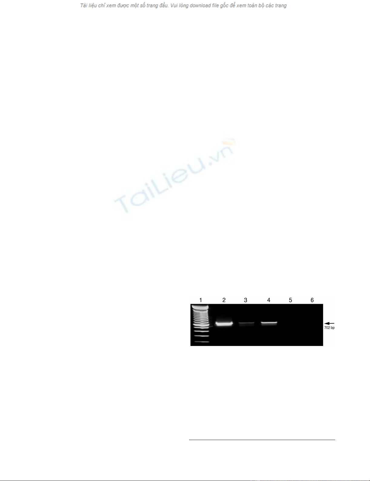
Conventional RT-PCR for strand-specific detection of DWV RNAFigure 1
Conventional RT-PCR for strand-specific detection of
DWV RNA. Total RNAs extracted from DWV-infected
bees. Both negative and positive-strands of DWV RNA were
specifically amplified by conventional RT-PCR using DWV-
specific forward (line 2) and reverse primer (line 3), respec-
tively, for initial reverse transcription. RT-PCR amplification,
was also conducted without inclusion of any primers for
reverse transcription (line 4). Negative controls containing
no template (line 5) and no reverse transcriptase (line 6)
yielded no products. The 100-bp ladder was loaded into lane
1. The arrow on the right indicates the expected 702 bp RT-
PCR products.

Virology Journal 2009, 6:221 http://www.virologyj.com/content/6/1/221
Page 3 of 9
(page number not for citation purposes)
RNAs, the synthesized cDNA could be amplified by PCR
not only with the primer pair, tag-primer and reverse
primer, but also with a single reverse primer. Meanwhile,
when tag-reverse primer was used to reverse transcribe the
positive-strand of DWV RNAs, the synthesized cDNA
could be amplified by PCR with both primer pairs, tag-
primer and forward primer, and with a single forward
primer. No amplification was detected in the negative
control (no template).
In order to achieve highly strand-specific detection of
RNA for DWV, strand-specific RT-PCR was conducted
using a biotinylated primer for cDNA generation and
magnetic separation to purify synthesized cDNA prior to
PCR amplification. As shown in Figure 3, without the
magnetic separation step, cDNA generated using bioti-
nylated forward, reverse or lack of primers for reverse tran-
scription were all amplified by subsequent PCR, just like
with conventional RT-PCR. However, the purification of
biotinylated cDNA using streptavidin-coated magnetic
beads excluded the non-specific amplification cDNAs that
were spontaneously formed without the addition of prim-
ers for reverse transcription, which occurred in both con-
ventional RT-PCR and Tagged RT-PCR.
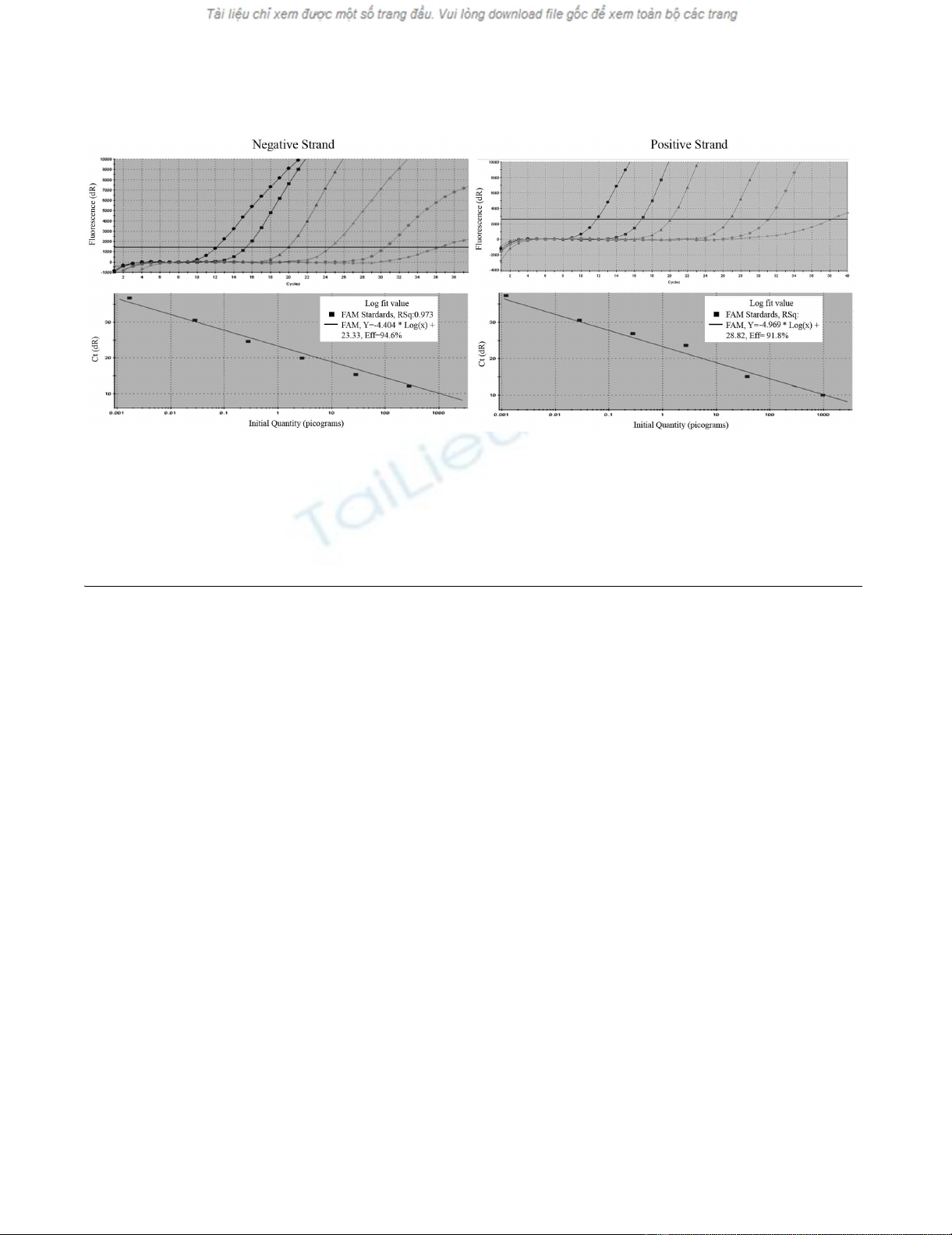
Further, the strand-specific TaqMan real-time qRT-PCR
incorporated with biotinylated primer and magnetic sep-
aration was carried out for quantification of DWV replica-
tion in host tissues and parasitic mites of honey bees. To
ensure an accurate quantification as well as the highest
sensitivity of the assay, a standard curve was first estab-
lished by plotting seven 10-fold dilutions of DWV-specific
RNA in vitro transcribed from the pCR2.1 TA cloning vec-
tor against corresponding CT value. As shown in Figure 4A
and 4B, a linear progression of the RT-PCR amplification
was observed between the amount of input RNA ranging
from 103 pg to 1 fg and the corresponding CT values
within the concentration range. The detection limit of
positive and negative-strand DWV RNAs were the same.
The lowest limit of detection with qRT-PCR for DWV was
1 fg per reaction for both positive and negative-strand
Tagged RT-PCR for strand-specific detection of DWV RNAFigure 2
Tagged RT-PCR for strand-specific detection of
DWV RNA. Total RNAs extracted from bees with
deformed wings. The negative and positive-strand cDNAs
that were generated by tag-forward primers or tag-reverse
primers in reverse transcription, respectively, were consist-
ently amplified by PCR using a pair of tag-primer and reverse
primer (lane 2), a single reverse primer (lane 3), a pair of tag-
primer and forward primer (lane 4), or a single forward
primer (lane 5). Water was used as a negative control (lane
6) and a plasmid with DWV fragment was used as a positive
control (lane 7). A 100-bp ladder was loaded into lane 1. The
arrow on the right indicates the expected 702 bp RT-PCR
products.
RT-PCR incorporated with biotinylated primer and purification of magnetic beads for strand-specific detection of DWV RNAFigure 3
RT-PCR incorporated with biotinylated primer and purification of magnetic beads for strand-specific detec-
tion of DWV RNA. Total RNAs extracted from bees with deformed wings. The negative and positive-strand cDNAs that
were generated by biotinylated forward (Lane 2 and 6) and reverse primers (Lane 3 and 7) in reverse transcription, respec-
tively. The cDNA was generated by one step RT-PCR with both biotinylated forward and reverse primers (Lane 4 and 8).
Reverse transcription was conducted without the addition of primer (Lane 5 and 9). The biotinylated cDNAs were either
amplified directly by PCR (Lane 2-5) or subjected to magnetic bead purification before PCR amplification (Lane 6-9). Negative
control without template (Lane 10) and positive control with recombinant DWV plasmid DNA (Lane 11) were included in the
reaction. A 100-bp ladder was loaded into lane 1. The arrow on the right indicates the expected 702 bp RT-PCR products.
Virology Journal 2009, 6:221 http://www.virologyj.com/content/6/1/221
Page 4 of 9
(page number not for citation purposes)
RNA. The RNA concentration below 1 fg could not be
amplified in the reaction.
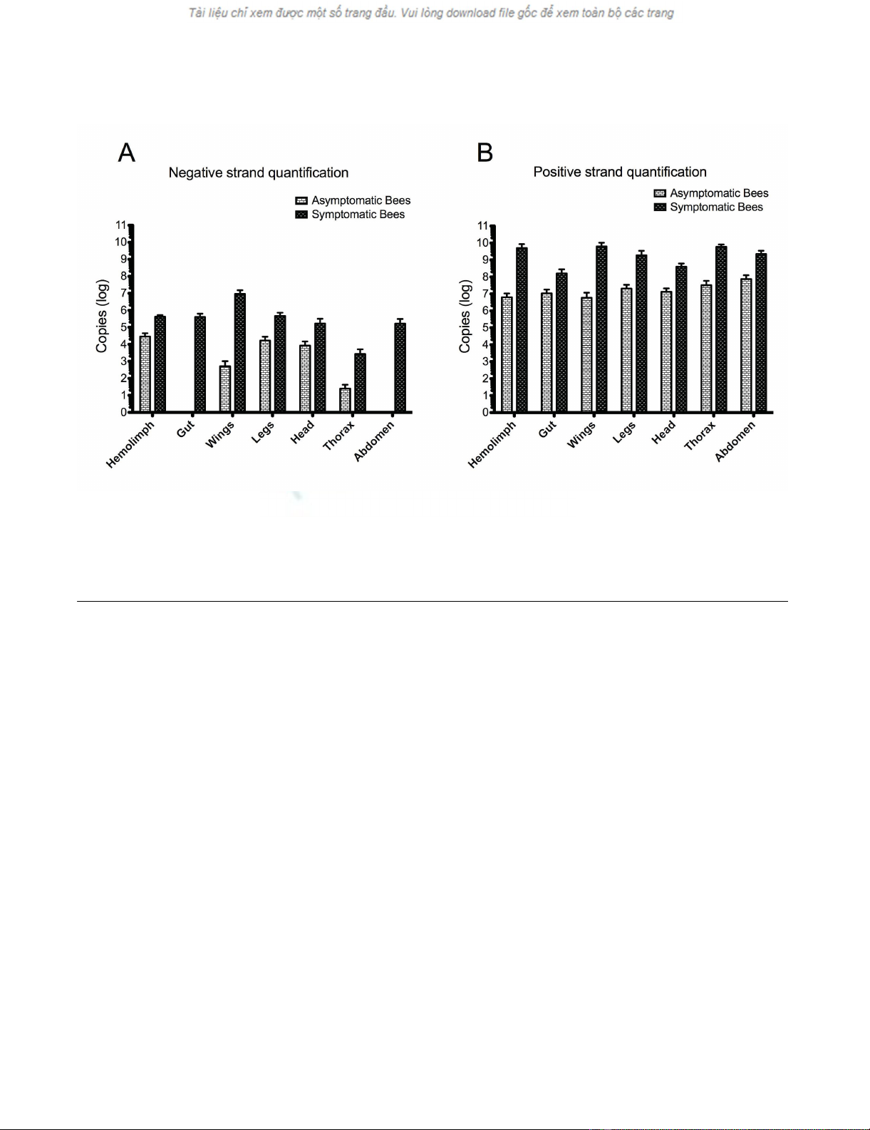
The absolute quantification of positive and negative-
strand DWV RNA in tissues of bees with symptomatic or
asymptomatic infection and individual Varroa mites was
carried out using a developed strand-specific assay. In bees
with deformed wings, negative-strand DWV RNA was
detected in all tissues examined. The concentration of neg-
ative-strand DWV RNA varied significantly among differ-
ent tissues of the deformed bees (p < 0.001) and
descended down in order of wings, hemolymph, legs, gut,
head, abdomen, and thorax (Figure 5A). Meanwhile,
except for the gut and abdomen, negative-strand DWV
RNA was also detected in tissues of bees with normal
wings. However, the average titer of negative-strand viral
RNA in bees with wing deformity was 14.6 times higher
than that in bees with normal wings in the hemolymph (p
value < 0.0001), 1.8 × 10 4 times higher in the wings (p <
0.0001), 27.8 times higher in the legs (p < 0.0001), 19.8
times higher in the head (p = 0.0015), 107.1 times higher
in the thorax (p = 0.0008) (Figure 5A).
Positive-strand DWV RNA showed predominant presence
and the average positive-strand DWV RNA being 3 × 10³
times more abundant than negative-strand DWV RNA in
the virus-infected bees (p = 0.007). Positive-strand DWV
RNA was detected in all tissues of bees with symptomatic
or asymptomatic infections even though the average con-
centration of positive-strand viral RNA in tissues of bees
with wing deformity was significantly higher than tissues
of bees with normal wings; 8 × 10² times more in the
hemolymph, 103 times more in the wings, 90 times more
in the legs, 29.8 times more in the head, 1.8 × 10³ times
more in the thorax, 15.2 times more in the gut (p <
0.0001) and 29.8 times more in the abdomen (p <
0.0001) (Figure 5B).
Using the same methodology, negative-strand RNA of
DWV was detected in 81% (17/21) while the positive-
strand RNA of DWV was found in 95.2% (20/21) of the
Varroa mites tested. Quantification of positive and nega-
tive-strand in the Varroa mites showed no significant dif-
ference (p = 0.07) between the titers of the negative-strand
and positive-strand RNAs as seen in the honey bees (p <
0.05).
Discussion
Replication is a key step in successful virus infections. The
replication strategies of positive-strand RNA viruses share
remarkable similarities: all replicate and express their
genomes through negative-strand RNA intermediates that
are used as templates for the production of positive-strand
progeny RNAs packaged in new virion particles [24].
Therefore, the presence of negative-strand RNA intermedi-
Amplification plot and standard curve of TaqMan quantitative RT-PCR (qRT-PCR) incorporated with biotinylated primer and magnetic beads purificationFigure 4
Amplification plot and standard curve of TaqMan quantitative RT-PCR (qRT-PCR) incorporated with bioti-
nylated primer and magnetic beads purification. In vitro transcribed DWV RNA was serially diluted and subjected to
RT-PCR to assess the sensitivity of the assay. A) Amplification plots were generated by using seven serial dilutions of the RNA,
ranging from 103 pg to 1 fg per reaction as the template for the qRT-PCR assays. The amplification plot shows the fluorescence
(dR) plotted against the cycle number for the standard dilution series of DWV. B) Standard curves were generated by plotting
the observed CT value (Y axis) against the initial quantities of 10-fold serial diluted RNA. CT values are the average of three rep-
etitions.
Virology Journal 2009, 6:221 http://www.virologyj.com/content/6/1/221
Page 5 of 9
(page number not for citation purposes)
ates should serve as a reliable marker for active virus rep-
lication in infected hosts.
In an attempt to develop an in vitro RNA replication assay
for honey bee viruses, we first evaluated the existing meth-
ods, including conventional RT-PCR and Tagged RT-PCR
for specificity of strand-specific detection of DWV RNA.
The results showed that DWV RNA could be amplified by
conventional RT-PCR without any primer for reverse tran-
scription. The reason for nonspecific cDNA synthesis by
conventional RT-PCR could be attributed to different
events, including false-priming by antigenomic viral RNA
or cellular RNAs, as well as self primering due to the sec-
ondary structure at the 5'UTR of viral RNA during reverse
transcription, as earlier reports suggested [19,25-27].
Tagged RT-PCR was developed to resolve the problem of
PCR amplification of falsely-primed cDNA associated
with conventional RT-PCR [28,29]. However, the finding
that cDNAs generated by tag-forward or tag-reverse prim-
ers were amplified by subsequent PCR with a single
reverse primer or forward primer, respectively, suggested
that the residue of tagged-primer from reverse transcrip-
tion possibly served in subsequent PCR and led to ampli-
fication of non-strand-specific products, making
necessary a purification step to assure total elimination of
remaining primer from RT products or primer concentra-
tion reduction in RT reaction [28]. The evidence that con-
ventional RT-PCR and Tagged RT-PCR without
purification, failed to discriminate between the two
strands of viral RNA in this study, suggests that caution
should be taken with regards to the strand specificity of
such methods.
In order to circumvent the problems of false priming and
contamination of residue primers from RT, it is worth-
while to develop improved procedures for analysis of
virus replication. We report here the development of a
novel strand-specific RT-PCR coupled with a biotinylated
primer for reverse transcription and purification of bioti-
nylated cDNA with magnetic beads. Using biotinylated
oligonucleotide primers during the reverse transcription
can lead to subsequent synthesis of biotinylated cDNAs,
which have a high binding affinity to the streptavidin-
coated magnetic beads. The purification step of streptavi-
din magnetic beads-cDNA complex, ensures the capture
of only biotinylated cDNAs. The disappearance of a posi-
tive signal for non-strand-specific amplification of mag-
netic-bead purified biotinylated cDNA in our study
suggests that the assay developed is a significant modifica-
tion of conventional or Tagged RT-PCR. The purification
TaqMan Real-Time qRT-PCR for quantification of negative and positive-strand DWV RNA in tissues of symptomatic and asymptomatic beesFigure 5
TaqMan Real-Time qRT-PCR for quantification of negative and positive-strand DWV RNA in tissues of symp-
tomatic and asymptomatic bees. The hemolymph, gut, wings, legs, head, thorax and abdomen were individually dissected
out from symptomatic and asymptomatic bees and subjected to TaqMan real-time qRT-PCR coupled with biotinylated primer
and magnetic beads purification. The virus titer of negative-strand (A) and positive-strand (B) DWV of each sample was quanti-
fied with the standard curve and expressed as copy numbers (log).


