
RESEARCH Open Access
Tat RNA silencing suppressor activity contributes
to perturbation of lymphocyte miRNA by HIV-1
Amy M Hayes
1
, Shuiming Qian
1,2
, Lianbo Yu
3
and Kathleen Boris-Lawrie
1,2*
Abstract
Background: MicroRNA (miRNA)-mediated RNA silencing is integral to virtually every cellular process including cell
cycle progression and response to virus infection. The interplay between RNA silencing and HIV-1 is multifaceted,
and accumulating evidence posits a strike-counterstrike interface that alters the cellular environment to favor virus
replication. For instance, miRNA-mediated RNA silencing of HIV-1 translation is antagonized by HIV-1 Tat RNA
silencing suppressor activity. The activity of HIV-1 accessory proteins Vpr/Vif delays cell cycle progression, which is a
process prominently modulated by miRNA. The expression profile of cellular miRNA is altered by HIV-1 infection in
both cultured cells and clinical samples. The open question stands of what, if any, is the contribution of Tat RNA
silencing suppressor activity or Vpr/Vif activity to the perturbation of cellular miRNA by HIV-1.
Results: Herein, we compared the perturbation of miRNA expression profiles of lymphocytes infected with HIV-
1
NL4-3
or derivative strains that are deficient in Tat RNA silencing suppressor activity (Tat K51A substitution) or
ablated of the vpr/vif open reading frames. Microarrays recapitulated the perturbation of the cellular miRNA profile
by HIV-1 infection. The miRNA expression trends overlapped ~50% with published microarray results on clinical
samples from HIV-1 infected patients. Moreover, the number of miRNA perturbed by HIV-1 was largely similar
despite ablation of Tat RSS activity and Vpr/Vif; however, the Tat RSS mutation lessened HIV-1 downregulation of
twenty-two miRNAs.
Conclusions: Our study identified miRNA expression changes attributable to Tat RSS activity in HIV-1
NL4-3
.The
results accomplish a necessary step in the process to understand the interface of HIV-1 with host RNA silencing
activity. The overlap in miRNA expression trends observed between HIV-1 infected CEMx174 lymphocytes and
primary cells supports the utility of cultured lymphocytes as a tractable model to investigate interplay between
HIV-1 and host RNA silencing. The subset of miRNA determined to be perturbed by Tat RSS in HIV-1 infection
provides a focal point to define the gene networks that shape the cellular environment for HIV-1 replication.
Background
MicroRNA (miRNA)-mediated RNA silencing is integral
to virtually every aspect of biology, including pluripo-
tency, development, differentiation, proliferation, and
antiviral defense [1-3]. The activity of miRNA has the
capacity to coordinate intricate gene expression net-
works [2]. Most coding genes exhibit one or many
miRNA recognition elements (MRE), and a single
miRNA may regulate dozens of genes in response to
viral infection or another environmental cue. The
mature miRNAs are processed from a primary transcript
to a precursor form that is subject to nuclear export. In
the cytoplasm, the activity of Dicer, Argonaute (Ago)
and TAR RNA-binding protein (TRBP) produces mature
miRNA, which is ~22 nt in length [4]. This ribonucleo-
protein complex (RNP) is loaded onto a multicompo-
nent RNA-induced silencing complex (RISC), and the
miRNA guides the interaction of RISC with one or
more partially complementary MRE. MRE interaction
with the cognate miRNA guide strand produces
sequence-specific RNA silencing by RISC. Virus modu-
lation of miRNA expression or RNA silencing activity
has the capacity to counteract antiviral restriction [5].
Collectively, viruses encode proteins and decoy RNAs
to counter innate restriction of endogenous and
* Correspondence: boris-lawrie.1@osu.edu
1
Department of Veterinary Biosciences; Center for Retrovirus Research; Center
for RNA Biology; Comprehensive Cancer Center, Ohio State University,
Columbus OH, USA
Full list of author information is available at the end of the article
Hayes et al.Retrovirology 2011, 8:36
http://www.retrovirology.com/content/8/1/36
© 2011 Hayes et al; licensee BioMed Central Ltd. This is an Open Access article distributed under the terms of the Creative Commons
Attribution License (http://creativecommons.org/licenses/by/2.0), which permits unrestricted use, distribution, and reproduction in
any medium, provided the original work is properly cited.

exogenous viruses. The interplay between viral infec-
tions and miRNA-mediated RNA silencing is best
understood in plants. Plant miRNA activity provides a
robust antiviral host restriction that is countered by
plant virus-encoded RNA silencing suppressors (RSS)
that are necessary for viral pathogenesis [6]. RSS have
also been found in animal viruses [7], and the list of
human viruses that encode an RSS is growing [8]. RSS
activity is exhibited by multifunctional RNA binding
proteins encoded by ebolavirus [9,10], influenza virus
[11], and human T-cell lymphotropic virus type 1 [12].
In the case of ebolavirus, RNA silencing suppressor
activity is exhibited by three viral proteins (VP30, VP35,
VP40), which suggests an effective counter strike to the
small RNA-based host defense is under strong positive
selection [10]. Adenovirus expresses abundant levels of
VA1 RNA that saturates pre-miRNA nuclear export and
pre-miRNA processing to potently reduce miRNA pro-
duction [13]. In contrast to the generalized downregula-
tion of RNA silencing by VA1, the activity of viral RSS
proteins on protein effectors of RNA silencing activity is
subtle and conceivably may target a subset of miRNA
[6,8,14,14].
Several lines of evidence indicate that small RNA
activity is important for HIV-1. Cell-encoded miRNA
attenuate virus replication in activated T lymphocytes
[15] and in latently infected resting T lymphocytes [16].
HIV-1 mRNA translation is attenuated by RNA silen-
cing [14], and HIV-1 mRNAs associate and co-localize
with components of the RISC [17]. Downregulation of
RNA silencing effectors (RCK/p54 or DGCR8) in
PBMCs of HIV-1 infected patients on HARRT results in
virus reactivation [17]. While RISC activity suppresses
HIV-1 replication in at least some circumstances, the
small RNA pathway appears to be harnessed to alter cel-
lular gene expression to foster virus replication [18-20].
HIV-1-encoded RNA silencing suppressor activity has
been controversial, given differences in experimental
conditions [21,22]. Consensus is emerging of an intricate
and multifaceted relationship between the human
miRNA-mediated silencing pathway and HIV-1 [23] that
operates in a strike-counterstrike manner [24]. A cor-
nerstone of this complex relationship is the essential
viral transcriptional trans-activator Tat and its cis-acting
trans-activation responsive element, TAR. TAR is a
structured RNA element within the 5’terminus of all
HIV-1 transcripts that forms a stem-bulge-stem RNA
structure that is recognized by Tat and cellular factors
TRBP and P-TEFb to robustly activate productive viral
gene transcription. Bennasser and colleagues identified
RSS activity in Tat that requires the arginine-rich dou-
ble-stranded RNA binding domain [21]. Tat RSS activity
is genetically separable from Tat transcriptional activity
by K51A substitution in the double-stranded RNA
binding domain [21]. HIV-1 Tat functions across the
plant and animal kingdoms to suppress a common step
in RNA silencing that is downstream of small RNA
maturation [14]. Translation of virion structural protein
is exacerbated by K51A substitution in the Tat RNA
binding domain (HIV-1
NL4-3
RSS) [14]. The delay in
HIV-1 replication by Tat K51A substitution can be
complemented by TBSV P19 [14] and rice hoja blanca
virus non-structural protein 3 (NS3) [25]. Thus, virus
interplay with miRNA-mediated RNA silencing is con-
served across the plant and animal kingdoms, and Tat
RSS activity is important in biology of the human retro-
virus, HIV-1.
The potential for RSS activity by TAR RNA was initi-
ally identified by Bennasser and colleagues [26]. Similar
in principle to adenovirus VA1 RNA, TAR squelches
the activity of host protein required for RNA silencing
activity. In cells transfected with TAR RNA, TAR acts
to occlude TRBP from Dicer and thereby interferes with
dsRNA-processing [26]. TAR interaction with TRBP
exerts several activities in HIV-1 biology [27-30]. TRBP
was originally identified in a cDNA screen for proteins
necessary for TAR/Tat transcriptional trans-activation
[31,32]. Subsequently, TRBP was identified to inhibit the
activity of protein kinase R (PKR) that is directed to
double stranded features of viral RNA [33]. The poten-
tial for TAR to sequester TRBP and downregulate
miRNA maturation or RISC activity [26] is attributable
to structural features of the HIV-1 RNA that are pro-
cessed to viral miRNA [18-20] or to early HIV-1 viral
transcripts that are prematurely terminated [34]. In sum,
Tat and TAR have the potential to manipulate the RNA
silencing pathway in a strike-counter-strike manner
[23,24]. The resulting alteration of the cellular environ-
ment may tip the balance to favor virus replication or
favor viral latency. The identification of the miRNA
affected by HIV-1 RSS activity and future determination
of the MRE targeted by these miRNA, are strategic mile-
stones in the process to understand the viral interface
with host RNA silencing.
MiRNAs contribute to physiological control of the cell
cycle [35]. Hsa-miR-17-5p modulates the G1/S transi-
tion by targeting over twenty genes that regulate pro-
gression of the cell cycle [36]. The broadly conserved
miRNA let-7 family controls exit from the cell cycle in
Caenorhabditis elegans [37]. Human fibroblasts arrest in
G2/M by overexpression of let-7 family members [38].
In human cancers, tumor progression is attributable to
dysregulation of cell cycle control by miRNA [39,40].
G2/M delay is a feature of HIV-1 infected cells that is
attributable to the HIV-1 accessory proteins Vpr and Vif
[41-43]. Ablation of vpr/vif restores cell cycle profiles to
be similar to uninfected cells [43]. A primary role for
Vpr is to trans-activate viral gene expression during
Hayes et al.Retrovirology 2011, 8:36
http://www.retrovirology.com/content/8/1/36
Page 2 of 13
virus-induced G
2
/M delay [41,44,45]. A primary role of
Vif is to combat antiviral restriction by APOBEC pro-
teins [46,47]. Vif additionally contributes to downregula-
tion of Vpr, which would reduce transcription trans-
activation [48]. The possibility remains to be addressed
that Vpr and Vif contribute to perturbation of cellular
miRNA by HIV-1, perhaps by trans-activation. A neces-
sary step in the process to understand interplay of the
virus with host RNA silencing is the definition of
miRNA expression differences during infection with
HIV-1 or Vpr/Vif-deficient HIV-1.
Herein, we have evaluated the perturbation of miRNA
signature of cultured lymphocytes by HIV-1 and HIV-1
derivatives deficient in Vpr/Vif (∆VV) or Tat RSS (RSS).
Our results indicate that the miRNA signature is per-
turbed by HIV-1 infection, and a subset of miRNA is
differentially expressed by elimination of the HIV-1 Tat
RNA silencing antagonist. Additionally, we observed
~50% overlap between the miRNA signatures of cul-
tured lymphocytes infected with HIV-1 and clinical sam-
ples from HIV-1 infected individuals. The outcomes are
a list of candidate miRNAs that interface with cellular
genes important to HIV-1 replication, and a tractable
model to investigate the interplay between HIV-1 and
cellular miRNA that alters the cellular environment dur-
ing virus infection.
Results
Comparison of miRNA expression profiles produced by
HIV-1 and strains deficient in Tat RSS or Vpr/Vif
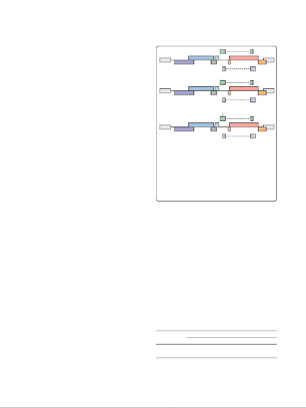
Three strains of HIV-1
NL4-3
were propagated by trans-
fection of provirus (Figure 1) into HEK293 cells, and
cell-free virus was used to generate HIV-1/CEMx174
lymphocytes. HIV-1 infection by cell-free HIV-1 is rela-
tively inefficient unless enhanced by spinoculation
[49,50], whereas HIV-1 infection by co-culture is effi-
cient [51]. All experiments were carried out by co-cul-
ture infection of CEMx174 lymphocytes to minimize the
confounding signal from uninfected cells. We monitored
the progression of the infection by FACS of intracellular
Gag at several intervals. The benchmark criterion for
lymphocyte harvest was set at ≥80% infection in order
to minimize the background signal from residual unin-
fected cells. Comparison of HIV-1
NL4-3
to the derivative
strains ∆VV and RRS revealed differences in replication
kinetics, similar to previous observations [21,52]. The
FACS of intracellular Gag at ~12 h intervals determined
that HIV-1
NL4-3
and ∆VV reached ≥80% infection by 40
to 48 hr, while RSS reached ≥80% infection by 60 hr
(Table 1). Cell viability was monitored by trypan blue
exclusion and was determined to be ≥90% at time of
harvest. Total cellular RNA was harvested from replicate
infections and subjected to bioanalyzer analysis to verify
integrity. The RNA samples were treated with reverse
transcriptase and random hexamer primer, and biotiny-
lated cDNA was generated for hybridization by the
miRNA microarray shared resource of the Ohio State
University Comprehensive Cancer Center. Two replicate
experiments used miRNA microarray chips printed with
906 duplicate probes that measure levels of 518 mature
miRNA and 332 precursor miRNA [53]; four probes
were excluded because they have been deleted from
miRBase. Signal intensity from two independent infec-
tions per virus was quantified with GenePix Pro 6 image
analysis software, and the data were evaluated for back-
ground correction, log base 2 transformation, and quan-
tile normalization. Microsoft Excel pivot tables were
used to manage comparative expression trends for viral
strains. Signal intensities in log
2
values ranged from 0.3
to 16.0; and a signal intensity of log
2
value of 5 or
x
x
LTR gag
vif
nef
tat
vpu
vpr
rev
tat
rev
tat
K51A
r
e
v
pol env
HIV-1NL4-3
LTR gag
vif
nef
vpu
vpr
pol env
6VV
LTR
LTR
LTR
LTR
gag
vif
nef
vpu
vpr
pol env
RSS
Figure 1 Host miRNA expression levels were compared
between HIV-1
NL4-3
, Vif/Vpr-deficient or Tat K51A RSS-deficient
strains. CEMx174 lymphocytes were infected by co-culture with
HIV-1
NL4-3
, HIV-1
NL4-3
∆VV that contains a premature stop codon in
vif and frameshift in vpr, or HIV-1
NL4-3
RSS that contains the K51A
substitution that eliminates Tat RSS activity. Total cellular RNA was
reverse transcribed and hybridized to miRNA microarray chips with
two or three independent biological replicates to determine relative
expression levels of 518 mature miRNA and 336 precursor miRNA
that were monitored by 906 human miRNA probes spotted in
duplicate [53].
Table 1 Percentage of CEMx174 infected cells at time of
RNA harvest
Percentage of Virus Infected Cells
a
Experiment Mock HIV-1 RSS ∆VV
Replicate 1 0 90 83 80
Replicate 2 0 95 87 90
a
CEMx174 cells were infected by co-culture and the progression of infection
was monitored by FACS of intracellular Gag. Values indicate the percentage of
Gag
+
cells at time of harvest. Total cellular RNA was prepared in Trizol,
integrity verified by bioanalyzer and processed for the miRNA microarrays.
Hayes et al.Retrovirology 2011, 8:36
http://www.retrovirology.com/content/8/1/36
Page 3 of 13
below was considered below minimally detectable limits.
Signal intensities in log
2
values greater that 16 corre-
sponded to saturation of signal. MiRNA expression was
considered changed if upregulated 2-fold or downregu-
lated by a factor of 2 or more. Four categories of
miRNA expression were enumerated: Up; Down; No
change (levels remained within a factor of 2 of unin-
fected control); or Less than the minimum detectable.
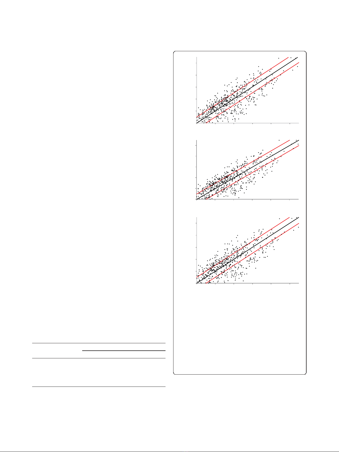
The miRNA signature is perturbed by HIV-1 and
derivatives deficient in vpr/vif or Tat RSS
HIV-1 perturbed the expression of ~200 of the 518
mature miRNAs on the chip; ~70 miRNAs were upregu-
lated and ~100 miRNAs were downregulated (Table 2).
The number of up- or down-regulated miRNAs was
similar between HIV-1
NL4-3
,∆VV and RSS (Table 2).
Scatterplot analysis of the expression changes relative to
mock infection revealed the range of expression differ-
ences was similar among the infections (Figure 2). Fifty-
two miRNAs were upregulated by all three strains, and
eighty-three miRNAs were downregulated by all three
strains.
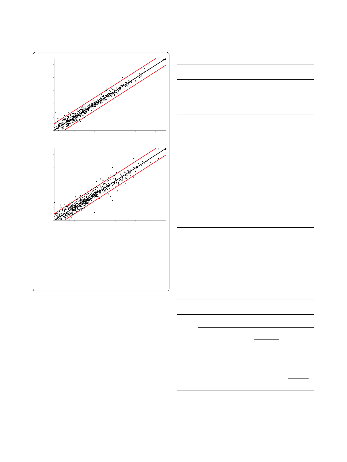
We examined the data for miRNAs that exhibited ≥2-
fold expression change between the viral strains. As
shown in scatterplot analysis between HIV-1 and ∆VV,
five miRNAs fall outside the two-fold change lines (Fig-
ure 3); HIV-1 exhibited ≥2-fold greater expression of
hsa-miR-32, hsa-miR-194, hsa-miR-199a, hsa-miR-496,
and expression of hsa-miR-450 was reduced. The results
indicate that ablation of vif/vpr modestly alters miRNA
profile. We expected this minor difference is attributable
to experimental variation, and this issue would be
resolved by additional experiments. By comparison, the
scatterplot analysis unveiled nineteen miRNAs that
exhibited expression differences between HIV-1 and
RSS (Figure 3, Table 3). The results indicate that pertur-
bation of the cellular miRNA signature by HIV-1 infec-
tion is largely independent of the activity of vpr/vif or
Tat RSS.
Tat RSS mutation affects the steady state of a subset of
miRNA
HIV-1 exhibited 2 to 3-fold greater expression of fifteen
miRNA relative to RSS (Table 3). Four miRNA were
downregulated in HIV-1 relative to RSS by a factor of 2
Table 2 Distribution of changes in mature miRNA
expression relative to uninfected lymphocytes for
infection with indicated viral strain
Infection Relative to Mock
a
Expression Trend
b
HIV-1 RSS ∆VV
Up 72 74 74
Down 106 104 111
No change 157 153 146
<MD 234 238 238
a
Human CEMx174 lymphocytes infected by co-culture with indicated virus
were screened by miRNA microarray. The number of mature miRNA probes
present on the chip was 518 after exclusion of four probes removed from
miRBase. Values represent number of probes affected.
b
Up: upregulated (≥2.0
×); Down: downregulated (≤0.5×); No change: between 0.5-2.0 ×; <MD: less
than minimum detectable limits.
Log2 Mock
Log2 RSSLog2 6VV Log2 HIV-1
Log2 Mock
Lo
g
2 Mock
5
7
9
11
13
15
5 7 9 111315
5 7 9 111315
5 7 9 111315
5
7
9
11
13
15
5
7
9
11
13
15
A
B
C
Figure 2 Host miRNA expression is changed by infection with
HIV-1, Vif/Vpr -deficient or RSS-deficient viral strains. Scatterplot
analysis of miRNA mature and precursor probes expression changes
observed on microarrays hybridized with RNA of human CEMx174
lymphocytes unexposed to virus or infected with HIV-1, or ∆VV, or
RSS. Each data point represents one unique probe sequence. The
black line at x = y illustrates baseline of no change. The red lines
illustrate change by a factor of 2. Axes are truncated at log
2
=5to
eliminate measurement uncertainty at lower signal intensities. Log
2
expression values of human miRNA probes in the mock sample are
shown on the x-axis and the corresponding values for the HIV-1
sample are shown on the y-axis. (a) HIV-1 versus mock infection; (b)
RSS versus mock infection; (c) ∆VV versus mock infection.
Hayes et al.Retrovirology 2011, 8:36
http://www.retrovirology.com/content/8/1/36
Page 4 of 13
to 4 (Table 3). Of the 145 miRNA perturbed by the
three viral infections relative to cells without virus infec-
tion (mock), Tat RSS activity in HIV-1 correlated with
higher steady state for 15 of the 18 and lower steady
state for 3 miRNA (Table 4). These differences may be
attributable to direct effects of Tat RSS activity on RNA
stability or by secondary effects elicited through
upstream genes. In sum, the observed generalized per-
turbation of miRNA expression by HIV-1 infection of
cultured lymphocytes is consistent with previous micro-
arrays of HIV-1 infected cells [15,54,55]. The compari-
son of the three derivative viruses determined that the
generalized perturbation of miRNA expression levels by
HIV-1 is largely independent of the ablation of Vpr/Vif
and Tat RSS.
The miRNA that were downregulated by all three viral
infections (n = 83) were filtered to ascertain possible dif-
ferences in the level of downregulation. Twenty-two
miRNA exhibited less downregulation by 10% or more
in RSS compared to HIV-1 or ∆VV infection (p =
≤0.0001) (Table 5). Subsequent investigations are war-
ranted to evaluate the possibility that these miRNA have
conserved features and to determine the MRE that are
Lo
g
2 RSS
Log2 6VV
Log2 HIV-1 Log2 HIV-1
5 7 9 11 13 15
5 7 9 11 13 15
5
7
9
11
13
15
5
7
9
11
13
15
A
B
Figure 3 Ablation of Tat RSS alters miRNA expression trends
relative to HIV-1 and Vif-/Vpr-deficient HIV-1. Scatterplot analysis
of miRNA mature and precursor probes expression changes
observed on microarrays hybridized with RNA from human
CEMx174 lymphocytes infected with HIV-1, ∆VV, or RSS. Log
2
expression values of human miRNA probes in the HIV-1 infections
are shown on the y-axis, log
2
expression values for miRNA probes in
either RSS or ∆VV infection are shown on the x-axis. (a) HIV-1 versus
∆VV infection; (b) HIV-1 versus RSS infection.
Table 3 Mature miRNAs that exhibit expression change
by a factor of ≥2 for RSS relative to HIV-1 infection
MiRNAs differing in expression by ≥2 between RSS and HIV-1
MiRNA Probe Ratio RSS/HIV-1
Upregulated
hsa-miR-105 2.1
hsa-miR-550 2.1
hsa-miR-32 2.2
hsa-miR-33b 2.2
Downregulated
hsa-miR-30e-3p 0.3
hsa-miR-194 0.3
hsa-miR-494 0.3
hsa-miR-500 0.3
hsa-miR-20a 0.4
hsa-miR-20b 0.4
hsa-miR-21 0.4
hsa-miR-26b 0.4
hsa-miR-106a 0.4
hsa-miR-215 0.4
hsa-miR-219 0.4
hsa-miR-453 0.4
hsa-miR-17-5p 0.5
hsa-miR-499 0.5
hsa-miR-658 0.5
Table 4 Mature miRNAs that exhibit expression change
by a factor of ≥2 between RSS and HIV-1 infection
standardized to mock
RSS Relative to Mock
a
Up Unchanged Down
Up hsa-miR-494 hsa-miR-194
hsa-miR-500
-
Relative Unchanged -hsa-miR-33b
hsa-miR-105b
hsa-miR-453
hsa-miR-499
hsa-miR-17-5p
hsa-miR-20a
hsa-miR-20b
hsa-miR-30e-3p
hsa-miR-106a
hsa-miR-219
Mock Down - - hsa-miR-21
hsa-miR-26b
hsa-miR-32
hsa-miR-215
hsa-miR-658
a
Nineteen miRNAs exhibited expression differences between the indicated
strains relative to mock infection. The miRNAs indicated in plain font
exhibited reduced expression by a factor of 2 or more for RSS compared to
HIV-1. The three miRNAs in underlined font exhibited increased expression by
2-fold or more for RSS compared to HIV-1. Notably, miR550 upregulation by
HIV-1 was attenuated in RSS infection (Table 3) but is excluded from Table 4
because miR550 was not detectable in cells lacking virus (mock infection).
Hayes et al.Retrovirology 2011, 8:36
http://www.retrovirology.com/content/8/1/36
Page 5 of 13


![Vaccine và ứng dụng: Bài tiểu luận [chuẩn SEO]](https://cdn.tailieu.vn/images/document/thumbnail/2016/20160519/3008140018/135x160/652005293.jpg)


%20--%3e%3cdefs%3e%3cstyle%3e%20.st0%20{%20fill:%20%23fff;%20}%20.st1%20{%20fill:%20%237800fa;%20}%20%3c/style%3e%3c/defs%3e%3cpath%20class='st1'%20d='M117.78,12.18H43.11c2.9,3.47,4.65,7.94,4.65,12.82,0,5.6-2.3,10.66-6.01,14.29h76.02l7.22-13.56-7.22-13.56Z'/%3e%3cg%3e%3cpath%20class='st0'%20d='M53.58,26.17h-.59v-1.46h.59v-4.96h2.83c1.78,0,2.67.94,2.67,2.82v5.76c0,1.87-.89,2.81-2.67,2.81h-2.83v-4.96ZM55.36,21.37v3.34h1.1v1.46h-1.1v3.34h1.01c.61,0,.91-.37.91-1.1v-5.93c0-.74-.3-1.1-.91-1.1h-1.01Z'/%3e%3cpath%20class='st0'%20d='M65.99,31.14h-1.8l-.31-2.07h-2.19l-.31,2.07h-1.64l1.82-11.39h2.62l1.82,11.39ZM65.28,18.04c-.25.46-.51.77-.75.94-.21.15-.47.22-.79.22-.26,0-.57-.07-.92-.22l-.38-.15c-.14-.05-.26-.07-.37-.07-.3,0-.53.18-.71.54l-.91-.68c.25-.46.51-.77.75-.94.21-.14.48-.21.79-.21.26,0,.57.07.92.21l.38.15c.14.05.26.07.37.07.3,0,.53-.18.71-.54l.91.68ZM61.91,27.52h1.73l-.87-5.76-.87,5.76Z'/%3e%3cpath%20class='st0'%20d='M74.53,26.89v1.52c0,1.91-.89,2.86-2.67,2.86s-2.67-.95-2.67-2.86v-5.93c0-1.91.89-2.86,2.67-2.86s2.67.95,2.67,2.86v1.11h-1.69v-1.22c0-.75-.31-1.12-.93-1.12s-.93.37-.93,1.12v6.15c0,.74.31,1.11.93,1.11s.93-.37.93-1.11v-1.63h1.69Z'/%3e%3cpath%20class='st0'%20d='M81.4,31.14h-1.8l-.31-2.07h-2.19l-.31,2.07h-1.64l1.82-11.39h2.62l1.82,11.39ZM75.9,19.2l1.52-1.91h1.71l1.51,1.91h-1.61l-.76-.95-.75.95h-1.61ZM77.32,27.52h1.73l-.87-5.76-.87,5.76ZM83.1,15.99l-1.76,1.91h-1.26l1.17-1.91h1.86Z'/%3e%3cpath%20class='st0'%20d='M84.86,19.75c1.78,0,2.67.94,2.67,2.82v1.48c0,1.87-.89,2.81-2.67,2.81h-.85v4.28h-1.79v-11.39h2.64ZM84.01,21.37v3.86h.85c.58,0,.87-.36.87-1.08v-1.71c0-.71-.29-1.07-.87-1.07h-.85Z'/%3e%3cpath%20class='st0'%20d='M93.51,19.75c1.78,0,2.67.94,2.67,2.82v1.48c0,1.87-.89,2.81-2.67,2.81h-.85v4.28h-1.79v-11.39h2.64ZM92.66,21.37v3.86h.85c.58,0,.87-.36.87-1.08v-1.71c0-.71-.29-1.07-.87-1.07h-.85Z'/%3e%3cpath%20class='st0'%20d='M98.8,31.14h-1.79v-11.39h1.79v4.88h2.03v-4.88h1.83v11.39h-1.83v-4.88h-2.03v4.88Z'/%3e%3cpath%20class='st0'%20d='M105.36,24.55h2.46v1.62h-2.46v3.34h3.09v1.63h-4.88v-11.39h4.88v1.63h-3.09v3.18ZM108.17,17.29l-1.76,1.91h-1.26l1.17-1.91h1.86Z'/%3e%3cpath%20class='st0'%20d='M112.2,19.75c1.78,0,2.67.94,2.67,2.82v1.48c0,1.87-.89,2.81-2.67,2.81h-.85v4.28h-1.79v-11.39h2.64ZM111.35,21.37v3.86h.85c.58,0,.87-.36.87-1.08v-1.71c0-.71-.29-1.07-.87-1.07h-.85Z'/%3e%3c/g%3e%3ccircle%20class='st1'%20cx='25'%20cy='25'%20r='20'/%3e%3cpath%20class='st0'%20d='M32.78,19.27c2.92,0,4.43,2.55,5.28,5.33l.71,2.17c.14.38-.33.75-.71.75h-5.61c.19-.33.24-.71.09-1.08l-.75-2.45c-.43-1.32-.99-2.64-1.79-3.77.75-.57,1.65-.94,2.78-.94h0ZM25,18.38c3.25,0,4.9,2.78,5.89,5.89l.76,2.45c.14.42-.33.8-.8.8h-11.69c-.42,0-.94-.38-.8-.8l.75-2.45c.99-3.11,2.64-5.89,5.89-5.89h0ZM25,11.35c1.74,0,3.11,1.37,3.11,3.11s-1.37,3.11-3.11,3.11-3.11-1.41-3.11-3.11,1.41-3.11,3.11-3.11h0ZM17.27,19.27c1.08,0,1.98.38,2.73.94-.8,1.13-1.37,2.45-1.74,3.77l-.8,2.45c-.14.38-.05.75.09,1.08h-5.56c-.42,0-.9-.38-.75-.75l.71-2.17c.9-2.78,2.41-5.33,5.33-5.33h0ZM17.27,12.91c1.51,0,2.78,1.27,2.78,2.83s-1.27,2.83-2.78,2.83-2.83-1.27-2.83-2.83,1.27-2.83,2.83-2.83h0ZM32.78,12.91c1.56,0,2.78,1.27,2.78,2.83s-1.23,2.83-2.78,2.83-2.83-1.27-2.83-2.83,1.27-2.83,2.83-2.83h0ZM27.07,28.56v.09c0,.57-.24,1.08-.61,1.46h0v.05c-.38.33-.9.57-1.46.57s-1.08-.24-1.46-.61h0c-.38-.38-.61-.9-.61-1.46v-.09h1.41v.09c0,.19.05.38.19.47v.05c.09.09.28.19.47.19s.38-.09.47-.19v-.05c.14-.09.24-.28.24-.47t-.05-.09h1.41ZM30.99,28.56v.09c0,1.65-.66,3.16-1.74,4.24-1.08,1.08-2.59,1.79-4.24,1.79s-3.16-.71-4.24-1.79l-.05-.05c-1.04-1.08-1.7-2.55-1.7-4.2v-.09h1.41v.09c0,1.27.47,2.4,1.27,3.25h.05c.85.85,1.98,1.37,3.25,1.37s2.4-.52,3.25-1.37c.85-.8,1.37-1.98,1.37-3.25v-.09h1.37ZM34.99,28.56v.09c0,2.78-1.13,5.28-2.92,7.07-1.79,1.79-4.29,2.92-7.07,2.92s-5.23-1.13-7.07-2.92c-1.79-1.79-2.92-4.29-2.92-7.07v-.09h1.41v.09c0,2.4.94,4.53,2.5,6.08,1.56,1.56,3.72,2.5,6.08,2.5s4.52-.94,6.08-2.5c1.56-1.56,2.5-3.68,2.5-6.08v-.09h1.41Z'/%3e%3c/svg%3e)