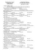
Công nghệ chuyển protein và sự tổng hợp một peptid
Runx 3 có thể di chuyển trực tiếp vào tế bào
Pham Thanh Van, Suk-Chul Bae
Viện nghiên cứu Ung thư, trường Đại học Quốc Gia Chung
Buk, Hàn Quốc.
Sự vận chuyển những phân tử lớn như protein hay
polipeptid vào tế bào động vật vốn gặp nhiều khó khăn do
sự cản trở của màng tế bào. Tuy nhiên trong 10 năm trở lại
đây, một phương pháp mới đã được đề xuất bởi nhóm
nghiên cứu Steven Dowdy để giải quyết khó khăn trên, đó
là công nghệ chuyển protein (protein transduction
technology). Công nghệ chuyển protein cho phép vận
chuyển trực tiếp protein hay các cao phân tử vào trong tế
bào bằng cách dung hợp phân tử cần nghiên cứu với một
trình tự đặc biệt có khả năng vượt qua màng tế bào vào nội
bào. Những trình tự này đã được phát hiện đầu tiên trên
protein Tat của HIV 1, Antp của ruồi giấm và HSV VP22
của virus HSV, và được đặt tên là "vùng chuyển protein"
(protein transduction domains).
Dựa trên các kết quả thực nghiệm, các nhà khoa học cho
rằng nếu protein bị biến tính trước khi đưa vào môi trường
nuôi cấy tế bào thì quá trình chuyển protein sẽ xảy ra nhanh
hơn và hiệu quả lớn hơn rất nhiều so với việc vận chuyển
protein tự nhiên. Sau khi đã vào tế bào, hệ thống chaperone
trong tế bào sẽ đưa protein biến tính trở về dạng tự nhiên
ban đầu và hoạt động bình thường. Tuy nhiên, vì cơ chế
hoạt động của chaperone vẫn chưa được xác định rõ ràng,
cách thức này có một trở ngại là với những protein chưa rõ
chức năng, sẽ khó có thể kết luận được sự bất hoạt của nó
trong tế bào là do nó không có được chức năng như ta
phỏng đoán hay do chaperone của tế bào đã không hồi phục
được protein từ dạng biến tính trở về cấu trúc tự nhiên ban
đầu.
Bài báo này muốn giới thiệu thử nghiệm đưa protein ở dạng
tự nhiên, không bị biến tính vào một số dòng tế bào động
vật, mặc dầu phương pháp này đã được đánh giá là sẽ vấp
phải nhiều khó khăn. Đối tượng nghiên cứu là trình tự gồm
30 axit amin ở đầu cuối của protein Runx3 - một thành viên
trong gia đình Runx đóng vai trò như những tác nhân điều
hoà nghiêm ngặt liên quan tới sự hình thành mô mới và sự
chết của tế bào. Bằng cách dung hợp đoạn peptid cần
nghiên cứu với "vùng chuyển protein" Tat, chúng tôi đã
đưa được protein vào 2 dòng tế bào nguyên bào sợi
NIH/3T3 và nguyên bào cơ C2C12. Sự chuyển protein đạt
hiệu quả cao nhất ở nồng độ protein tối thiểu là 400ug/ml.
esearch, Chungbuk National University
A. An introduction of Protein Transduction
Technology.
The delivery of large, hydrophilic molecules such as
proteins and oligonucleotides to the cytoplasm and nucleus
of cells is problematic due to their poor plasma membrane
permeability. Manipulation of protein expression in
mammalian cells has been achieved by microinjection,
electroporation or transfection of expression vectors. While
these approaches have been somewhat successful, the
classic manipulation methods are not easily regulated and
can be laborious due to the limitations of each method.
One approach to circumvent these problems is Protein
Transduction Technology, a new method that allows the
direct entry of protein into mammalian cells.
Protein Transduction Technology.
Protein transduction was first reported in 1988 by Green [1]
and Frankel [2], who independently demonstrated that the
full-length (86-amino-acid) TAT protein from the HIV-1
virus was able to enter cells when added to the surrounding
media and afterwards transactivate the viral LTR promoter.
Consequently, in 1991, Frankel suggested that TAT might
prove a useful vehicle to deliver proteins or peptides into
cells and later, in 1994, HRP and -galactosidase
chemically crosslinked to a 36-amino-acid domain of TAT
were shown to transduce into cells. Subsequent to the TAT
discovery, other proteins processing the ability to transduce
have been identified, including Drosophila Antennapedia
(Antp) homeotic transcription factor and the herpes-
simplex-virus-1 DNA-binding protein VP22.
In all of these above proteins, the activity of translocating
across cellular membranes is confined to a short stretch of
less than 20 amino acids. These sequences are called
"protein transduction domains" (PTDs) [3] (Table.1). The
minimal TAT transduction domain is the basic residues 47-
57, whereas residues 267-300 of VP22 and the third alpha
helix (residues 43-58) of the ANTP homeodomain are
required for transduction.
Table 1. Amino acid sequence of characterized PTDs
(S.Dowdy, 2000). HIV-1 TAT: transcription-activating
factor involved in the replication of HIV-1; HSV VP22:
herpes-simplex-virus-1 DNA-binding protein VP22; Antp:
Drosophila Antennapedia (Antp) homeotic transcription
factor.
Significant efforts have been taken to advance this
phenomenon into a broadly applicable method that allows
for the rapid introduction of full-length proteins into
primary and transformed cells. The first convenient method
to apply the protein delivery potential of Tat was developed
by the group of Steven Dowdy [4]. The technology requires
the synthesis of a fusion protein, linking the TAT
transduction domain to the molecule of interest using a
bacterial expression vector, followed by the purification of
this fusion protein through a series of affinity and desalt
columns. The purified fusion protein is made soluble in an
aqueous buffer and can be directly added to mammalian
cell culture medium.
One of the main advantages of protein transduction is that,
by varying the amount of protein added to the culture
medium, researchers can control the final intracellular
protein concentration. In addition, the process is specific
since only peptides fused with a PTD can cross the plasma
membrane. Finally, whereas some mammalian cells are
notoriously difficult to transfect, all mammalian cells tested
to date are receptive to protein transduction.
With the above advantages, protein transduction
technology has the potential of becoming a useful and
broadly applicable tool in biological research and
especially, in molecular medicine. It is possible that protein
delivery will have some application to current gene therapy
protocols, in cases where the direct delivery of the gene
product itself may be more beneficial than the delivery of
the gene. In addition, the technology has potential drug
delivery applications and may be applied in vaccine
development. With the completion of the human genome
project, protein transduction technology promises the
ability of determining the function of newly identified
proteins.
B. Synthesis and transduction of native Runx3
terminal peptide into some mammalian cell lines.
I. Introduction.
Despite new possibilities promised by PTDs, the exact
mechanism of these domains across membrane remains
poorly understood. It has been reported that because of
reduced structural constraints, high energetic, denatured
proteins may transduce much more efficiently into cells
than low energetic, correctly folded proteins [4,5]. Once
inside the cells, denatured proteins may be correctly folded
by chaperones such as HSP 90 [6]. Although this
hypothesis seems experimentally true and as a matter of
fact, most of studies up to now have prepared denatured
proteins for their transduction assays, the mechanism of
chaperone activity is yet unestablished and it is not
guaranteed that chaperones operate appropriately every
time. It leads to an obstacle that in experiments to identify
the function of a protein, it may be difficult to confirm
whether the protein does not have the function we assumed,
or denatured protein has not been folded precisely.
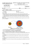
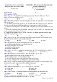
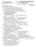
Figure 1. Internalization model of denatured protein into
cells.
(a) The positively charged protein transduction domain
makes contact with the negatively charged outer
membrane; (b) The protein translocates through the
membrane in an unfolded state; (c) Once inside the cell,
members of HSP 90 family refold the protein into an active
conformation (S.Dowdy 2000).
Runx3 is a member of Runx family that are critical
regulators in inducing new tissues or determining cell fate.
Runx 3 is expressed in normal gastrointestinal epithelial
cells and has been suggested as a tumour suppressor
involved in gastric cancer. The molecular mechanism of
Runx3 activity, however, is still poorly understood. In this
paper, we synthesized a cell-permeable Runx3 terminal
peptide for future study. By fusing the peptide with Tat
transduction domain, we tried transducing the peptide
under native conditions to preserve its structure and
function, despite its difficulty mentioned above. The fusion
protein has been applied into NIH/3T3 fibroblast and C2C12
myoblast cell lines.
II. Materials and Methods.
1. Plasmid construction.
The 90bp RUNX3 cDNA fragment from pT3-C41 was
amplified by PCR and ligated into peGFP-C1 treated with
XhoI and EcoRI. The eGFP cDNA and eGFP-RUNX3
fusion fragment were inserted into NcoI and EcoRI sites of
pTAT (given by Dr. Steven Dowdy) to generate the in-
frame expression constructs: pTAT-eGFP and pTAT-
eGFP-Runx3 term., respectively.
2. Expression and purification of Tat-eGFP, Tat-
eGFP-Runx3 term. fusion proteins.
Two constructs were transformed into BL21(DE3)LysS by heat shock (420C in 50 seconds). The expression of proteins
was induced by IPTG. The proteins were purified through
Ni-NTA column using native lysis, wash and elution
buffers (containing NaH2PO4, NaCl and imidazole, pH 8.0).
These purification buffers were made according to Qiagen's
instruction.
3. Invitro intracellular transduction assays.
The mouse C2C12 myoblasts and NIH/3T3 fibroblasts were
grown in DMEM medium supplemented with 20% (C2C12)
or 10% FBS (NIH/3T3).
Intracellular transduction assays were performed in 24-well
plates (NUNC). Cells treated with Tat fusion proteins at
various concentrations were quickly rinsed once in PBS
after different time intervals and mounted in PBS under
coverslips. The fluorescence was detected and recored with
fluorescent microscopy connecting with a computer that
possesses a micrograph-recording software.
III. Results and Discussion
1. Construction of expressing plasmids using pTAT
plasmid.
In order to amplify the 90bp fragment of RUNX3 terminal,
specific primers were designed based on human RUNX3
sequence from NCBI gene bank (Figure 2B). The primers
contain XhoI and EcoRI restriction sites to provide sites for
the ligation of RUNX3 into peGFP-C1 plasmid at next step.
The fusion of Runx3 with eGFP (enhanced green
fluoresence protein) made possible the live observation of
the transduction process, and is a sign that the protein's
function is preserved during transduction process.
Figure 2A shows the pTAT vector (~ 3kb) containing a T7
polymerase promoter, an ATG start codon, a 6 histidine
leader for Ni affinity purification, followed by the 11
amino-acid-TAT domain fused to the 5'-end of the HA tag.
For the constructs, either eGFP or eGFP-RUNX3 term. was
cloned into the multiple cloning site (NcoI/EcoRI)
downstream of the HA sequence. The pTAT-cDNA
plasmids were then transformed into DH5 bacterial strain
which yields a high-copy plasmid number. The individual
clones were isolated, the DNA sequence was confirmed by
automated DNA sequencing.
Figure 2. Plasmid construction.
A- pTAT vector; B- specific primers for making Runx3
term. expressing construct;
C- Two constructs that have been made.
2. Expression and purification of the Tat fusion
proteins under native conditions.
The constructed plasmids were transformed into high-
expressing BL21(DE3)LysS bacterial strain. The
expression was under the control of the IPTG-induced T7
promoter, in the presence of 1mM IPTG added to the
baterial culture.
Molecular weight of each protein is as follows: TAT-eGFP:
37 kDa; TAT-eGFP-Runx3 term.: 37.3 kDa. Purification of
the proteins through a Ni-NTA affinity column was
performed under native conditions (described above) to
preserve their structure and function before introduced into
the cells. The purification produced more than 95% pure
yields at a final concentration of 0.2 to 6.5 mg/ml. (Figure
3).
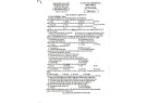
Figure 3. SDS-polyacrylamide gel electrophoresis analysis
of purified native proteins.
(a): Fractions collected during purification procedure: M:
protein marker; Ctrl: non-expressing bacteria; I(-): IPTG-
non induced bacteria; I(+): IPTG-induced bacteria; St:
crude bacterial lysate; Fl: flow-through; W1, W2: wash; E:
eluate; E¬¬¬de: desalted eluate. (b): Distinguishing
between pTAT-eGFP and pTAT-eGFP-Runx3 term.
Samples were applied on 15% SDS-polyacrylamide gel and
visualized by Coomassie blue staining.
3. Intracellular localization of the TAT-eGFP-Runx3
fusion protein in cultured cell lines.
To access the transduction capacity, we added the Tat
fusion protein to the cultured media of NIH/3T3 and C
cells at various concentrations of 10, 20, 50, 100, 190, 400
and 800ug/ml. The transduction process was observed
under fluorescent microscopy. Intracellular green
fluorescence was first detected in the cytoplasm of
NIH/3T3 when cells were incubated with the protein at
concentration of 190ug/ml in 12h. The number of cells
uptaking the protein and intracellular green fluorescence
were enhanced when the protein concentrations increased,
indicating a concentration dependency for protein
transduction which is consistent with previous studies, and
thus, the ability to modulate intracellular concentration.
At the protein concentration of 400ug/ml, 100% of cells
showed green fluorescence. This demonstrates that the
highest efficiency of protein transduction may be reached at
the minimum concentration of 400 ug/ml. As a result, we
were able to optimize the appropriate concentration of
proteins to be applied efficiently for future experiments.
(Figure 4).
When we tested transduction assays on C2C12 mouse
myoblasts, transduction efficiency definitely fell down,
showing the dependency of transduction efficiency on cell
type. (Figure 5).
Figure 4: Intracellular delivery of Tat-eGFP-Runx3 term. in
NIH/3T3 fibroblasts.
Fluoresence micrographs of NIH/3T3 treated with Tat-
eGFP-Runx3 term. at (A) before transduced; (B) 190ug/ml;
(C) 400ug/ml or (D) 800ug/ml and corresponding phase-
constrast images (A', B', C' and D'). Cells were incubated
for 12h, quickly washed and then mounted in PBS before
examined.
Figure 5. Comparison of transduction efficiency between
NIH/3T3 fibroblasts and C2C12 myoblasts.
Fluoresence micrographs of NIH/3T3 treated with
800ug/ml of (A) Tat-eGFP or (C) Tat-eGFP-Runx3term.;
C2C12 treated with 800ug/ml of (B) Tat-eGFP or (D) Tat-
eGFP-Runx3term. Cells were incubated for 12h, quickly
washed and then mounted in PBS before examined.
IV. Conclusion.
1. Cell-permeable Runx3term. maining native structure
were synthesized.
2. Highest efficiency of transduction into NIH/3T3 was
obtained at the minimum protein concentration of
400ug/ml.
3. Transduction efficiency varies in two different cell
lines studied.
Acknowledgement.
The authors would like to thank Dr. Kwang-Youl Lee and
colleagues at Institute for Tumor Research, Chungbuk
National University for their useful help and suggestions.
Main references:
1. Green, M. and Loewenstein, P.M. (1988). "Autonomous
functional domains of chemically synthesized human
immunodeficiency virus TAT trans-activator protein." Cell
55: 1179-1188.
2. Frankel, A.D. and Pabo, C.O. (1988). "Cellular uptake of
the TAT protein from human immunodeficiency virus."
Cell 55: 1189-1193.
3. Paul A. Wender, D.J.M., Kanaka Pattabiraman, Erin T.
Pelkey, Lawrence Steinman (2000). "The design, synthesis,
and evaluation of molecules that enable or enhance cellular
uptake: peptoid molecular transporter." PNAS 97: 13003-
13008.
4. Michelle Becker-Hapak, S.S.M, S.F.D. (2001). "TAT-
mediated protein transduction into mammalian cells".
Methods 24: 247.
5. Neris Bonifaci, R.S., Anna Rubartellie (1995). "Nuclear
translocation of an exogenous fusion protein containing
HIV Tat requires unfolding." AIDS 9: 995-1000.
6. Christine Schneider, L. S.-L., Elmar Nimmesgern,
Ouathek Ouerfelli, Samuel Danishefsky, Neal Rosen, F.
Ulrich Hartl (1996). "Pharmacologic shifting of a balance
between protein refolding and degradation mediated by
Hsp90". Proc. Natl. Acad. Sci. USA 93: 14536-14541.
oa học: GS. Lê Đình Lương.