
JOURNAL OF MEDICAL
CASE REPORTS
Revision of a nonunited subtrochanteric femoral fracture
around a failed intramedullary nail with the use of RIA
products, BMP-7 and hydroxyapatite: a case report
Tzioupis et al.
Tzioupis et al.Journal of Medical Case Reports 2011, 5:87
http://www.jmedicalcasereports.com/content/5/1/87 (1 March 2011)

CAS E REP O R T Open Access
Revision of a nonunited subtrochanteric femoral
fracture around a failed intramedullary nail with
the use of RIA products, BMP-7 and
hydroxyapatite: a case report
Christopher Tzioupis
1
, Pavlos Panteliadis
1
, Zakareya Gamie
1
, Eleftherios Tsiridis
1,2*
Abstract
Introduction: Femoral subtrochanteric fractures are commonly treated using intramedullary devices. Failure of the
implant and subsequent nonunion is still an issue, however, and limited evidence exists regarding the most
appropriate treatment.
Case presentation: We report the case of an 80-year-old Caucasian woman with a subtrochanteric fracture
originally treated using a trochanteric gamma nail which failed, resulting in a nonunion and fracture of its proximal
end. The nonunion was revised with the removal of the broken trochanteric gamma nail, application of a condylar
blade plate, ipsilateral Reamer/Irrigator/Aspirator autografting, recombinant human bone morphogenetic protein-7
and injectable hydroxyapatite cement. The fracture united fully at ten months following revision surgery, with no
signs of femoral head avascular necrosis at 18-month follow-up.
Conclusion: The essential requirements for success when revising a nonunited fracture are to provide anatomical
reduction, mechanical stability, bone defect augmentation and biological stimulation to achieve healing. Current
advances in molecular biology, such as recombinant human bone morphogenetic protein-7, and biotechnology
such as the Reamer/Irrigator/Aspirator system and hydroxyapatite injectable cement can improve patient outcomes
over the use of our traditional revision techniques.
Introduction
Most fractures of the subtrochanteric region of the
femur heal when treated using contemporary methods
of internal fixation [1]. Improved understanding of the
biomechanics of this region has shifted treatment
toward the use of intramedullary devices (IMD) as the
shorter-levered arm on the proximal fixation results in
greater load sharing and less bending movement across
thefractureandimplant[2,3],reducingtherateof
implant failure [2,4]. The overall incidence of failure for
any type of fixation and subsequent nonunion of subtro-
chanteric fractures varies from 7% to 20% [5]. Complica-
tions occur mainly in patients with poor bone quality,
unfavorable fracture patterns and suboptimal positioning
of the fixation implant [1,5]. IMD complications include
femoral shaft fracture below the tip of the IMD, collapse
of the fracture and cutting out of the femoral neck
screw, for which reoperation is required [6]. For extra-
medullary devices such as the sliding hip screw or the
dynamic condylar screw, failure often occurs following
screw cutout [2,3].
There is limited evidence regarding the most appro-
priate method of treating a nonunion of a subtrochan-
teric fracture [1,3]. Debridement of fibrous tissue,
correction of varus malalignment, autografting and frac-
ture compression are essential to achieve union [5]. It
has been reported that subtrochanteric nonunions trea-
ted with the condylar blade plate (CBP) are associated
with good healing rates [1,5]. Autograft harvesting from
the iliac crest, however, is related to comorbidities [7],
increasing the need for autograft substitution. The
* Correspondence: etsiridis@doctors.org.uk
1
Academic Department of Trauma and Orthopaedics, School of Medicine,
University of Leeds, Leeds General Infirmary, Leeds Teaching Hospitals NHS
Trust, Clarendon Wing A, Great George Street, Leeds, LS1 3EX, UK
Full list of author information is available at the end of the article
Tzioupis et al.Journal of Medical Case Reports 2011, 5:87
http://www.jmedicalcasereports.com/content/5/1/87 JOURNAL OF MEDICAL
CASE REPORTS
© 2011 Tzioupis et al; licensee BioMed Central Ltd. This is an Open Access article distributed under the terms of the Creative Commons
Attribution License (http://creativecommons.org/licenses/by/2.0), which permits unrestricted use, distribution, and reproduction in
any medium, provided the original work is properly cited.
Reamer/Irrigator/Aspirator (RIA) system (Synthes North
America, Inc., West Chester, PA, USA) is a recently
developed device used to perform corticocancellous
intramedullary autografts containing human mesenchy-
mal stem cells (hMSCs) to stimulate bone healing [8].
In addition, recombinant human bone morphogenetic
protein-7 (rhBMP-7) has been introduced with success
for the treatment of nonunions [9]. Biocompatible mate-
rials such as hydroxyapatite (HA) have also been tested
in combination with rhBMP-7 in vivo to induce osteo-
genic differentiation of hMSCs [10]. We report the case
of a patient with a subtrochanteric fracture originally
treated using a trochanteric gamma nail (TGN)
(Gamma 3 IM nailing system; Stryker Biotech, Hopkin-
ton, MA, USA) which failed and resulted in a nonunion
and fracture of the proximal end of the TGN device.
The nonunion was revised with the removal of the
broken TGN, application of a CBP, ipsilateral RIA auto-
grafting, and use of BMP-7 and HA injectable cement,
with success and healing achieved at 10 months follow-
ing revision surgery.
Case presentation
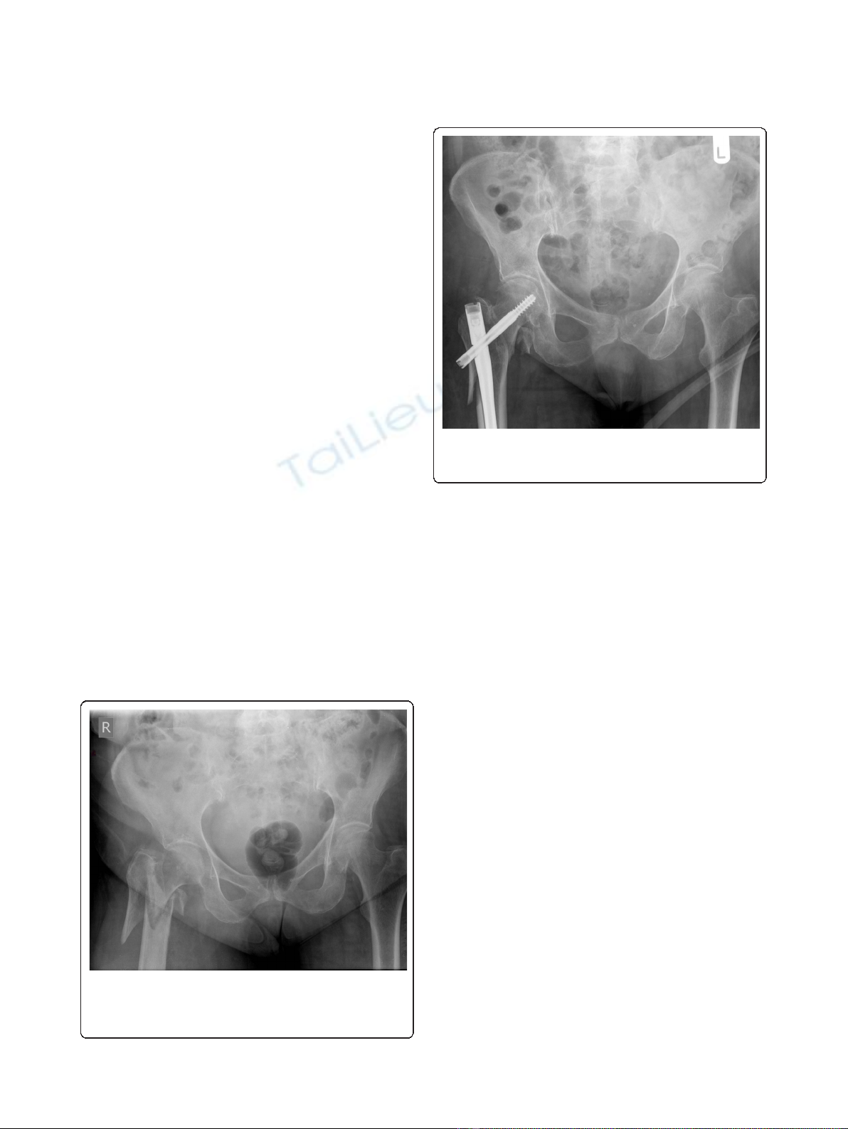
An 80-year-old Caucasian woman sustained a right sub-
trochanteric femoral fracturefollowingadomesticfall,
classified according to the AO Foundation (AO)/Ortho-
paedic Trauma Association (OTA) fracture classification
system as 31-A3.3 (Figure 1). The fracture was reduced
and stabilized with a TGN (Figure 2). The patient had
an uncomplicated recovery and was discharged to home.
After three months, the patient reported pain on ambu-
lation, and radiographs failed to demonstrate sufficient
callus formation. Subsequent radiographs obtained at
four and six months revealed delayed union; therefore,
the nail was dynamized by removing the two distal lock-
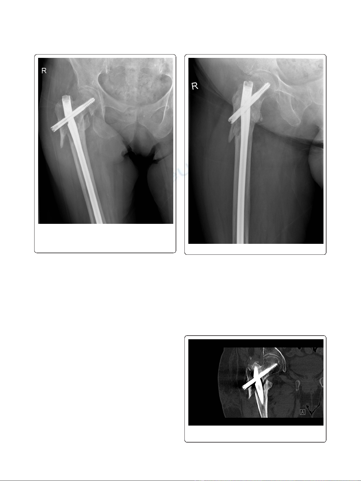
ing screws to promote union. At 10 months following
revision surgery, the patient’s pain had increased, mak-
ing her unable to bear weight, and at that time a further
radiograph revealed failure of the TGN with fracture of
the proximal end of the nail, nonunion of the fracture
site and varus deformity of the proximal femur (Figures 3
and 4). A computed tomographic scan confirmed the
diagnosis of nonunion (Figure 5), and revision surgery
was planned to remove the failed TGN and to stabilize
the fracture with an extramedullary device and graft.
The patient was placed in a lateral decubitus position
without traction on a radiolucent table. Four hundred
milligrams of teicoplanin were administered preopera-
tively according to the standard antibiotic prophylaxis
protocol for revision trauma surgery at our institution.
The old incision was incorporated and extended distally
into a straight lateral approach to the femur with the
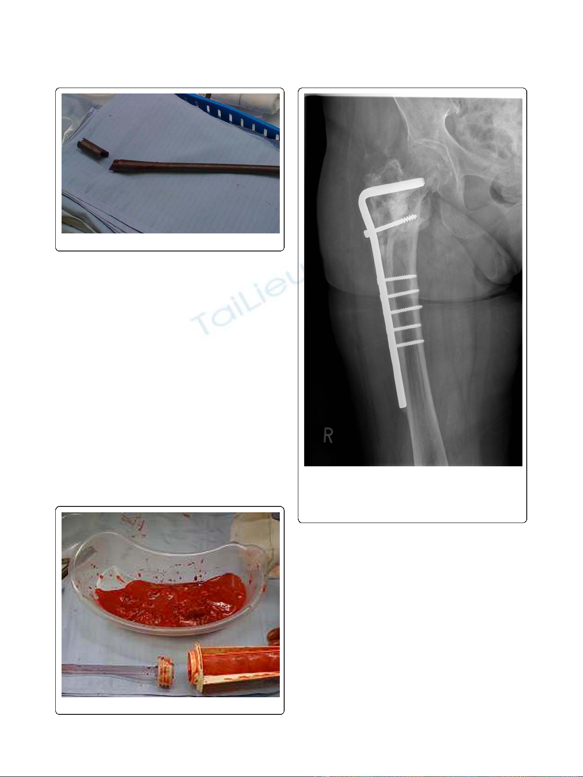
fracture site fully exposed. The broken TGN was
removed through the fracture site, and the fibrous non-
union tissue was taken out until bleeding bone was
exposed (Figure 6). Care was taken to protect the vascu-
larsupplytothefracturesitebyminimalmusclestrip-
ping. Six tissue samples were sent for microbiological
testing to exclude infection according to revision surgery
protocol. The fracture was then aligned over an intrame-
dullary guidewire for reaming. The RIA reamers were
used to ream and irrigate the endosteal bone-implant
interface, and thereafter intramedullary corticocancellous
Figure 1 Anteroposterior radiograph of the pelvis demonstrating
a right subtrochanteric femoral fracture classified as 31-A3.3
under the AO Foundation (AO)/Orthopaedic Trauma Association
(OTA) fracture classification system.
Figure 2 Anteroposterior radiograph demonstrating reduction
and stabilization of the fracture with a trochanteric gamma
nail (TGN).
Tzioupis et al.Journal of Medical Case Reports 2011, 5:87
http://www.jmedicalcasereports.com/content/5/1/87
Page 3 of 7
reaming autograft was collected following the standard
RIA protocol (Figure 7). Reduction forceps were then
used to accurately reduce the fracture in the desired ana-
tomical position, and guidewires were placed to deter-
mine the direction and starting point for the CBP
insertion. A 90° CBP was inserted, restoring the proper
shaft-neck hip angle compared to the contralateral site
(Figures 8 and 9). Prior to CBP insertion, the femoral
neck was filled with injectable HA cement (BoneSource
BVF; Stryker Biotech) to fill the void created by the
removal of the proximal TGN screw and augment its
mechanical strength. The RIA autograft was mixed with
the rhBMP-7 implant (Stryker Biotech) and added onto
the fracture site.
Postoperatively, the patient was administered low-
molecular-weight heparin prophylaxis for six weeks. Par-
tial weight bearing was commenced from the second
postoperative week onward as dictated by the patient’s
tolerance of pain. Clinical and radiographic follow-up
was arranged at 6 weeks and 3, 6, 12 and 18 months.
The fracture united fully at 10 months following revi-
sion surgery, with no sign of femoral head avascular
necrosis at the 18-month follow-up examination. The
patient achieved a full range of hip movement, scoring
80 on the Charnely D’Aubigne Postel scale [11].
Discussion
There has been controversy in the literature regarding
the best type of implant for the fixation of subtrochan-
teric femoral fractures [2]. Both intramedullary and
extramedullary devices have been advocated for the man-
agement of subtrochanteric fractures [3]. Less favorable
results and implant failure occur in patients with osteo-
porotic bone, complex fracture patterns, suboptimal
Figure 3 Anteroposterior radiograph demonstrating failure of
the TGN with fracture of the proximal end of the nail,
nonunion of the fracture site and varus deformity of the
proximal femur.Figure 4 Lateral radiograph demonstrating failure of the TGN.
Figure 5 Coronal computed tomographic scan confirming the
diagnosis of nonunion at the fracture site.
Tzioupis et al.Journal of Medical Case Reports 2011, 5:87
http://www.jmedicalcasereports.com/content/5/1/87
Page 4 of 7
implant positioning, shaft medialization and varus malre-
duction, for which revision fixation may be recom-
mended [1,2,5,12]. The biomechanical advantages of
IMD are often diminished by suboptimal fracture reduc-
tion and false entry point prior to nail insertion [5]. The
incidence of neck screw cutout and fracture below the
nail was found to be 4% and 3.2%, respectively, for the
TGN nail in a comparison study with the proximal
femoral nail (PFN) [13]. The PFN was associated with
varus malreduction in 7.2% of patients and screw migra-
tion resulting in fracture collapse in 8% of patients; how-
ever, with a lower incidence of shaft fractures and neck
screw cut-out incidence, compared to TGN [13]. In a
prospective study comparing the success rate of TGNs,
PFNs and dynamic hip screws for unstable trochanteric
fractures, the TGN group had four failures in 40 patients
attributed to screw cutout and nonunion, which was
greater than the number of failures in the other groups
studied [6].
In a recent systematic review, pooled analysis of level I
studies suggested a nonsignificant lower risk of failure in
the IMD group compared with extramedullary devices
and no difference in the rate of nonunion [2]. Modes of
failure included femoral fracture in the IMD group and
screw cutout in the extramedullary device group.
Another frequent mode of failure in the dynamic condy-
lar screw (DCS) implant group was fracture of the plate
through the proximal screw hole due to inadequate
restoration of the medial calcar and fatigue loading of
the DCS implant [2]. It is therefore important to restore
the medial column to prevent cyclical loading of the
plate on the tension side of the femur and potentially
implant failure. This study also highlighted a lack of
agreement regarding the definition of a subtrochanteric
Figure 6 TGN with fracture of the proximal end of the nail.
Figure 7 Reamer/Irrigator/Aspirator aspirate.
Figure 8 Anteroposterior radiograph demonstrating the 90°
condylar blade plate (CBP) restoring the proper shaft-neck hip
angle and union of the fracture site at 10 months following
revision surgery with no signs of avascular necrosis of the
femoral head.
Tzioupis et al.Journal of Medical Case Reports 2011, 5:87
http://www.jmedicalcasereports.com/content/5/1/87
Page 5 of 7


![Báo cáo seminar chuyên ngành Công nghệ hóa học và thực phẩm [Mới nhất]](https://cdn.tailieu.vn/images/document/thumbnail/2025/20250711/hienkelvinzoi@gmail.com/135x160/47051752458701.jpg)


