
Cleavage site analysis of a serralysin-like protease, PrtA,
from an insect pathogen Photorhabdus luminescens and
development of a highly sensitive and specific substrate
Judit Marokha
´zi
1
, Nikolett Mihala
2
, Ferenc Hudecz
2,3
, Andra
´s Fodor
1
,La
´szlo
´Gra
´f
1,4
and
Istva
´n Venekei
1
1 Department of Biochemistry, Eo
¨tvo
¨s Lora
´nd University, Budapest, Hungary
2 Department of Organic Chemistry, Eo
¨tvo
¨s Lora
´nd University, Budapest, Hungary
3 Research Group of Peptide Chemistry, Hungarian Academy of Sciences, Budapest, Hungary
4 Biotechnology Research Group, Hungarian Academy of Sciences, Budapest, Hungary
Of the various enzymes that microorganisms secrete
for defence as well as for invasion and bioconversion
of their environment, proteases have the most diverse
functions. Exploration of the enzymatic properties
and functions of these proteases may contribute to a
better understanding of the pathomechanism and
gaining control over the infection process. Few
such proteases have been characterized enzymatically
and even less is known about their role in the patho-
mechanism.
Keywords
cleavage site; serralysin; specific substrate;
metalloprotease; PrtA of Photorhabdus
Correspondence
I. Venekei, Department of Biochemistry,
Eo
¨tvo
¨s Lora
´nd University, Budapest,
Pa
´zma
´ny Pe
´ter se
´ta
´ny, 1 ⁄C., 1117,
Hungary
Fax: +36 1 381 2172
Tel: +36 1 209 0555 ⁄8777
E-mail: venekei@cerberus.elte.hu
(Received 5 December 2006, revised 9
February 2007, accepted 12 February 2007)
doi:10.1111/j.1742-4658.2007.05739.x
The aim of this study was the development of a sensitive and specific
substrate for protease A (PrtA), a serralysin-like metzincin from the entomo-
pathogenic microorganism, Photorhabdus. First, cleavage of three biological
peptides, the A and B chains of insulin and b-lipotropin, and of 15 synthetic
peptides, was investigated. In the biological peptides, a preference for the
hydrophobic residues Ala, Leu and Val was observed at three substrate posi-
tions, P2, P1¢and P2¢. At these positions in the synthetic peptides the pre-
ferred residues were Val, Ala and Val, respectively. They contributed to the
efficiency of hydrolysis in the order P1¢>P2>P2¢. Six amino acids of the
synthetic peptides were sufficient to reach the maximum rate of hydrolysis, in
accordance with the ability of PrtA to cleave three amino acids from both
the N- and the C-terminus of some fragments of biological peptides. Using
the best synthetic peptide, a fluorescence-quenched substrate, N-(4-[4¢
(dimethylamino)phenylazo]benzoyl–EVYAVES)5-[(2-aminoethyl)amino]
naphthalene-1-sulfonic acid, was prepared. The 4·10
6
m
)1
Æs
)1
specificity
constant of PrtA (at K
m
5·10
)5
mand k
cat
2·10
2
s
)1
) on this sub-
strate was the highest activity for a serralysin-type enzyme, allowing precise
measurement of the effects of several inhibitors and pH on PrtA activity.
These showed the characteristics of a metalloenzyme and a wide range of
optimum pH, similar to other serralysins. PrtA activity could be measured in
biological samples (Photorhabdus-infected insect larvae) without interference
from other enzymes, which indicates that substrate selectivity is high towards
PrtA. The substrate sensitivity allowed early (14 h post infection) detection
of PrtA, which might indicate PrtA’s participation in the establishment of
infection and not only, as it has been supposed, in bioconversion.
Abbreviations
Dabcyl, N-(4-[4¢(dimethylamino)phenylazo]benzoyl; Dabcyl-OSu, N-(4-[4¢(dimethylamino)phenylazo]benzoyloxy)succinimide; Edans,
5-[(2-aminoethyl)amino]naphthalene-1-sulfonic acid; OpdA, oligopeptidase A; Php-C, Photorhabdus protease C; PrtA, protease A.
1946 FEBS Journal 274 (2007) 1946–1956 ª2007 The Authors Journal compilation ª2007 FEBS

The roles played by secreted proteases of two ento-
mopathogenic bacterium groups, Photorhabdus and
Xenorhabdus, might be of special interest because: (a)
Photorhabdus and Xenorhabdus strains are highly
pathogenic, and may serve as an excellent pathogen
component for an infection model; (b) in nature, sur-
vival of these bacteria is strictly dependent on their
symbiosis with entomopathogenic nematodes from the
families Heterorhabditidae and Steinernematidae,
respectively; and (c) bacterium–nematode complexes
might be exploited in environmentally friendly insect
biological control technologies. Secretion of three
proteases has been detected in Photorhabdus [1], which
is better characterized at the molecular level than
Xenorhabdus. Two of these, Photorhabdus protease C
(Php-C) and protease A (PrtA), were identified by
their sequences [1–3]; Php-C is a metallopeptidase
from the M4 (thermolysin) family, whereas PrtA (first
found in Erwinia chrysantemi), belongs to the 50 kDa
bacterial metallo-endopeptidases, the serralysins, a
subfamily of the interstitial collagenase family (M10).
The intensively studied proteases in the latter sub-
family, beside the 56 kDa metallo-endoprotease of
Serratia marcescens (serralysin), are the alkaline protei-
nase of Pseudomonas aeruginosa, the ZapA metallo-
protease of Proteus mirabilis and proetases A, B, C,
G and W of various Erwinia strains. One function of
these proteases is thought to be as virulence factors.
However, their contribution to pathogenesis cannot be
properly assessed because of a lack of information
about the dynamics of their production during infec-
tion and their proteolytic systems [comprising the
protease as well as its natural substrate(s) and inhibi-
tor(s)]. Several potential natural substrates have been
found for ZapA of P. mirabilis and the 56 kDa prote-
ase of S. marcescens (IgA and IgG proteins, some
defenesins, cytoskeletal proteins, complement system
components, extracellular matrix molecules) [4–10],
but the in vivo significance of cleavage of these pro-
teins remains to be established. According to sub-
strate-specificity studies on synthetic peptides,
serralysin, ZapA and alkaline proteinase exhibited
relaxed side-chain discrimination at substrate positions
P3–P3¢[11–15]. (The scissile bond is between the P1
and P1¢sites, Schechter and Berger’s notation [16].)
Consistent with this finding was the observation that
these enzymes cleaved (denatured) oligopeptide sub-
strates of biological origin at numerous sites in var-
ious sequence environments [8,12,17]. These properties
do not indicate proteases that have specific sets of
natural substrates, and make difficult the development
of selective and sensitive substrates for measuring
enzyme activity during infection. To date, the best
synthetic substrates for serralysin-like enzymes are
between six and eight amino acids long and contain
mostly hydrophobic P2 and P2¢residues [11–13,15].
Although both the relatively small number of peptide
sequence variants and their amino acid composition
limit the conclusions that can be drawn about side-
chain discrimination in these enzymes, some of the
kinetic data on these substrates seem interpretable by
the structure of the enzymes’ active site [18–21]. It is
also important to mention that the usability of these
substrates was not tested on biological samples.
For an exploration of the proteolytic system of
PrtA, and an understanding of its role in the infection
process of Photorhabdus, we needed a highly sensitive
and specific substrate to selectively measure activity in
biological samples. Here we describe the development
of such a substrate based on analysis of PrtA cleavage
site specificity, and kinetic characterization of PrtA
activity on the new substrate.
Results and Discussion
Identification of PrtA cleavage sites in biological
peptides
To obtain an initial view of the cleavage-site specificity
of PrtA, we analysed the sequence of PrtA hydrolysis
sites in three biological peptides, insulin A and B
chains, and b-lipotropin. We were able to draw two
conclusions from the data (Figs 1–3):
(a) Alignment of the cleavage sites (Fig. 3) showed a
preference for hydrophobic amino acids at substrate
positions P2, P1¢and P2¢, a property that is not
pronounced in the case of other serralysins of known
specificity. A simple probability analysis of amino acid
frequencies (not shown) indicated a slightly higher
frequency of Leu and Val at position P2¢, which is in
accordance with the presence of a conserved Leu
(Leu3, a position equivalent to P2¢) of the known bac-
terial inhibitors of serralysin-like proteases [20,22–24].
Because an even longer peptide inevitably samples only
a small fraction of all the possible sequence combina-
tions around potential cleavage sites (usually spanning
between six and eight amino acids) which might, addi-
tionally, be biased by the unique frequency of amino
acids in the peptide, the predictive power of such clea-
vage site analysis on (biological) peptides is restricted.
Nonetheless, from our results it could be concluded
that PrtA cleavage sequences are rich in the aliphatic
amino acids Ala, Leu and Val.
(b) From the dynamics of hydrolysis (estimated
from the change in the amount of some fragments)
(Figs 1A,2A), it was evident that most of the cleavage
J. Marokha
´zi et al. Substrate specificity of a serralysin-like enzyme
FEBS Journal 274 (2007) 1946–1956 ª2007 The Authors Journal compilation ª2007 FEBS 1947
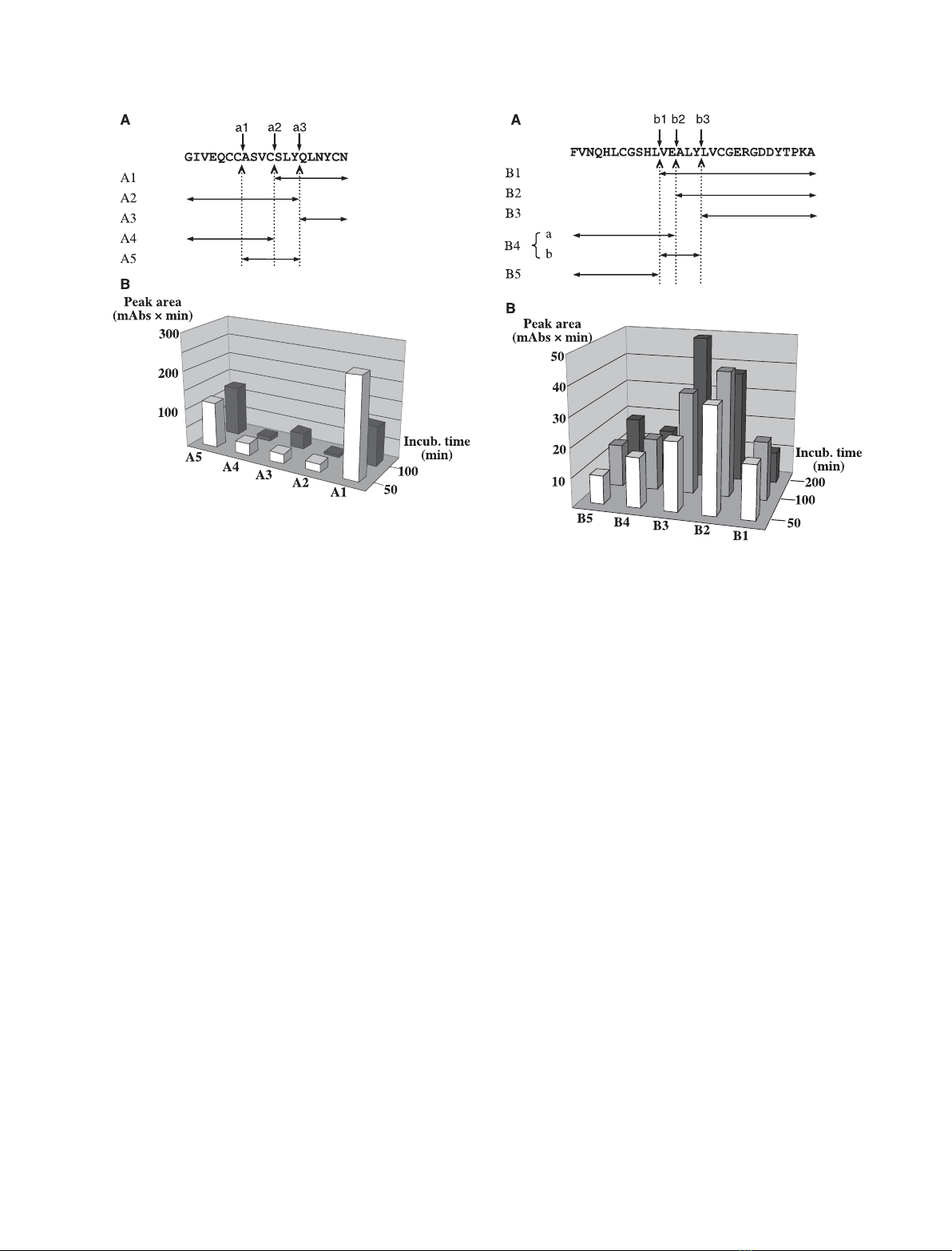
sites could serve as sites of secondary cleavage, even if
they were only three amino acids from either the C- or
the N-terminus. This suggests that PrtA might be able
to cleave peptides as short as six amino acids.
Optimization of peptide sequence and length
Supposing that hexapeptides were bound by PrtA such
that they span the S3–S3¢enzyme sites in an N- to
C-terminal (i.e. P3–P3¢) orientation and would be
cleaved between amino acids 3 and 4 (peptide positions
P1 and P1¢, respectively), the amino acids at positions
P2, P1¢and P2¢were selected for variation for the
following reasons:
(a) They are among the four inner sites (P2–P2¢) that
contribute most significantly to the proper positioning
of the scissile bond in almost every protease.
(b) We found that the side-chain discrimination of
PrtA is the most restricted in these positions, with a
preference for the aliphatic residues Ala, Leu and Val.
As for the three other positions, we took advantage
of the apparent relaxed side-chain preference of PrtA
to increase the solubility of the peptides (by choosing
Glu at positions P3 and P3¢), and Tyr at the (sup-
posed) P1 position, which rendered the peptide seg-
ment, N-terminal to the scissile bond, distinguishable
at 280 nm. Thus 12 hexapeptides (Pa1–Pa12) were syn-
thesized which contained, in every possible combina-
tion, each of the amino acids chosen to vary at
positions P2, P1¢and P2¢(Fig. 3).
The results of PrtA hydrolysis of the hexapeptide
library are summarized in Table 1 and Fig. 4. For each
peptide only two hydrolysis products were observed,
showing that they were cleaved at only one bond. With
the exception of Pa6 and Pa12 (see Experimental pro-
cedures and the legend to Table 1), identification of
the cleavage products and determination of the cleaved
bond were possible using only the retention times
(Table 1). One of the products always absorbed at
280 nm, which identified it as an N-terminal (Tyr-
containing) one. There were only two retention times
(either 26.2 or 28.8 min), showing that the products
were variants of only two sequences. This was possible
only if the products differed at position P2, i.e. if the
Fig. 2. Cleavage site analysis of PrtA on oxidized insulin chain B.
(A) The position of cleavage sites (vertical arrows, b1–b3) and clea-
vage fragments (horizontal double arrows, B1–B5) in the sequence
of insulin chain B. (B) Change over time in the chromatographic
peak area of cleavage fragments. Note, that fragments B1, B2 and
B4 show a temporary accumulation. Fragments B4a and B4b did
not separate under the applied conditions of reverse-phase HPLC.
(For details see Experimental procedures.)
Fig. 1. Cleavage site analysis of PrtA on oxidized insulin chain A.
(A) The position of cleavage sites (vertical arrows, a1–a3) and clea-
vage fragments (horizontal double arrows, A1–A5) in the sequence
of insulin chain A. (B) Change over time in the chromatographic
peak area of cleavage fragments. Note that the amount of frag-
ments A1, A2 and A4 decreases on longer exposure to PrtA clea-
vage. (For details see Experimental procedures.)
Substrate specificity of a serralysin-like enzyme J. Marokha
´zi et al.
1948 FEBS Journal 274 (2007) 1946–1956 ª2007 The Authors Journal compilation ª2007 FEBS
P1–P1¢peptide bond (on the C-terminal side of Tyr)
was cleaved in each case. The same conclusion could
be reached for the cleavage of these peptides if the
retention times of C-terminal hydrolysis fragments and
the possible sequences were coupled.
When library peptides were ranked in the order of
degree of hydrolysis (Fig. 4), groups and subgroups
became evident depending on the amino acid at
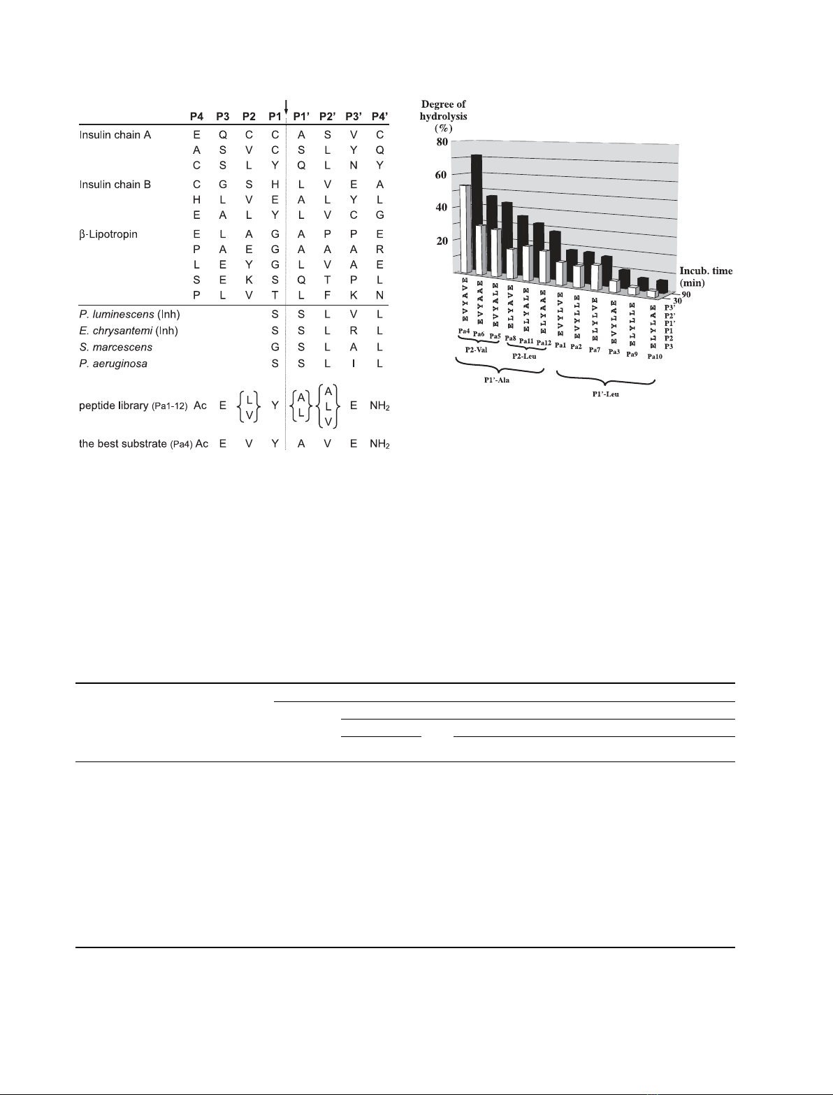
Fig. 3. Alignment of PrtA cleavage sites in three biological peptides
and the N-terminal (inhibitory) peptide segment of four inhibitors of
serralysin-type enzymes. The sequence variants of the synthetic
hexapeptide library (Pa1–Pa12) are also shown aligned in the expec-
ted and observed cleavage positions (indicated with a dashed line
and a vertical arrow). Inh, is a PrtA inhibitor from Photorhabdus.
Table 1. Reverse-phase HPLC analysis of cleavage of the hexapeptide library. nd, not detectable under the chromatographic conditions
used.
Substrates
Substrate position
3211¢2¢3¢
Retention times (min)
Peptide
Products
P3–P1 P1¢–P3¢
EVY ELY LVE LLE LAE ALE AVE AAE
Pa1 Ac-EVYLVE-NH
2
32.1 26.2 22.0
Pa2 Ac-EVYLLE-NH
2
34.6 26.2 25.0
Pa3 Ac-EVYLAE-NH
2
30.6 26.2 20.2
Pa4 Ac-EVYAVE-NH
2
28.2 26.2 nd
Pa5 Ac-EVYALE-NH
2
30.9 26.2 21.0
Pa6 Ac-EVYAAE-NH
2
25.5
a
25.1
a
nd
Pa7 Ac-ELYLVE-NH
2
34.0 28.9 22.0
Pa8 Ac-ELYAVE-NH
2
30.4 28.9 nd
Pa9 Ac-ELYLLE-NH
2
36.3 28.9 25.0
Pa10 Ac-ELYLAE-NH
2
32.3 28.9 20.2
Pa11 Ac-ELYALE-NH
2
32.8 28.9 21.0
Pa12 Ac-ELYAAE-NH
2
28.4
a
28.8
a
nd
a
Retention times of hydrolysis fragments of these peptides are not comparable with those of the others because different chromatography
conditions had to be applied (see Experimental procedures).
Fig. 4. Variants of the hexapeptide library ranked by the degree of
hydrolysis. The ranking is according to the degree of peptide hydro-
lysis after 90 min incubation at 0.25 mMpeptide and 0.36 nMPrtA
concentrations. Links indicate groups (P1¢Ala or Leu) and
subgroups (P2 Leu or Val). (For further details see Experimental
procedures.)
J. Marokha
´zi et al. Substrate specificity of a serralysin-like enzyme
FEBS Journal 274 (2007) 1946–1956 ª2007 The Authors Journal compilation ª2007 FEBS 1949

positions P1¢and P2, respectively. This allowed assess-
ment of the contribution the three positions and their
amino acids made to hydrolysis efficacy. Also, within
the limits of the library sequence set, it provided infor-
mation about the preferred cleavage site sequence. For
example, each of the first six, best cleaved, peptides
have Ala at the P1¢site (P1¢-Ala group), whereas each
of the three best substrates within this group have Val
at the P2 site (P2-Val subgroup). Analysis of the data
in Fig. 4 suggests that if P1¢is Ala then Val is better
than Leu at the P2 position, regardless of the amino
acid at position P2¢. This preference for Val over Leu
at the P2 site can also be seen in the P1¢-Leu group,
but here, the fact that Val is the best residue at the P2¢
site has some influence on the preferred residue at P2
(peptide Pa7 is better than Pa3). Thus, of the three
positions varied in our hexaeptide library, the contri-
bution of P1¢to cleavage efficacy is the strongest and
that of P2¢is the weakest, with an Ala, Val and Val
preference at positions P1¢, P2 and P2¢, respectively.
Of the 14 residues at sites S1–S3¢that contact the
inhibitor in the crystal structure of inhibitor enzyme
complexes of serralysin and alkaline protease, only
three differ in PrtA: Ser132, Tyr133 and Phe217, but
only the latter two appear to be significant. (These are
Gln ⁄Ala and Trp, respectively, in other serralysins, ser-
ralysin numbering.) Because these positions are
involved mainly in formation of the S1¢and S2¢sites
[20,25], in PrtA the differences may cause an increase
in hydrophobicity and some reshaping at these sites.
This may explain the higher preference of PrtA for ali-
phatic segments in biological peptides, and the prefer-
ence for Val over Leu at the P2¢substrate position,
relative to other serralysins [11–13,15].
Because the best peptide, Pa4, was cleaved almost
twice as fast as the second best (Pa6), we chose Pa4 to
construct a chromogenic substrate. Keeping its
sequence, we made extensions to the C-terminus by the
addition of one (Ser or Tyr) or two (Ser–Tyr) amino
acids to examine the effect of a longer peptide chain
on cleavage. Neither extension influenced the rate of
hydrolysis (data not shown) indicating that PrtA is
able to cleave three amino acids from the peptide ends,
and also that a length of six amino acids is enough for
efficient substrate binding and hydrolysis.
It was evident from the peptide hydrolysis that for
efficient cleavage PrtA requires interactions with the
substrate on both sides of the scissile bond. To allow
all such interactions to form, we designed a fluores-
cence-quenched substrate. Linkage of a quencher and
a fluorophore to Pa4 hexapeptide would have been
their closest positioning, ensuring the most efficient
fluorescence quenching, and thereby the highest
possible sensitivity of activity measurement. However,
to reduce the possibility of interference of the chro-
mophores with binding of the peptide to the enzyme,
which could not be excluded in this case and
might have compromised the specificity of the substrate,
we conjugated the quencher N-(4-[4¢(dimethylamino)
phenylazo]benzoyl (Dabcyl) and the fluorophore 5-[(2-
aminoethyl)amino]naphthalene-1-sulfonic acid (Edans)
to one of the extended forms of Pa4 hexapeptide, and
prepared the Dabcyl–EVYAVES–Edans substrate.
When PrtA hydrolysis of this substrate was followed
using HPLC and mass spectrometry (see Experimental
procedures), it was found that conjugation of the quen-
cher and the fluorophore influenced neither the rate nor
the site of hydrolysis of the peptide.
Sensitivity and selectivity of the Dabcyl–
EVYAVES–Edans substrate and the activity
of PrtA
After determining the optimal excitation and emission
wavelengths, the molar fluorescence value and the
calibration of the inner filter effect (see Experimental
procedures), the kinetic parameters of four PrtA
preparations (the two isoforms, PrtAi and PrtAii, their
mixture and the recombinant form of PrtA) were
determined along with those of several other enzy-
mes (Table 2). The PrtA preparations exhibited app-
roximately the same, high-specificity constants
(2.3 ·10
6
m
)1
Æs
)1
), which were one order of magni-
tude higher than the highest constant for a serralysin-
like enzyme measured to date (ZapA of P. mirabilis)
[14], and 100-fold higher than the specificity constants
Table 2. Kinetic parameters of PrtA and comparison of the specific
activity of PrtA to several other enzymes on Dabcyl–EVYAVES–
Edans substrate.
k
cat
(·10
2
s
)1
)
K
M
(·10
)5
M)
k
cat
⁄K
M
(·10
6
s
–
1ÆM
)1
)
Substrate
specificity
a
PrtA
b
2.10 ± 0.3 9.0 ± 0.2 2.34 1.00
PrtAi 1.67 ± 0.3 7.0 ± 0.3 2.39 –
PrtAii 1.27 ± 0.2 5.0 ± 0.1 2.54 –
Recombinant
PrtA
2.30 ± 0.6 11.0 ± 4.0 2.09 –
OpdA 0.023 0.01
Php-C 0.024 0.01
Clostridium
collagenase
0.0044 0.002
Trypsin 0.0056 0.0024
Chymotrypsin 0.026 0.01
a
The specificity of the substrate was calculated as the ratio of spe-
cific activities of the different enzymes relative to PrtA.
b
A PrtA
preparation containing both PrtAi and PrtAii variants.
Substrate specificity of a serralysin-like enzyme J. Marokha
´zi et al.
1950 FEBS Journal 274 (2007) 1946–1956 ª2007 The Authors Journal compilation ª2007 FEBS


