* Corresponding author.
E-mail address: kanakapurabasavaiah@gmail.com (K. Basavaiah)
2018 Growing Science Ltd.
doi: 10.5267/j.ccl.2018.03.002
Current Chemistry Letters 7 (2018) 45–56
Contents lists available at GrowingScience
Current Chemistry Letters
homepage: www.GrowingScience.com
Spectrophotometric assay of pioglitazone hydrochloride using permanganate in
acidic and basic media
Kanakapura Basavaiaha* and Nagaraju Rajendraprasadb
aDepartment of Chemistry, University of Mysore, Manasagangothri, Myssuru-570 006, Karnataka, India
bPG Department of Chemistry, JSS College of Arts, Commerce & Science, B N Road, Mysuru-570 025, Karnataka, India
C H R O N I C L E A B S T R A C T
Article history:
Received December 22, 2017
Received in revised form
March 21, 2018
Accepted March 25, 2018
Available online
March 25, 2018
Pioglitazone hydrochloride (PGH) is an oral anti-hyperglycemic agent used in the treatment
of type-2 diabetes mellitus. Potassium permanganate was found to oxidize PGH both in acidic
and basic conditions, based on which two simple and sensitive methods were developed for its
determination in bulk sample and tablets, and validated. In the first method (indirect method),
PGH was reacted with a measured excess of standard permanganate in H2SO4 medium, and
the residual oxidant was determined by measuring its absorbance at 550 nm. The second
method (Direct method) entails treating PGH with permanganate in NaOH medium, followed
by the measurement of the resulting bluish-green manganite at 610 nm. Experimental variables
affecting the reactions were studied and optimized. Under optimum conditions, linear
relationships with good correlation coefficients were found between absorbance and
concentration in the ranges, 1.25 – 25 µg mL-1 (Indirect method) and 1-12 µg mL-1 (Direct
method) with respective molar absorptivity values of 1.10 × 10⁴ and 2.77 × 10⁴ l mol-1 cm-1.
The limits of detection (LOD) and quantification (LOQ) were 0.36 and 1.08 (Indirect method)
and 0.23 and 0.69 µg mL-1 (Direct method). Intra-day and inter-day precisions were
satisfactory, with %RSD values of ≤2.11, and the respective accuracies were excellent with
%RE values of ≤2. The methods were also validated for robustness, ruggedness and selectivity.
The methods were applied to the determination of PGH in its tablets with good accuracy and
precision, and no interference from the tablet additives was encountered. The results were also
compared with those obtained by a reference method.
© 2018 Growing Science Ltd. All rights reserved.
Keywords:
Pioglitazone
Determination
Permanganate
Spectrophotometry
Pharmaceuticals
1. Introduction
Pioglitazone hydrochloride (PGH), chemically known as 5-[[4-[2-(5-ethyl-2-pyridinyl) ethoxy]
phenyl] methyl]-2,4-thiazolidinedione monohydrochloride (Fig. 1),1 is an oral anti-hyperglycemic
agent.2It addresses the main pathophysiological defects, i.e., insulin resistance, and hence used alone
or in combination with insulin, metformin or sulphonyl ureas (glipizide and glibenclamide), as an agent
to treat diabetes.3
No monographs are available in any pharmacopeia for assay of this drug. Various analytical
methods developed for its determination in pharmaceuticals and biological samples have recently been

46
reviewed.
4
Various techniques such as uv-spectrophotometry,
5-23
potentiometry,
224-26
voltammetry,
27-28
high-performance liquid chromatography,
29-39
ultra-performance liquid chromatography,
40
high-
performance thin layer chromatography
41-46
and capillary electrophoresis
47
have been reported for the
determination of PGH in bulk sampleand tablets.
Fig. 1. Structure of PGH
Visible spectrophotometry is still the most sought-after technique in industrial quality control and
research laboratories because of its speed, cost-effectiveness, ease of performance, and fair selectivity
and sensitivity. Despite these advantages, technique has been scantily applied for the determination of
PGH, with only two reports being found in the open literature
9, 10
. PGH is reported to form ion-pair
complexes with acidic dyes: methyl orange and bromocresol green, in chloroform and these complexes
were measured at 267 and 297 nm, respectively, allowing its determination in 2.5 – 20 µg mL
-1
range.
9
In a similar method
10
using bromocresol green as ion-pair agent, the coloured species formed in
phthalate buffer of pH 2.4 was extracted into chloroform and absorbance measured at 419 nm. Beer’s
law was obeyed over 2.5 -14 µg mL
-1
concentration range and the method was applied to tablets. In the
first case,
9
absorbance is measured in the uv region where the interference from co-formulated
substances is expected to be high and the extractive method
9
has several disadvantages such as need
for pH adjustment, liquid-liquid extraction step and use of large quantities of organic solvents. Thus
arises, need for simple, facile and reliable visible spectrophotometric methods for the determination of
PGH in pharmaceuticals.
In the presence of reducing agent, potassium permanganate gets reduced to different oxidation states
in both acidic and basic media. In acidic solution, manganese(VII) is reduced to manganese(II), whereas
in basic medium, it gets reduced to manganese(VI) as shown below:
48
MnO
4-
+ 8H
+
+ 5e
-
= Mn
2+
+ 4H
2
O
MnO
4-
+ e
-
= MnO
42-
The innate intense purple color of permanganate solution which absorbs in the vicinity of 550 nm
and the bluish-green color of manganite ion
48
with a λ
max
at 610 nm, the reduced form of permanganate
in alkaline medium have been exploited for the spectrophotometric determination of many
pharmaceutical compounds.
49-60
In spite of its extensive application in pharmaceutical analysis,
permanganate, as per the literature, has not been used for the determination of PGH. In this work,
permanganate was used as an oxidimetric agent for developing two spectrophotometric methods. In the
Indirect method, the residual permanganate was measured at 550 nm, after allowing the reaction
between PGH and known amount of oxidant in H
2
SO
4
medium. Whereas in the direct method, the
bluish-green color of manganite, the product of reaction between drug and permanganate in alkaline
medium, was measured at 610 nm, which served as the basis of the Direct method. The methods were
found to be much simpler and more sensitive than the existing spectrophotometric methods.
2. Results and discussion
The proposed methods are based on the redox reaction between permanganate and PGH in acid
(Indirect method) or in basic (Direct method) medium. In Indirect method, a known excess of standard
KMnO
4
was added to PGH in acid medium followed by the determination of the residual oxidant by
measuring its absorbance at 550 nm. The decrease in absorbance at 550 nm with respect to water blank
was taken as the measure of PGH concentration. In Direct method, K
2
MnO
4
resulting from the
K. Basavaiah and N. Rajendraprasad / Current Chemistry Letters 7 (2018)
47
reduction of KMnO
4
by PGH in alkaline medium was measured at 610 nm and related to PGH
concentration. The possible reaction pathways and basis of assays are given in Scheme 1.
PGH + KMnO
4
H
+
OH
-
Oxidation product of PGH + Mn
2+
+ Unreacted KMnO
4
Absorbance measured at 550 nm
(Indirect method)
Oxidation product of PGH + MnO
42-
Bluish-green colour, measured at 610 nm
(Direct method)
Scheme 1. Reaction pathways and basis of determination
1.1. Optimization of experimental conditions
1.1.1. Indirect Method
Preliminary experiments were performed to determine the permanganate concentration which
would give a reasonable maximum absorbance at 550 nm in H
2
SO
4
medium; and this was found to be
60 μg mL
-1
. Hence, different concentrations of PGH were reacted with 1 mL of 600 μg mL
-1
KMnO
4
in acid medium, and after the elapsed contact time, the absorbance of the residual permanganate was
measured and related to PGH concentration. When a fixed concentration of KMnO
4
(60 μg mL
-1
) was
reacted with varying concentrations of PGH, the former was consumed in proportion to PGH
concentration and there occurred a concomitant fall in the concentration of KMnO
4
as shown by the
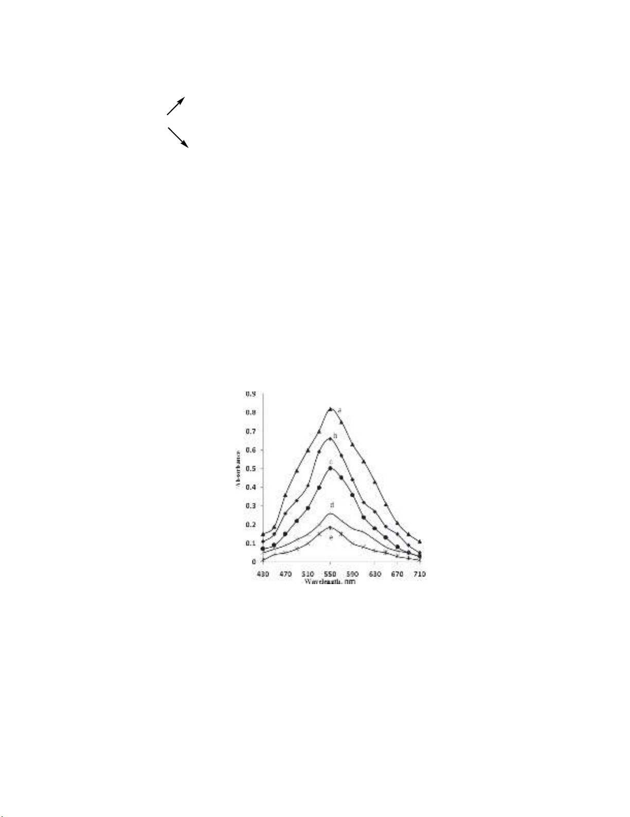
decreasing absorbance values at 550 nm with increase in the PGH concentration. This is depicted in
Fig. 2. This facilitated the evaluation of the linear range over which the method could be applicable to
the determination of PGH.
Fig. 2. Absorption spectra of KMnO
4
(60 μg mL
-1
) after reacting with PGH (μg mL
-1
):
a) 0.0, b) 5.0, c) 10.0, d) 17.5 and e) 20.0
Sulphuric acid is the most suitable acid, since it has no action upon permanganate in dilute solution.
With hydrochloric acid, there is the likelihood of the reaction taking place and some permanganate may
be consumed in the formation of chlorine.
61
Hence, the reaction of the oxidant with the drug was carried
out in H
2
SO
4
medium. Experiments were performed with 0.5-3.0 mL of 2 M H
2
SO
4
and it was found
that constant and reproducible absorbance readings were obtained in the range studied. Hence, 2 mL of
2M H
2
SO
4
in a total volume of 10 mL was fixed as the optimum. The redox reaction with 10 μg mL
-1
PGH was complete in 15 min, and the absorbance of residual oxidant remained constant for the next
45 min at room temperature (28±2 ᴼC).
48
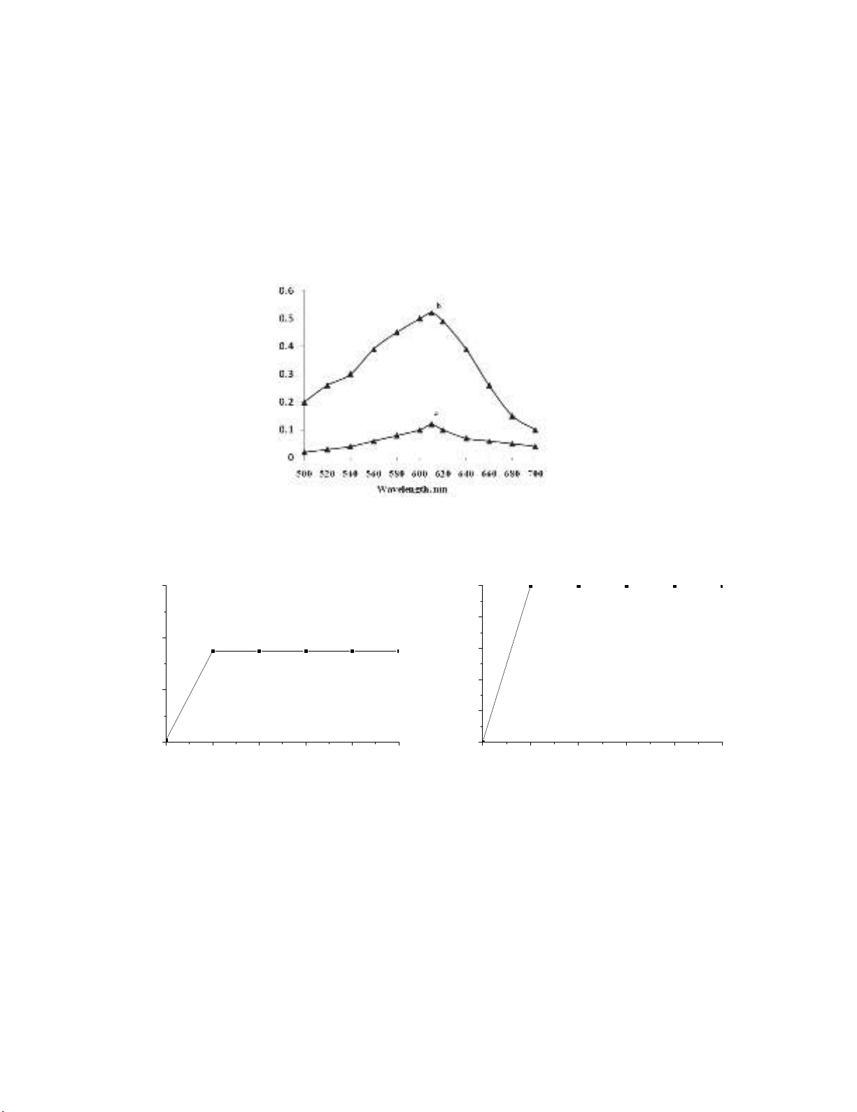
2.1.2. Direct method
This method is based on the reduction of permanganate to manganite by PGH in the presence of
NaOH, the bluish-green colored chromogen
62
having the absorption maximum at 610 nm (Fig. 3). The
formation of the colored product and the sensitivity of the reaction were found to be influenced by the
alkali and permanganate concentrations. Maximum and constant absorbance readings were observed
with 1 mL of 0.5M NaOH in a total volume of 10 mL (Fig. 4a). The reaction took 10 min for
completion, and the bluish-green manganite color was stable for 40 min thereafter (Fig 4b). When a
separate experiment was conducted to study the effect of permanganate concentration, it was found that
maximum absorbance associated with a minimum blank reading was obtained when 1 mL of 0.1%
KMnO
4
in a total volume of 10 mL was used. Higher concentrations of permanganate resulted in
increased sensitivity, but the blank absorbance showed an increasing trend simultaneously.
1-
uced for 6 μg mLcolor prod green-) bluishb) Blank, a: ofAbsorption spectra 3. .Fig
PGH, in Direct method
(a) (b)
Fig. 4. Effects of: a) NaOH concentration, b) reaction time and stability of colored species (6 μg mL
-1
PGH)
2.2. Method validation
2.2.1 Linearity, sensitivity, limits of detection and quantification
A linear correlation was found between absorbance at λmax and concentration of PGH (Fig. 5).
The slope (m), intercept (b) and correlation coefficient (r) for each system were evaluated using the
method of least squares. Optical characteristics such as Beer’s law limits, molar absorptivity and
012345
0.44
0.48
0.52
0.56
Absorbance
mL NaOH (0.5 M)
0 1020304050
0.0
0.1
0.2
0.3
0.4
0.5
Absorbance
Time, minutes
K. Basavaiah and N. Rajendraprasad / Current Chemistry Letters 7 (2018)
49
Sandell sensitivity values are presented in Table 1. The limits of detection (LOD) and quantitation
(LOQ) are also calculated according to ICH guidelines
63
and these data are presented in Table 1.
(a) (b)
Fig. 5. Calibration curves: a) Indirect method and b) Direct method
Table 1. Sensitivity and regression parameters
Parameter Indirect method Direct method
max
, nm 550 610
Linear range, µg mL
-1
1.25 – 25.0 1.0-12.0
Molar absorptivity(ε), L mol
-1
cm
-1
1.16 × 10
4
2.77
× 10
4
Sandell sensitivity
*
, µg cm
-2
0.0315 0.0128
Limit of detection (LOD), µg mL
-1
0.36 23
Limit of quantification (LOQ), µg mL
-1
1.08 0.69
Regression equation, y
*
Intercept (b) 0.821 0.0024
Slope (m) -0.0318 0.0804
Standard deviation of b (S
b
) 0.0017 0.0009
Standard deviation of m (S
m
) 7.52 × 10
-4
1.53 × 10
-3
Correlation coefficient (r) -0.9972 0.9991
*y=mx+b, where y is the absorbance, x concentration in μg mL
-1
, b intercept, m slope.
2.2.2. Precision and accuracy
To check the precision and accuracy of the proposed methods, the assays described under “general
procedures” were repeated seven times within the day (intra-day precision and accuracy) and five times
on five different days (inter-day precision and accuracy). These assays were performed at three levels
of analyte. The RSD values were ≤2.06% (intra-day) and ≤ 2.11% (inter-day) indicating high precision
of the methods. %RE values of ≤ 2.0% demonstrate the fair accuracy of the proposed methods. The
results of this study are summarized in Table 2.
Table 2. Results of accuracy and precision study
Method
PGH
taken,
μg mL
-1
Intra-day accuracy and
precision (n=7)
Inter-day accuracy and
precision (n=5)
PGH
found,
μg mL
-1
%RE %RSD
PGH
found,
μg mL
-1
%RE %RSD
Indirect
15
20
25
15.15
20.19
25.23
1.00
0.95
0.92
1.89
1.45
2.06
15.19
20.21
25.26
1.27
1.05
1.04
2.11
1.35
1.48
Direct
4
6
8
4.06
6.10
8.13
1.50
1.67
1.62
1.67
1.48
1.88
4.08
6.11
8.14
2.00
1.83
1.75
1.25
0.95
1.50
RE-Relative error, RSD-Relative standard deviation


![Hướng dẫn giải chi tiết bài tập phân li, phân li độc lập: Tài liệu [mới nhất]](https://cdn.tailieu.vn/images/document/thumbnail/2025/20251204/lethu2868@gmail.com/135x160/84711764814448.jpg)


![Bài tập Đa dạng thế giới sống [kèm đáp án/ hướng dẫn giải]](https://cdn.tailieu.vn/images/document/thumbnail/2025/20251123/thaohoang9203@gmail.com/135x160/5861763951302.jpg)