
RESEARCH Open Access
T cell subpopulations in lymph nodes may not
be predictive of patient outcome in
colorectal cancer
Roslyn A Kemp
1,5*
, Michael A Black
2
, John McCall
3
, Han-Seung Yoon
4
, Vicky Phillips
3
, Ahmad Anjomshoaa
1,6
and
Anthony E Reeve
1
Abstract
Background: The immune response has been proposed to be an important factor in determining patient
outcome in colorectal cancer (CRC). Previous studies have concentrated on characterizing T cell populations in the
primary tumour where T cells with regulatory effect (Foxp3+ Tregs) have been identified as both enhancing and
diminishing anti-tumour immune responses. No previous studies have characterized the T cell response in the
regional lymph nodes in CRC.
Methods: Immunohistochemistry was used to analyse CD4, CD8 or Foxp3+ T cell populations in the regional
lymph nodes of patients with stage II CRC (n = 31), with (n = 13) or without (n = 18) cancer recurrence after 5
years of follow up, to determine if the priming environment for anti-tumour immunity was associated with clinical
outcome.
Results: The proportions of CD4, CD8 or Foxp3+ cells in the lymph nodes varied widely between and within
patients, and there was no association between T cell populations and cancer recurrence or other
clinicopathological characteristics.
Conclusions: These data indicate that frequency of these T cell subsets in lymph nodes may not be a useful tool
for predicting patient outcome.
Background
Colorectal cancer is estimated to cause 639,000 deaths
world wide per year [1]. The prognosis following surgery
depends on disease stage, and this also determines the
need for additional treatment. However clinico-patholo-
gical stage characteristics alone provide imperfect prog-
nostic information. For example, approximately 25% of
patients with disease localised to the primary site (UICC
Stage I and II) relapse after surgery and may have bene-
fited from adjuvant therapy [2], whereas 25% of patients
with regional lymph node metastases (UICC Stage III)
are cured by surgery alone [3]. Various ways to improve
the prognostic accuracy of staging include increasing the
number of lymph nodes analysed [4,5], increasing the
sensitivity of the tests used to detect lymph node
metastases [6] and using microarray technology to ana-
lyse gene expression [7,8]. However these methods do
not take onto account potentially important host-related
factors such as the immune response.
Theimmuneresponsehaslong been associated with
eradication of tumours [9]. More recently, it has become
clear that T cells in the tumour are positively associated
with good patient prognosis [10,11] in colorectal cancer.
CD4 or CD8+ T cells expressing IFNg,ortheIFNg
inducing transcription factor Tbet, are the cells most
likely involved at the tumour site [12,13].
In immune responses to infection, the effector CD4
and CD8 T cell populations are held in check by a third
population of cells - regulatory T cells (Tregs). While
there are numerous subtypes of T cells with regulatory
function, the majority of suppressive function is
mediated by Foxp3+ CD4+ Tregs. As expected, low
numbers of these Foxp3+ Tregs have been associated
* Correspondence: roslyn.kemp@otago.ac.nz
1
Cancer Genetics Laboratory, University of Otago, Dunedin, New Zealand
Full list of author information is available at the end of the article
Kemp et al.Journal of Experimental & Clinical Cancer Research 2011, 30:78
http://www.jeccr.com/content/30/1/78
© 2011 Kemp et al; licensee BioMed Central Ltd. This is an Open Access article distributed under the terms of the Creative Commons
Attribution License (http://creativecommons.org/licenses/by/2.0), which permits unrestricted use, distribution, and reproduction in
any medium, provided the original work is properly cited.

with improved patient outcome in breast and colorectal
cancers [14-16]. However, some authors report an asso-
ciation between high numbers of Tregs and positive
patient outcome [17,18], although Salama et al found a
negative association between patient outcome and high
frequency of Tregs in the non-tumour associated tissue
[18]. More recently, Chaput et al identified a population
of CD8+Foxp3+ T cells in a cohort of colorectal cancer
patients that had suppressive activity and were proposed
to mediate tumour escape [19].
The immune response is initiated in the lymph nodes,
and although analyses of T cell subsets in the lymph
nodes of breast cancer patients have been performed
[20], the effect of these T cell subsets on colorectal can-
cer patient outcome had not been explored. We
hypothesised that the priming environment of an anti-
tumour immune response would be a useful predictor
of patient outcome. In this study we examined the
lymph nodes of Stage II colorectal cancer patients to
identify CD4+, CD8+ and Foxp3+ cell populations and
correlated these with patient outcome, alone, and in
combination with other clinico-pathological variables.
Methods
Patients
Patients with UICC stage II colon cancer were included
in this study. Stage II patients were chosen because they
have no tumour metastases in lymph nodes. The num-
ber of lymph nodes retrieved from patients for staging is
indicated in Table 1. Approximately 50% of the lymph
nodes obtained from each patient were randomly
selected for immunohistochemical analysis.
All patients underwent elective surgery for colon can-
cer at Dunedin Hospital, New Zealand. Pathological sta-
ging was verified by the study pathologist (HSY). In
addition to colon cancer, patients with inflammatory
boweldiseasewereusedascontrols.Thestudywas
approved by the Lower South Regional Ethics Commit-
tee and patients gave signed informed consent to parti-
cipate. All patients were prospectively followed up for a
minimum of five years from the date of surgery.
Immunohistochemical Analysis
Formalin fixed paraffin embedded (FFPE) lymph nodes
recovered at surgery were used for immunostaining. 4
um serial sections were stained for T cell markers using
two methods. Tonsil tissues were used as positive and
negative controls.
CD4 and CD8
Sections were dried for 30 min after cutting, then
dewaxed on the Bond™(Leica Microsystems, Germany)
after manual drying. Heat induced epitope retrieval was
performed using ER2 (Bond™)atpH9.0for20minat
100°C. After blocking with 3% peroxide block for 5 min,
the sections were incubated with the specific antibody
(anti-human CD4 (NCL-L-CD4-368; Novocastra, Leico
Microsystems; 1:40 dilution) or anti-human CD8 (NCL-
CD8-4B11; Novocastra, Leico Microsystems; 1:100 dilu-
tion)) for 20 min at RT. Unbound antibody was
removed by 3 washes in Bond™Wash Solution before
adding polymer for 10 min at RT. After washing
unbound labeled polymer in Bond™Wash Solution 3
times, peroxidase staining in tissue sections was revealed
by DAB solution (Bond™). After stopping the reaction
in running water, sections were counter-stained with a
rinse in hematoxylin solution. After dehydration, the
sections were mounted with DPX.
Foxp3
According to published methods[21],slideswereincu-
bated with rat anti-human Foxp3 antibody (clone
Table 1 Clinical characteristics of patients
CRC - recurrent CRC - non recurrent IBD controls
Number patients 13 18 9
Age (years, mean (SD)) 70.84 (8.922) 72.24 (11.032)
Gender %
M3928
F6172
Differentiation Poor 1 3
Moderate 11 14
Well 1 1
Tumour Site Right 8 13
Left 5 2
Rectum 0 1
Number lymph nodes used for staging (mean (SD)) 20 (12) 19 (8)
Number lymph nodes analysed (mean (SD)) 10 (6) 11 (8) 5 (3)
Kemp et al.Journal of Experimental & Clinical Cancer Research 2011, 30:78
http://www.jeccr.com/content/30/1/78
Page 2 of 7

PCH101, dilution 1:200, eBioscience, San Diego, CA) for
1 h at room temperature, followed by goat anti-rat anti-
body (dilution 1:50, Zymed) and ABC peroxidase detec-
tion system (Vector Vectastain ABC Elite kit, Vector
Laboratories, Burlingame, CA).
Between 1 and 33 lymph nodes per patient (Table 1)
were analysed with a Zeiss microscope (Carl Zeiss Co.,
Oberkochen, Germany) in their entirety to eliminate
regional variation due to the complex architecture of
lymph nodes. Each field was recorded using SpotOn
software (Brookvale, Australia) and CD4, CD8 and
Foxp3+ cells quantified using Image J software (NIH,
USA). Frequency of positively stained cells compared
with total cells was acquired for each field. All samples
were analysed in a double-blinded fashion.
Statistical analysis
Frequency counts of CD4, CD8 and Foxp3 stained cells
from each field were logged to reduce data skewness,
with an offset used to adjust zero counts. For each T-
cell marker the R statistical software [22] was used to fit
a linear mixed model to the logged count data, with a
fixed effect term used to represent clinical variables, and
random effects for patient number and lymph node. A
separate model was used for each of the available clini-
cal variables: (disease status, differentiation, lymphatic
invasion, margin, tumour site). In each model linear
contrasts were used to assess the presence of differences
in logged counts between each of the three disease sta-
tus groups for each T-cell marker. An identical
approach was taken in the analysis of log-ratio data for
pairs of T-cell markers (CD4:Foxp3, CD8:Foxp3), with
the log-ratios of counts derived using matched fields
from within each lymph node.
Results
Thirty three patients with stage II colon cancer were
included; 13 with and 18 without recurrence after 5
years of follow up. Of the 13 patients with recurrent dis-
ease, four recurred locally and nine had systemic disease
(seven liver, one lung, and one lung and brain). Patient
characteristics are summarised in Table 1. For each
patient, between 1 and 33 lymph nodes were available
for analysis (median = 10). Within each lymph node,
between one and 15 sections were examined for CD4,
CD8 and FoxP3 percentage (median = 10). For those
nodes for which multiple sections were available, the
“within-node”standard deviation was calculated to
assess the consistency of immunological signal being
obtained. Similarly, for those patients from whom multi-
ple lymph nodes were sampled, the “within-patient”(i.e.,
“between-node”for the same patient) standard deviation
was calculated. Finally the average immunological “sig-
nal “was calculated for each patient (for each of FoxP3,
CD8 and CD4) and used to assess inter-patient variabil-
itybydeterminingthe“between patient”standard
deviation.
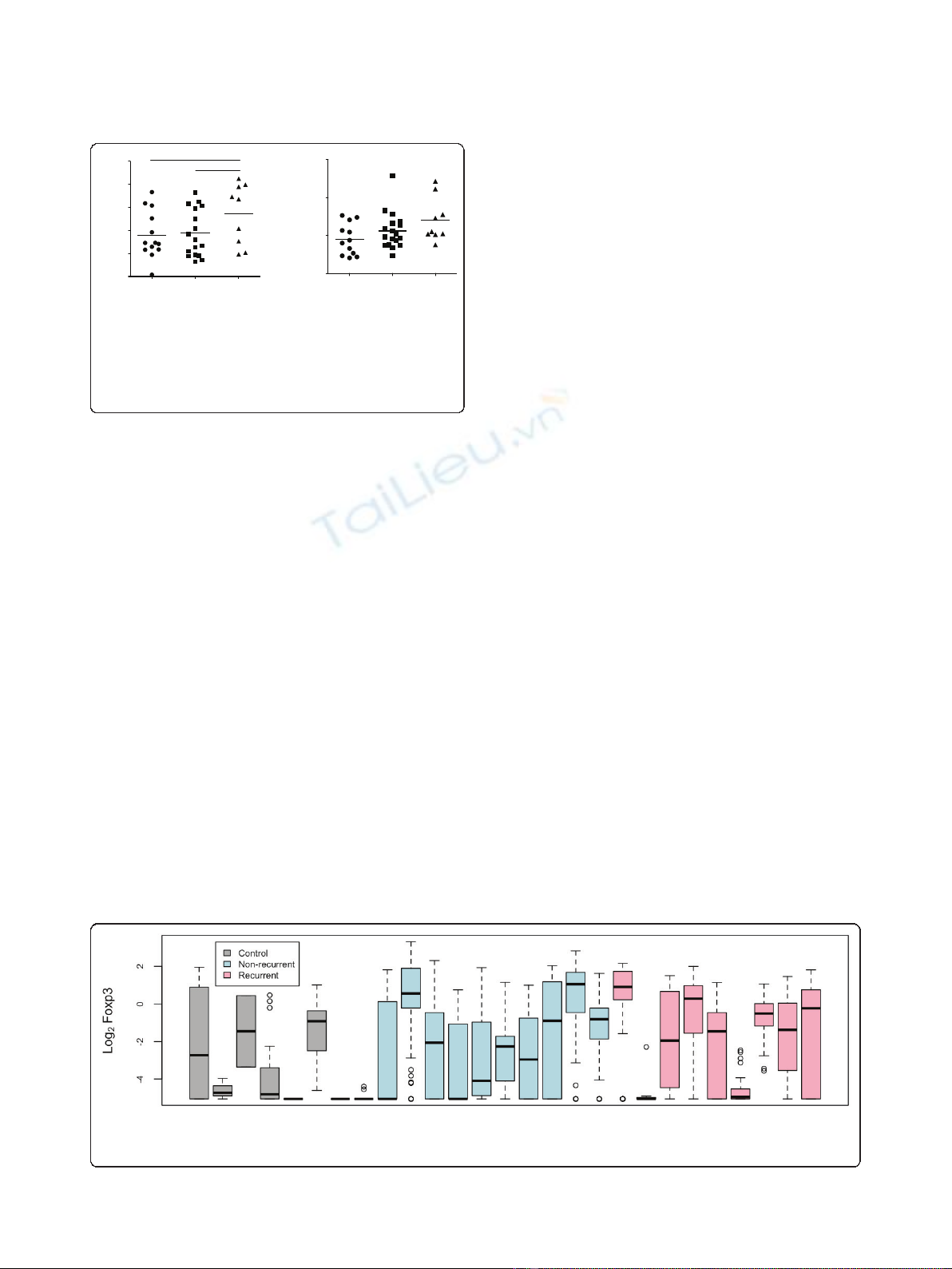
Figure 1 shows immunohistochemical staining for
CD4, CD8 and Foxp3 respectively. For all three mea-
sures of immunological activity (CD4, CD8 and FoxP3),
the within-node variability was around half the level of
the within-patient (between-node) variability (CD4:
5.81% vs 10.40%, CD8: 2.25% vs 4.24%, FoxP3: 0.24% vs
0.63%), indicating that replicate measurements obtained
from the same node were relatively consistent in all
cases. The same was not true, however, of nodes taken
from the same patient, with the between-node standard
deviation approximately the same as the between-patient
standard deviation for all three measures of immunolo-
gical activity (CD4: 10.40% vs 9.12%, CD8: 4.24% vs
4.15%, FoxP3: 0.63% vs 0.68%). That is, the variation in
CD4, CD8 and FoxP3 percentages between nodes from
the same patient was as great as the variation observed
from one patient to another.
Given the large amount of within-patient variability
that was observed across multiple lymph nodes from the
same patient, the task of identifying differences in
immunological activity between different groups of
patients could be expected to be very challenging, as is
reflected in the results presented below.
No association between T cell frequency in the lymph
nodes and patient outcome
There was no association between the frequency of
either CD4+ or CD8+ cells and cancer recurrence
(Figure2).Therewasadifferenceinthefrequencyof
CD4 cells in the inflammatory bowel disease control
Figure 1 Sections from representative regional lymph nodes
showing positive staining for CD4, CD8 or Foxp3. Lymph node
sections were stained for CD4 (A), CD8 (B) or Foxp3 (C) as outlined
in Materials and Methods. Foxp3 staining was optimised using tonsil
tissue - negative (D) and positive (E) control samples are shown.
Representative samples are shown.
Kemp et al.Journal of Experimental & Clinical Cancer Research 2011, 30:78
http://www.jeccr.com/content/30/1/78
Page 3 of 7
cohort (mesenteric lymph nodes from healthy controls
were unavailable). This was not unexpected given that
these patients have a chronic inflammatory disease that
involves CD4 T cells [23].
No association between Foxp3+ cells in the lymph nodes
and patient outcome
Although there was no difference in the percentage of T
cells between patients with and without cancer recur-
rence, it was possible a subpopulation of cells was asso-
ciated with disease. Because Tregs are important in
tumour immune responses, we analysed the frequency
of this cell population in the lymph nodes. Both CD4
and CD8 Tregs can express Foxp3 [15,19], and so we
used this marker to measure the frequency of Tregs in a
subset of patients from each group (control, recurrent
and non-recurrent) in Figure 2; these patients were
selected on availability of lymph node samples. No asso-
ciation was found between frequency of CD4+Foxp3+
or CD8+Foxp3+ cells and cancer patient outcome
(Figure 3). Furthermore, no association was found
between frequency of CD4+Foxp3+ or CD8+Foxp3+
cells in cancer patients and control IBD patients. This
last finding was interesting considering previous work
that suggests Tregs are decreased in IBD patients com-
pared to healthy controls [24]. It is possible that the
cancer patients are also presenting with an inflammatory
phenotype, but we were unable to make a comparison
with lymph nodes from healthy control subjects.
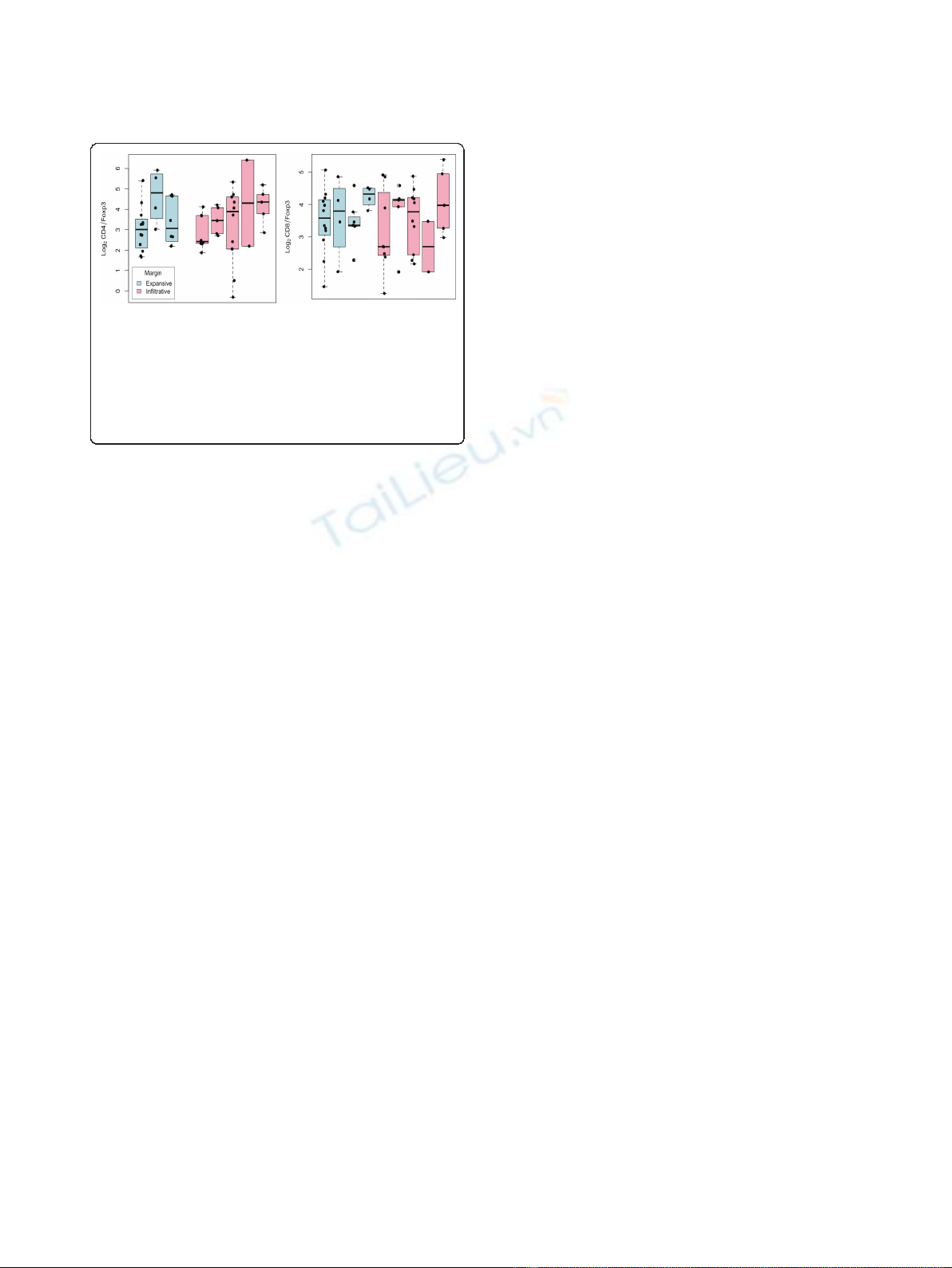
Association between T cell populations and other
clinico-pathological variables
The relationship between CD4, CD8 or Foxp3 positive
cells with clinico-pathological variables was examined
(differentiation, lymphatic invasion, tumour margin,
tumour site, vascular invasion). No significant associa-
tions between T cell subsets and these other variables
were identified (data not shown). However, it seemed
possible that the frequency of Foxp3 cells as a subset of
CD4+ or CD8+ cells could correlate with clinical para-
meters. Analysis of this ratio and tumour margin
showed no association (Figure 4).
Discussion
In this paper, we have described the analysis of T cell
populations in the lymph nodes of Stage II colorectal
cancer patients. We were unable to find any association
between CD4, CD8 or Foxp3+ (presumed Tregs) and
cancer recurrence or with other clinico-pathological
variables.
T cells have long been known to play a role in eradicat-
ing tumours. Colorectal cancer has been particularly well
studied, with several laboratories showing a positive asso-
ciation between patient survival and effector (IFNg+) T
cell infiltration into the tumour [10,11]. It was expected
that the regulatory T cell infiltration into the tumour
would be negatively associated with patient outcome;
however, regulatory (FoxP3+) T cells have been shown to
have a protective role in colorectal cancer, in contrast to
their negative role in many other cancers [17]. The posi-
tive effect of FoxP3+ T cells has been proposed to be a
result of their effects on other T cells that are promoting
tumour growth [25].
recurring non recurring control
0
10
20
30
40
50
*
**
% CD4+ cells
patient outcome
recurring non recurring control
0
10
20
30
% CD8+ cells
patient outcome
Figure 2 No association between CD4+ or CD8+ cells and
patient outcome. Between 1 and 20 lymph nodes per patient
(Table 1) were analysed for CD4 or CD8+ cells as indicated. Control
lymph nodes came from patients diagnosed with inflammatory
bowel disease. Data are represented as mean +/- SEM. * P = 0.095,
** p = .0669.
Figure 3 No association between Foxp3+ cells and patient outcome. Between 1 and 20 lymph nodes per patient (Table 1) were analysed
for Foxp3+ cells. Control lymph nodes came from patients diagnosed with inflammatory bowel disease. Data are represented as logged (base
two) cell counts, with each boxplot representing the distribution of mean log
2
Foxp3 cell counts for each lymph node of a single patient.
Kemp et al.Journal of Experimental & Clinical Cancer Research 2011, 30:78
http://www.jeccr.com/content/30/1/78
Page 4 of 7
T cell immune responses are initiated in the lymph
nodes by cells, such as dendritic cells, presenting
tumour antigens to responding specific T cells. These
activated T cells then migrate to the tumour and specifi-
cally destroy it. Munn et al proposed that the tumour
draining lymph node is a unique immunological envir-
onment where the presence of regulatory T cells could
mediate a suppressive effect on anti-tumour immune
responses [26]. Indeed, depletion of Tregs enhances
effector T cell responses in tumour draining lymph
nodes [27]. Recent data also indicated that the presence
of Foxp3+ T cells in tumour draining lymph nodes of
colorectal cancer patients correlated with disease pro-
gression [28]. Given the associations between Treg infil-
tration in primary colorectal tumours and patient
outcome [18], we questioned whether Tregs in the
regional lymph nodes could be predictive of patient
survival.
Our data is in contrast to Khort et al [20], who
described a population of CD4 cells in the axillary
lymph node could predict outcome in breast cancer
patients. Although our sample was smaller, there were
no apparent trends in the data to indicate that a larger
sample would be likely to yield significant results. In
fact, given the amount of variation in immunological
activity that we observed in lymph nodes taken from the
same patient, the use of lymph nodes for prognostic
purposes would seem to be extremely challenging. Even
if a difference in activation existed between patients
with “good”and “poor”prognosis, detection of a statisti-
cally significant difference would require collection of
large numbers of both patients and nodes. For per-
patient prognosis, the inter-node variability would make
accurate prediction almost impossible, with the good
and poor responders likely to be indistinguishable from
one another. This is likely due to the background of
non tumour-specific T cell overshadowing the presence
of tumour specific responses - indeed, the majority of
studies looking at T cells as predictors of outcome in
this disease have been restricted to the tumour tissue
[11,12,17,18,21,29].
We did not identify the sentinel nodes, which are
believed to be the primary priming site for the anti-
tumour immune response, however data exists to indi-
cate that there is often more than one sentinel node and
it’s spatial relationship to the tumour can vary consider-
ably [30].
Immunotherapy of cancer patients is difficult due to
the specific nature of the adaptive immune response
and the absence of easily identifiable tumour specific
antigens. The current study looked only at total T cell
populations in the lymph node, and it may be that
tumour specific T cell populations were present in dif-
ferent frequencies in patients with and without recur-
rence, but not able to be identified as such.
A further complication is the lack of healthy control
tissue. Studies comparing immune response in color-
ectal cancer patients have used blood of healthy
patients [14,15]; however the scope of our study was
to investigate the role of lymph nodes for predicting
patient outcome, and mesenteric lymph nodes from
healthy controls were not obtainable. We compro-
mised by using matching lymph node tissue from IBD
patients, as has been previouslypublished[15]butare
aware of the difficulties of using immune tissue from
patients with an immune mediated inflammatory
disease.
However, an interesting finding was the difference
between colorectal cancer patients and inflammatory
bowel disease patients with respect to CD4 expression.
IBD patients had a higher CD4 frequency that is not
surprising given the inflammatory nature of IBD and the
proven role for CD4 cells in driving this disease [23].
However, no difference was seen between cancer
patients and IBD patients in Foxp3+ cells. This indicates
that the Treg population was not diminished in IBD
patients, a finding in direct contrast to Clarke et al. We
are currently investigating this further to examine the
role of other T cell subpopulations.
Foxp3 is recognised as the most specific Treg marker;
however, there are reports of Foxp3 expression in effec-
tor T cells, especially in humans [31]. It is possible that
the Foxp3 cells detected in our study were effector
rather than regulatory cells. Studies are underway to
further characterise these cells, using a panel of regula-
tory markers. Clarke et al found that Foxp3+ cells
recovered from mesenteric lymph nodes of CRC
patients exhibited regulatory activity against CD4 T cells
[15], so it seems likely that Foxp3+ cells in our study
have regulatory function.
Figure 4 No association between Foxp3+ cells as a subset of
CD4 T cells and tumour clinical features. Between 1 and 20
lymph nodes per selected patients with data available regarding
tumour margin were analysed for Foxp3+ cells as a ratio of CD4+
(A) or CD8+ (B) cells. Data are represented as logged (base two) cell
count ratios, with each boxplot representing the distribution of
mean log
2
ratios for each lymph node of a single patient. Solid
circles indicate actual log-ratio values.
Kemp et al.Journal of Experimental & Clinical Cancer Research 2011, 30:78
http://www.jeccr.com/content/30/1/78
Page 5 of 7


![PET/CT trong ung thư phổi: Báo cáo [Năm]](https://cdn.tailieu.vn/images/document/thumbnail/2024/20240705/sanhobien01/135x160/8121720150427.jpg)


