Journal of military pharmaco-medicine n
o
8-2019
115
TWO TRITERPENOIDS ISOLATED FROM ETHYL ACETATE
FRACTION OF Huperzia Serrata (Thunb.) Trevis
Le Dinh Manh
1
; Nguyen Van Thu
2
; Trinh Nam Trung
2
Nguyen Duy Bac
2
; Pham Duc Thinh
2
SUMMARY
Objectives: To extract, isolate, determine the structure of compounds from Huperzia serrata
(Thunb.) Trevis. Subjects and methods: The samples were collected in Tamdao National Park.
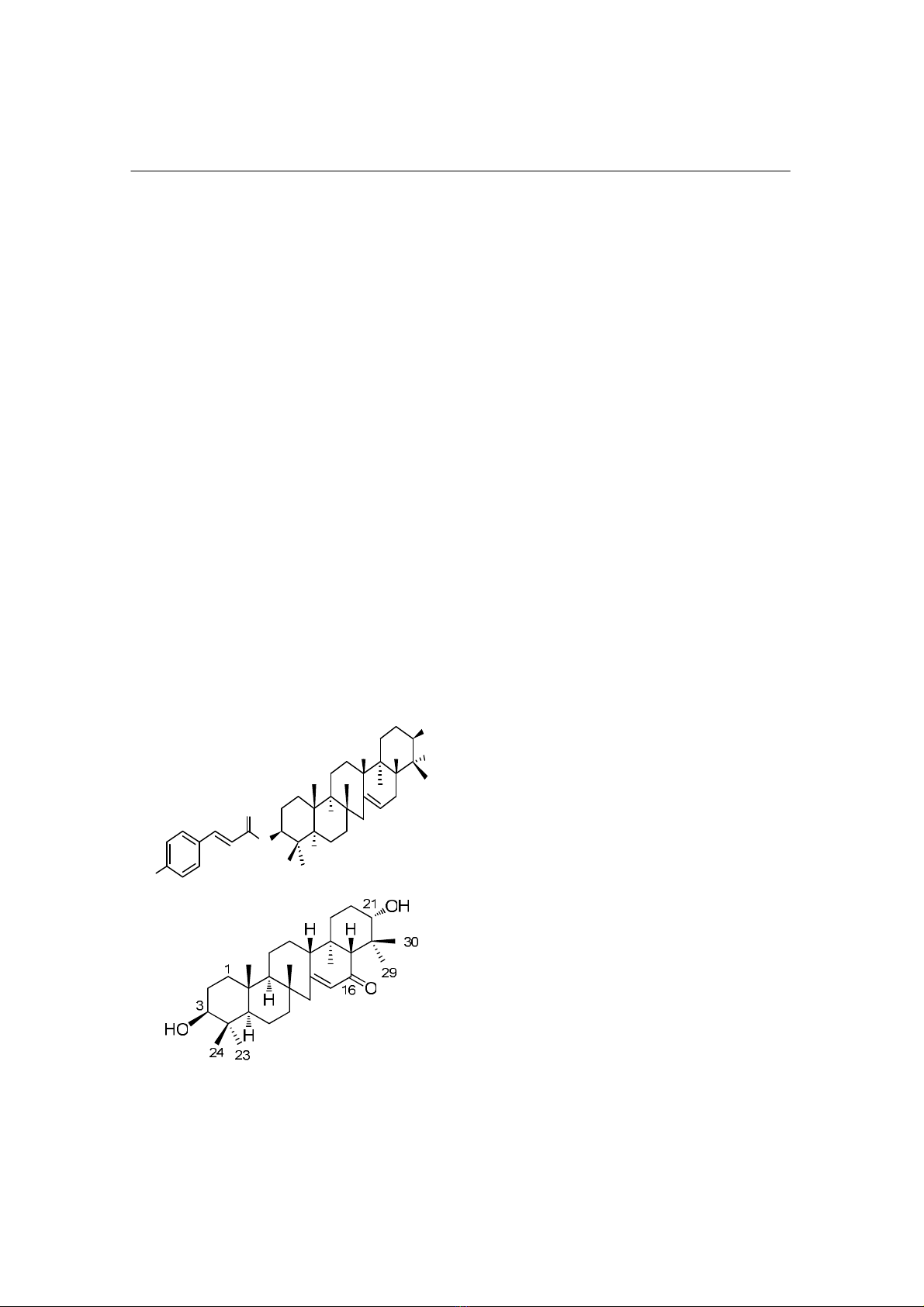
The structure of substances was determined by spectral methods. Results and conclusions: 2
compounds of terpenoid group were isolated, namely: 3β, 21β, 29-trihydroxyserrat-14-en-24-oic
acid-3β-yl- (7’-hydroxycinnamate); 16-oxo-3α-hydroxyserrat-14-en-21β-ol, the substances were
confirmed by the structure of NMR, MS, HMBC, HSQC.
* Keywords: Huperzia serrata (Thunb.) Trevis; Terpenoid; 3β, 21β, 29-trihydroxyserrat-14-
en-24-oic acid-3β-yl-(7’-hydroxycinnamate); 16-oxo-3α-hydroxyserrat-14-en-21β-ol.
INTRODUCTION
Huperzia serrata (Thunb.) Trevis belongs
to the family of Lycopodiaceae, which was
used as traditional medicinal plant in
treatment of types of lesions, hematemesis,
hematuria, hemorrhoids [1, 2]. In the 1980s,
scientists discovered an alkaloid compound,
huperzine A. The alkaloid compound has
been proven to be a potent, highly
specific, and reversible inhibitor of
acetylcholinesterase, and potential to
develop as a drug, which is used in
treatment of Alzheimer symptoms [3].
At Tamdao National Park (Vinhphuc
province), some plants belong to genus of
Huperzia were discovered, one of them is
identified as Huperzia serrata (Thunb.)
Trevis. Moreover, this genus is distributed
in wide area from north western midland
and mountainous, central, to central
highlands of Vietnam. This study was
conducted: To extract, isolate,
determine the structure of compounds
from Huperzia serrata (Thunb.) Trevis.
Results of our study provided more
information about phytochemical of
Huperzia serrata (Thunb.) Trevis.
MATERIALS AND METHODS
1. Materials.
The sample was collected at Tamdao
National Park (Vinhphuc province) and
was identified by Dr. Do Van Hai,
Department of Botany, Institute of
Ecology and Biological Resources,
Vietnam Academy of Science and
Technology. The sample was deposited
at the same Institute.
1. Military Medical College N
o
1
2. Vietnam Military Medical University
Corresponding author: Le Dinh Manh (manh.le40@gmail.com)
Date received: 08/09/2019
Date accepted: 10/10/2019
Journal of military pharmaco-medicine n
o
8-2019
116
2. Solvents and equipments.
- Ethanol was purchased from Vietnam.
Analytical solvent such as n-hexane,
methanol, dichloromethane, ethyl acetate
was purchased from Taian Health Co. Ltd,
China and used without further purification;
NMR solvents: pyridine-d
5.
- The 1D and 2D nuclear magnetic
resonance (NMR) spectra were obtained
by using a Bruker Advance 500 MHz
spectrometer (Germany). ESI-MS of the
compound was recorded on AGILENT
1,200 series LC-MSD Ion Trap. Evaporation
was performed on Rotavapor R-200
(BUCHI). Automatic collector was performed
on Eyela DC-1200 from Eyela, Japan.
3. Extraction and isolation of
compounds.
The air-dried whole plants (300 g) of
Huperzia serrata were powdered and
extracted by Soxhlet extraction method
with ethanol 96% (3 × 500 mL, 2 hours
each). The solvent was evaporated and
crude extract was suspended in hot water
(70
o
C). The solution was partitioned with
n-hexane and ethyl acetate, respectively.
Ethyl acetate layer was evaporated to
give 9.0 g of crude ethyl acetate extract
(HSE).
10 mg of HSE extract was dissolved
into 5 mL of methanol to determine the
content by thin layer chromatography
(TLC). The best solvent system was
selected as n-hexane/ethyl acetate 3/1
(v/v). Thus, solvent system was chosen
as n-hexane/EtOAc (15/1 to 5/1, v/v) as
eluent in column chromatography.
HSE extract was applied to silica gel
column chromatography, eluted with
n-hexane/EtOAc (15/1 to 5/1, v/v), and
monitored by TLC analysis. Five sub-
fractions (HSEI - HSEV) were collected
based on their TLC profiles.
Fraction HSEII (3.6 g) was fractionated
by silica gel chromatography eluted with
n-hexane/acetone (25/1 to 0/1, v/v) to
yield three sub-fraction HSEII-1 (320 mg),
HSEII-2 (1.2 g), and HSEII-3 (528 mg),
respectively, based on their TLC profiles.
Fraction HSEII-2 was applied to silica
gel chromatography, eluted with CH-
2
Cl
2
/EtOAc (20/1, v/v) to give compound
HSE-1 (11.2 mg). Fraction HSEII-3
was subjected into MIC gel column
chromatography, eluted with 90% to
100% MeOH to give two sub-fraction
HSEII-3-1 and HSEII-3-2, respectively.
Fraction HSEII-3-1 was further purified by
silica gel chromatography, eluted with
CH
2
Cl
2
/MeOH (75/1, v/v) to obtain
compound HSE-2 (9.8 mg).
RESULTS AND DISCUSSIONS
1. Extraction and isolation of
compounds.
Compound HSE-1 was isolated as a
white powder, molecule formula C
39
H
54
O
7
,
molecular weight 634.2.
1
H NMR and
13
C
NMR data were showed in table 1.
Journal of military pharmaco-medicine n
o
8-2019
117
Table 1:
1
H and
13
C NMR data for compound HSE-1 and reference compound.
C δ
Cb,
δ
Cb
δ
Ha
(J in Hz) C δ
Cb,
δ
Cb
δ
Ha
(J in Hz)
1 39.4
39.31 1.11, m; 1.92, m 21 70.2 70.11 4.61, br s
2 27.1
27.00 1.99, m; 2.20, m 22 44.0 43.89
3 80.1 80.01 5.11, dd (12.0, 4.0) 23 23.5
23.39 1.61, s
4 49.0 48.86 24 177.5 178.0
5 57.1 57.01 1.20, m 25 14.6
14.47 1.15, s
6 21.6
21.51 1.99, m; 2.15, m 26 20.3
20.18 0.91, s
7 45.9
45.81 1.25, m; 1.45, m 27 57.1
57.01 1.87, m; 2.33, m
8 37.8 37.69 28 14.6
14.47 0.87, s
9 63.1 62.99 0.85, m 29 65.6
65.41 3.99, d (10.8)
10 39.6 39.51
4.22, d (10.8)
11 27.1
27.00 1.15, m; 1.85, m 30 23.5
23.39 1.60, s
12 28.2
28.07 1.94, m; 2.01, m 1’ 168.2 168.1
13 58.0 57.87 2.10, m 2’ 116.4 116.28 6.77, d (16.0)
14 139.3 139.22 3’ 145.7 145.6 8.12, d (16.0)
15 124.5 124.12 5.51, br s 4’ 126.7 126.6
16 28.5
28.07 2.14, m; 2.25, m 5’ 131.2 131.05 7.56, d (8.4)
17 45.3 45.12 2.40, m 6’ 117.3 117.15 7.16, d (8.4)
18 36.9 36.74 1.85, m; 2.11, m 7’ 161.9 161.76
19 32.3
32.20 1.70, m; 2.05, m 8’ 117.3 117.15 7,16, d (8,4)
20 25.0
24.84 1.85, m; 2.11, m
9’ 131.2 131.05 7.56, d (8.4)
(a:
1
H NMR measured in pyridine-d
5
; b:
13
C NMR measured in pyridinre-d
5
;
±: Deference compound)
Compound HSE-2 was obtained as a white powder; molecule formula C
30
H
48
O
3
,
molecular weight 457.2.
1
H NMR and
13
C NMR data were showed in table 2.
Table 2:
1
H and
13
C NMR data for compound HSE-2 and reference compound.
C δ
Cb,
δ
Cb
δ
Ha
(J in Hz) C δ
C b,
δ
Cb
δ
Ha
(J in Hz)
1 33.8 33.8 16 201.3 201.85
2 26.7 26.5 17 59.0 58.7 3.05 (1H, s)
3 75.9 76.3 18 44.7 44.3
4 38.6 36.7 19 32.0 31.4
5 49.4 50.4 20 25.9 22.9
6 19.1 18.5
21 75.0 78.3
Journal of military pharmaco-medicine n
o
8-2019
118
7 45.3 44.7 22 37.6 36.8
8 38.2 38.2 23 29.3 27.9 1.24 (3H, s)
9 62.5 62.4 24 22.6 21.7
10 38.2 38.0 25 16.0 14.8 0.83 (3H, s)
11 25.2 24.9 26 20.1 20.0 0.91 (3H, s)
12 26.7 26.4 27 56.1 55.8
13 59.5 58.5 28 15.2 15.3 1.06 (3H, s)
14 163.6 164.05 29 22.2 22.4 1.39 (3H, s)
15 128.9 129.48 5.97 (1H, s) 30 29.1 28.0 1.72 (3H, s)
(a:
1
H NMR measured in pyridine-d
5
; b:
13
C NMR measured in pyridinre-d
5
;
±
Deference compound)
2. Structural identification of isolated
compounds.
* Compound HSE-1:
The ESI-MS (positive mode) spectrum
of compound HSE-1 showed a quasi-
molecular ion at m/z 635.1 [M+H]
+
, which
was consistent with the molecular formula
C
39
H
54
O
7
. In
1
H NMR analysis, signal for
five methyl groups at δ
H
0.87 (3H, s), 0.91
(3H, s), 1.15 (3H, s), 1.60 (3H, s), and
1.61 (3H, s), two oxygenated methine
protons at δ
H
4.61 (1H, s), and 5.11 (1H,
dd, J = 12.0, 4.0 Hz), and three olefinic
protons at δ
H
8,11 (1H, d, J = 16 Hz), 7.55
(2H, d, J = 8.4 Hz), 7.15 (2H, d, J = 8.4
Hz), 6.77 (1H, d, J = 16 Hz), and 5.51
(1H, brs) were observed. The
13
C NMR
data of compound HSE-1 showed 39
carbon signals including five methyls, ten
methylene, five methines carbons, six
quaternary carbons, two oxymethines
carbons at δ
C
80.01 (C-3) and δ
C
70.11
(C-21), one oxymethylene at δ
C
65.41 (C-
29), and one carbonyl group at δ
C
178.0
(C-24), together with signals of eight
aromatic carbons, which belong to para-
hydroxycinnamoyl moiety (table 1). The
NMR data of HSE-1 were similar with
those of lycernuic acid A [4, 5], except the
data at position C-3. The para-
hydroxycinnamate moiety was located at
C-3 in HSE-1 instead of hydroxyl group.
To confirm the structure of HSE-1,
2D-NMR analysis (HMQC and HMBC) were
performed. The HMQC data were indicated
all carbons bearing proton. Detailed of
HMBC spectrum showed the correlation
between proton at δ
H
3.99 (1H, d, 10.8)
and 4.22 (1H, d, 10.8) and carbon at δ
C
70.2 (C-21). This correlation indicated the
location of -CH
2
OH moiety at carbon C-22.
Moreover, the correlation from proton at
δ
H
5.11 (H-3α) to carbon at δ
C
168.2
(C-1’) confirmed the location of para-
hydroxycinnamate moiety at C-3. Thus,
the structure of HSE-1 was identified as
3β, 21β, 29-trihydroxyserrat-14-en-24-oic
acid-3β-yl-(7-hydroxycinnamate) [6], which
was isolated from Lycopodiella cernua
(L.) Pie.-Serm, and the structure was
shown as in figure 1.
Journal of military pharmaco-medicine n
o
8-2019
119
* Compound HSE-2:
The molecular formula was determined
to be C
30
H
48
O based on positive ESI-MS
data at m/z 457.2 [M + H]
+
. In
1
H NMR
analysis, signal for seven methyl groups
at δ
H
0.74 (3H, s), 0.83 (3H, s), 0.91 (3H,
s), 1.06 (3H, s), 1.24 (3H, s), 1.39 (3H, s),
and 1.72 (3H, s), an oxygenated methine
proton at δ
H
3.05 (1H, s), and an olefinic
proton at δ
H
5.97 (1H, s) were observed.
The
13
C NMR data of HSE-2 showed the
presence of thirty carbons including a
ketone carbon at δ
C
201.9, two olefinic
carbons at δ
C
164.1, and 129.5, and two
oxygenated methine carbons at δ
C
76.3,
and 78.3. The NMR data of HSE-2 were
similar with those of 16-oxo-3α-
hydroxyserrat-14-en-21β-ol [7]. Thus, the
structure of HSE-2 was identified as 16-
oxo-3α-hydroxyserrat-14-en-21β-ol, and
the structure was shown in figure 1.
O
O
HO HOOC
H H
OH
CH
2
1
3
23
24
4
6
7
9
12
14
15
16
21
30
28
25 26
1'
2'
3'
7'
4'
H
H
Figure 1: Chemical structure of isolated
compounds (HSE-1 and HSE-2).
CONCLUSION
In this study, we successed in isolation
and purification of two triterpenoid
compounds from the EtOAc extract of
Huperzia serrata (Thunb.) Trevis by using
column chromatography. Structures of
isolated compounds were studied using
spectroscopic methods and were compared
with the literatures. Two compounds were
identified as 3β, 21β, 29-trihydroxyserrat-
14-en-24-oic acid-3β-yl-(7ʹ-hydroxycinnamate)
(HSE-1), and 16-oxo-3α-hydroxyserrat-
14-en-21β-ol (HSE-2). Compound 3β, 21β,
29-trihydroxyserrat-14-en-24-oic acid-3β-
yl-(7ʹ-hydroxycinnamat) (HSE-1) was first
isolated from this plant.
REFERENCES
1. Vo V.C. Dictionary of Vietnam medicinal
plants. Medical Publishing House. Hanoi. 1997,
p.1119.
2. Pham H.H. Vietnam Tre Publishing House.
1999, Vol I, p.22.
3. Ma X, Tan C, Zhu D et al. Huperzine A
from Huperzia species: An ethnopharmacology
review. J Ethnopharmacol. 2007, 113, pp.15-34.
4. Zhang Z, Elsohly H.N, Jacob M.R et al.
Natural products inhibiting Candida albicans
secreted aspartic proteases from Lycopodium
cernuum. J Nat Prod. 2002, 65, pp.979-985.
5. Tsuda Y, Isobe K, Sano T. Triterpenoid
chemistry. IX. Lycopodium triterpenoid. (6).
The structures of three new tetra-ols, lycocryptol,
21-epilycocryptol, and diepilycocryptol, and
two new acids, lycernuic acid-a and -b. Chem
Pharm Bull. 1975, 23, pp.264-271.
6. Nguyen V.T, To D.C, Tran M.H et al.
Isolation of cholinesterase and β-secretase 1
inhibiting compounds from Lycopodiella cernua.
Bioorg Med Chem. 2015, 23, pp.3126-3134.
7. Zhou H, Tan C.H, Jiang S et al.
Serratene-type triterpenoids from Huperzia
serrata. J Nat Prod. 2003, 66 (10), pp.1328-1332.