
Open Access
Available online http://ccforum.com/content/10/1/R38
Page 1 of 9
(page number not for citation purposes)
Vol 10 No 1
Research
Lung and 'end organ' injury due to mechanical ventilation in
animals: comparison between the prone and supine positions
George Nakos1, Anna Batistatou2, Eftychia Galiatsou1, Eleonora Konstanti1, Vassilios Koulouras1,
Panayotis Kanavaros3, Apostolos Doulis1, Athanassios Kitsakos1, Angeliki Karachaliou1,
Marilena E Lekka4 and Maria Bai2
1Department of Intensive Care Unit, University Hospital of Ioannina, Greece
2Department of Pathology, University of Ioannina, Greece
3Department of Anatomy-Histology-Embryology, University of Ioannina, Greece
4Department of Chemistry, University of Ioannina, Greece
Corresponding author: George Nakos, gnakos@cc.uoi.gr
Received: 2 Nov 2005 Revisions requested: 8 Dec 2005 Revisions received: 25 Jan 2006 Accepted: 3 Feb 2006 Published: 28 Feb 2006
Critical Care 2006, 10:R38 (doi:10.1186/cc4840)
This article is online at: http://ccforum.com/content/10/1/R38
© 2006 Nakos et al.; licensee BioMed Central Ltd.
This is an open access article distributed under the terms of the Creative Commons Attribution License (http://creativecommons.org/licenses/by/2.0),
which permits unrestricted use, distribution, and reproduction in any medium, provided the original work is properly cited.
Abstract
Introduction Use of the prone position in patients with acute
lung injury improves their oxygenation. Most of these patients die
from multisystem organ failure and not from hypoxia, however.
Moreover, there is some evidence that the organ failure is
caused by increased cell apoptosis. In the present study we
therefore examined whether the position of the patients affects
histological changes and apoptosis in the lung and 'end organs',
including the brain, heart, diaphragm, liver, kidneys and small
intestine.
Methods Ten mechanically ventilated sheep with a tidal volume
of 15 ml/kg body weight were studied for 90 minutes. Five
sheep were placed in the supine position and five sheep were
placed in the prone position during the experiment. Lung
changes were analyzed histologically using a semiquantitative
scoring system and the extent of apoptosis was investigated
with the TUNEL method.
Results In the supine position intra-alaveolar hemorrhage
appeared predominantly in the dorsal areas, while the other
histopathologic lesions were homogeneously distributed
throughout the lungs. In the prone position, all histological
changes were homogeneously distributed. A significantly higher
score of lung injury was found in the supine position than in the
prone position (4.63 ± 0.58 and 2.17 ± 0.19, respectively) (P <
0.0001). The histopathologic changes were accompanied by
increased apoptosis (TUNEL method). In the supine position,
the apoptotic index in the lung and in most of the 'end organs'
was significantly higher compared with the prone position (all P
< 0.005). Interestingly, the apoptotic index was higher in dorsal
areas compared with ventral areas in both the prone and supine
positions (P < 0.003 and P < 0.02, respectively).
Conclusion Our results suggest that the prone position
appears to reduce the severity and the extent of lung injury, and
is associated with decreased apoptosis in the lung and 'end
organs'.
Introduction
Mechanical ventilation has constituted an indispensable part
of basic life support in the intensive care unit for several dec-
ades and is undoubtedly essential for patients with acute lung
injury/acute respiratory distress syndrome (ALI/ARDS). In
recent years, however, it has become clear that mechanical
ventilation can also be injurious. Repeated application of
transalveolar pressures that exceed those corresponding to
the inflation capacity causes tissue stresses and disrupts the
lung. In animals, mechanical ventilation at high volumes and
high pressures can cause ventilator-induced lung injury (VILI)
with similar histological appearance to ALI/ARDS. These his-
tological disorders are due to injury of the alveolar epithelium,
basement membrane and microvascular endothelium and
accompanied by high-permeability pulmonary edema. Injurious
AI = apoptotic index; ALI = acute lung injury; ARDS = acute respiratory distress syndrome; FiO2 = fraction inspired oxygen; H&E = haematoxylin–
eosin; PCO2 = partial pressure of CO2; TUNEL= terminal deoxynucleotidyl-transferase-mediated dUTP nick end-labeling; VILI = ventilator-induced
lung injury.

Critical Care Vol 10 No 1 Nakos et al.
Page 2 of 9
(page number not for citation purposes)
mechanical ventilation exacerbates the damage in previously
injured lungs [1-3].
The damage to the lungs has been attributed to two overlap-
ping mechanisms, namely mechanical damage of tissues and
cells due to overdistention and shear stress (barotrauma or
volutrauma) as well as mechanical damage due to the produc-
tion, release and/or activation of cytotoxic and inflammatory
cascades (biotrauma). In addition to inducing or worsening
existing lung injury, the pulmonary production of inflammatory
mediators is likely to spill over into the systemic circulation,
also contributing to extrapulmonary end-organ failure [3,4].
Despite considerable progress, the death rate of patients with
ALI/ARDS remains quite high [5]. In fact, most patients die
from multisystem organ failure and not from hypoxia. However,
pathogenesis of multiorgan failure in ARDS/ALI remains a
dilemma. There is some evidence that multisystem organ fail-
ure is caused by increased apoptosis of the epithelial cells of
'end organs', such as the kidneys and small intestine [6,7].
Apoptosis is an active mechanism of cell death, which is
important for the development and homeostasis of tissues.
Environmental conditions or specific receptor/ligand interac-
tions activate intracellular signaling pathways that lead to DNA
cleavage and apoptotic cell death (for a review, see [8]).
As early as 1976 it was reported that placing patients with
ALI/ARDS in the prone position improves their oxygenation [9-
17]. Prone positioning improves secretion drainage from the
airways, relieving lung compression by the heart and abdo-
men. The transalveolar forces are redistributed so as to allow
expansion of the dorsal regions. All these events lead to an
increase in end-respiratory lung volume, to better ventilation-
perfusion matching and to alterations in chest-wall mechanics
leading to regional changes in ventilation. The effects of prone
ventilation on the cellular constituents of the lung alveoli have
not so far been studied.
Our working hypothesis was that VILI can lead to distant organ
damage through the increase in the circulation of mediators,
including proapoptotic soluble factors, such as soluble Fas lig-
and [6]. In this respect, using injurious tidal-volume-induced
lung damage, we studied the possible protective role of the
prone position through the reduction of atelectasis and/or
overdistention. In addition, we investigated whether cell apop-
tosis was related to the severity of tissue damage of the lung
and other organs induced by mechanical ventilation.
Materials and methods
Animal preparation
Protocols were approved by the University of Ioannina animal
research committee. We examined 10 sheep, each weighing
33 ± 5 kg. A peripheral vein was cannulated, and anesthesia
was induced with katanine, maintained by continuous intrave-
nous injection of midazolam and fentanyl citrate and paralyzed
with pancuronium bromide. The animals were tracheotomized,
and catheters were introduced into the carotid artery and the
external jugular vein. Mechanical ventilation was provided with
a Servo 900C ventilator (Siemens Elema, Solna, Sweden) in
the volume control mode with a tidal volume of 15 ml/kg body
weight for 90 minutes, with low positive end expiratory pres-
sure (3 cmH2O) and with FiO2 of 0.5 in both groups. The res-
piratory rate was adjusted appropriately to maintain
normocapnia at baseline measurements. Arterial pressure
from the carotid artery and airway was recorded throughout
the experiment. Blood gases, respiratory system compliance
(calculated as the end-inspiratory airway pressure minus the
end-expiratory pressure divided by the tidal volume) and bio-
chemistry were measured before, during and at the end of the
experiment. We continuously monitored the arterial blood
pressure, the central venous pressure, the heart rate and the
urine output. These parameters were kept stable by fluid infu-
sion (normal saline). The animal temperature was also kept sta-
ble.
Five animals were placed in the supine position and five in the
prone position during the whole experiment. The animals were
exsanguinated at the end of the experiment, which lasted 90
minutes from the beginning of mechanical ventilation, while
deeply anesthetized. The internal organs were removed and
representative sections from the lungs, the brain, the heart, the
diaphragm, the liver, the kidneys and the small intestine were
taken and fixed in 10% buffered formalin.
Histologic evaluation and TUNEL method
Paraffin sections, 5 µm thick, were stained with the standard
H&E stain and examined using light microscopy. Lung
changes were analyzed histologically using a semiquantitative
scoring system, as previously described elsewhere [18].
Briefly, six slides – two from the upper lobe (one from the dor-
sal area and one from the ventral area), two from the lower lobe
(one from the dorsal area and one from the ventral area) and
two from the middle lobe in the right lung and the middle area
in the left lung – were analyzed by two independent patholo-
gists. The pathologists were blinded to the assignment of the
animals. The slides were scanned in low power and the five
fields with the most pronounced changes were chosen. The
score given for each slide represented the mean score of
these fields.
Four parameters were examined: alveolar fibrin edema, alveo-
lar hemorrhage, septal thickening and intra-alveolar inflamma-
tory cells. The changes were scored according to their extent
(score 0, 1, 2 and 3 for an extent of 0%, <25%, 25–50% and
>50%, respectively) and the severity of the injury (score 0 for
no changes, score 1, 2 and 3 for more severe changes). The
injury score represents the sum of the extent and the severity
of injury.

Available online http://ccforum.com/content/10/1/R38
Page 3 of 9
(page number not for citation purposes)
Table 1
Gas exchange, respiratory system compliance and hemodynamics
Supine position Prone position P value 95% confidence interval of the
difference
PO2/FIO2 (mmHg)
Baseline 416 ± 23.6 412.4 ± 25.5 NS
90 minutes 105.6 ± 24.1 251.6 ± 56.1 <0.001 -208.9 to -83.0
P value <0.0001 <0.004
95% confidence interval of the difference 272.8–247.9 84.8–236.7
PCO2 (mmHg)
Baseline 38.8 ± 1.8 40.8 ± 1.3 NS
90 minutes 57.2 ± 1.5 43.0 ± 1.2 <0.001 2.2 to 6.1
P value <0.002 <0.04
95% confidence interval of the difference -5.0 to -6.9 -1.1 to -5.8
pH
Baseline 7.408 ± 0.013 7.398 ± 0.008 NS
90 minutes 7.322 ± 0.019 7.382 ± 0.018 0.0009 -0.08 to -0.03
P value 0.0005 NS
95% confidence interval of the difference 0.063–0.108
Static compliance of respiratory system (ml/cmH2O)
Baseline 30.4 ± 3.8 25.9 ± 2.1 NS
90 minutes 18.2 ± 2.8 22.8 ± 2.3 <0.02 -8.3 to -0.86
P value <0.001 <0.003
95% confidence interval of the difference -10.1 to -14.3 -1.7 to -4.5
Blood pressure (mmHg)
Baseline 81.80 ± 7.294 85.60 ± 9.476 NS
90 minutes 84.20 ± 5.167 86.00 ± 9.670 NS
P value NS NS
95% confidence interval of the difference
Heart rate (beats/minutes)
Baseline 117.2 ± 9.365 122.2 ± 6.140 NS
90 minutes 130.4 ± 4.722 132.8 ± 5.891 NS
P value 0.0074 0.0007
95% confidence interval of the difference -20.51 to -5.887 -13.72 to -7.484
Static compliance of respiratory system = (end inspiratory airway pressure – end-expiratory pressure)/tidal volume.
Apoptosis was detected with the terminal deoxynucleotidyl-
transferase-mediated dUTP nick end-labeling (TUNEL)
method (Apo-tag kit; Oncor, Craithersburg, MD, USA) in 5 µm
paraffin sections, as described in detail in previous studies
[19,20]. Positive and negative controls were included in every
staining. Positive staining in areas of lymphocytic infiltration
served as the internal positive control. No staining was noted
in negative controls.
Briefly, morphologically intact TUNEL-positive cells and apop-
totic cells in H&E-stained studies were considered positive
and are referred to as apoptotic cells. The number of apoptotic
cells and apoptotic bodies was recorded by using the 40×
objective lens, and at least 10 randomly selected fields were
counted. The apoptotic index (AI) was expressed as the
number of apoptotic cells/bodies per 10 high-power fields.
Care was taken to avoid areas with extensive inflammation.

Critical Care Vol 10 No 1 Nakos et al.
Page 4 of 9
(page number not for citation purposes)
The AI at the alveolar septum of the lungs, the neurons and
glial cells, the muscle cells of the diaphragm, the hepatocytes,
the glomerular and tubular renal cells, and the epithelial cells
of the small intestinal epithelium were estimated.
Statistical analysis
Statistical analysis was performed using the Statistical Pack-
age for Social Sciences (SPSS) version 12 for Windows
(SPSS Inc., Chicago, Illinois, USA). Data were tested for nor-
mality with the Kolmogorov-Smirnov test and are presented as
the mean ± SD. All variables were normally distributed. Com-
parisons between the prone and supine positions were made
using a t test. Comparisons between the ventral and dorsal
regions of the lungs in either the supine position or the prone
position were made using a paired t test.
Results
Lung mechanics and blood gases
Lung mechanics and blood gas alterations and the biochemi-
cal data are presented in Tables 1 and 2, respectively. Blood
gases and the compliance of the respiratory system deterio-
rated after 90 minutes of mechanical ventilation in both posi-
tions. The deterioration in blood gases as well as in the
compliance due to VILI was significantly less prominent in the
prone position. Transaminases (aspartate aminotransferase
and alanine aminotransferase) increased during mechanical
ventilation in the supine position, while they were both
unchanged in the prone position. γ-Glutamyl transpeptidase,
urea and creatinine were not altered during mechanical venti-
lation in both positions.
ALI score in the prone and supine positions
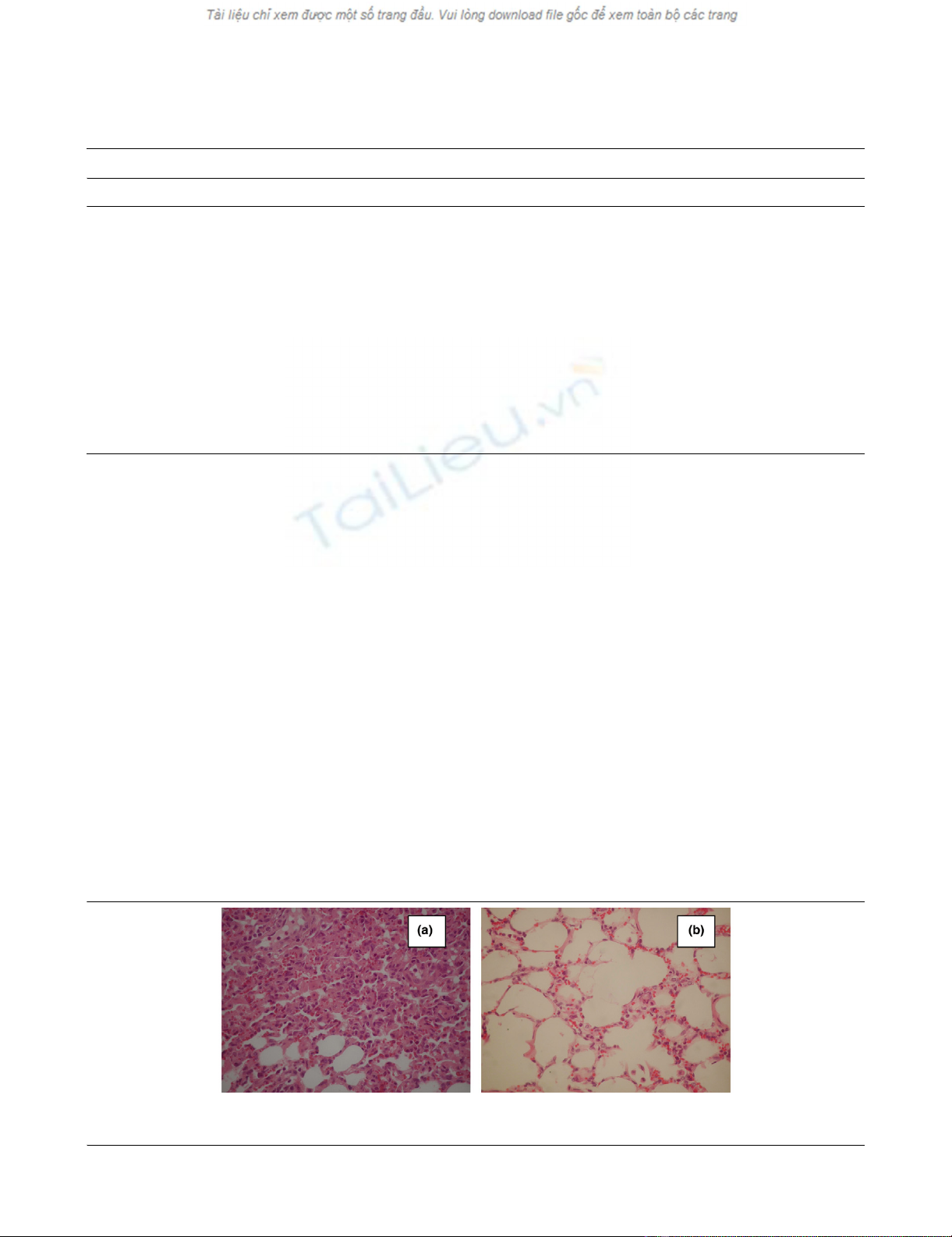
In the lungs of the animals placed in the supine position the
alveolar-septal membrane was thickened and there was con-
siderable intra-alveolar edema and eosinophilic material. Fur-
thermore, hemorrhage and increased numbers of inflammatory
cells (lymphocytes, plasma cells, macrophages and polymor-
phonuclear neutrophil granulocytes) were observed (Table 3).
Consolidated areas were frequently encountered (Figure 1a).
In animals placed in the prone position the lung injury was
milder (Table 3). There was considerably less inflammatory
infiltration, alveolar edema, hemorrhage thickening of the alve-
Table 2
Biochemistry at the beginning and the end of experiment
Supine position Prone position P value
Urea (mg/dl)
Baseline 34.9 ± 11.5 43.4 ± 6.5 NS
90 minutes 41.1 ± 7.3 37.1 ± 8.4 NS
P value NS NS
Creatinine (mg/dl)
Baseline 0.62 ± 0.1 0.48 ± 0.11 NS
90 minutes 0.55 ± 0.08 0.53 ± 0.1 NS
P value NS NS
aspartate aminotransferase (IU/l)
Baseline 94 ± 21 98 ± 25 NS
90 minutes 147 ± 19 84 ± 27 <0.05
P value <0.05 NS
alanine aminotransferase (IU/l)
Baseline 14 ± 6 16 ± 7 NS
90 minutes 27 ± 8 15 ± 9 <0.05
P value <0.05 NS
γ-Glutamyl transpeptidase (IU/l)
Baseline 26 ± 18 29 ± 24 NS
90 minutes 33 ± 22 25 ± 23 NS
P value NS NS
Available online http://ccforum.com/content/10/1/R38
Page 5 of 9
(page number not for citation purposes)
olar-septal membrane and consolidation. In addition, many
areas appeared uninjured or minimally affected (Figure 1b).
The differences between the supine and prone positions were
statistically significant (P < 0.0001). Interestingly, the overall
histological findings for each animal were consistent in all lung
areas – upper, middle and lower, ventral and dorsal (Table 3).
When alveolar hemorrhage was considered alone, however,
there was a significant difference between ventral and dorsal
samples in animals placed in the supine position. In these ani-
mals the mean score for alveolar hemorrhage was 4.8 ± 0.84
in the ventral areas and was 2.6 ± 0.55 in the dorsal areas of
both lungs (P < 0.01). This difference was not evident in ani-
mals placed in the prone position.
Apoptotic index in the prone and supine positions
TUNEL-positive nuclei/apoptotic bodies were observed in all
animals in the lungs, and the AI was increased in the supine
position group compared with the prone position group (Table
3 and Figure 2a,b). In both the supine position and the prone
position, the mean value of the AI was higher in areas dorsal
compared with ventral areas; the differences were statistically
significant (P = 0.04 and P = 0.046, respectively). Moreover,
the differences between the supine and prone positions were
statistically significant in the dorsal lung areas as well in the
ventral lung areas (P < 0.003 and P < 0.02, respectively)
(Table 3).
The AI in the liver was far less than that in the lungs. The liver
AI was increased in the supine position group (Figure 2c,d).
The difference was statistically significant (P < 0.05) (Table 3).
In the kidneys, particularly at the medulla, the nuclei of tubular
epithelial cells were TUNEL-positive without morphological
characteristics of apoptosis and were not included in the esti-
mation of the AI. Counts were performed at the cortex (Figure
2e,f). The mean values of the AI were higher in the supine posi-
Table 3
Acute lung injury score and apoptotic index in the supine and prone position
Supine position Prone position P value 95% confidence interval
Acute lung injury score 4.63 ± 0.58 2.17 ± 0.19 <0.0001 -3.9 to -1.82
Apoptotic index
Lung dorsal 112 ± 22 45.6 ± 28 0.003 -103.6 to -29.78
Lung ventral 80 ± 28 35 ± 22 0.02 -82.6 to -8.1
P value 0.04 0.046
95% confidence interval 2.37 to 61.09 0.29 to 20.5
Liver 56 ± 21 23 ± 10 0.05 -66.78 to -7.8.1
Kidney 31 ± 14 17 ± 10 NS
Small intestine 22 ± 11 16 ± 11 NS
Diaphragm 10 ± 0.5 0.5 ± 0.4 0.001 -10.6 to -9.01
Acute lung injury score corresponds to the sum of the extent (score 0, 1, 2 and 3 for an extent of 0%, <25%, 25–50% and >50%) and the
severity of lung injury (score 0 for no changes, score 1, 2 and 3 more severe changes). The apoptotic index was expressed as the number of
apoptotic cells/bodies per 10 high-power fields.
Figure 1
Histological changes of lungs (septal thickening, alveolar fibrin/edema, alveolar hemorrhage, intra-alveolar inflammatory cells) in animals placed in (a) the supine position and (b) the prone position (H&E, ×400)Histological changes of lungs (septal thickening, alveolar fibrin/edema, alveolar hemorrhage, intra-alveolar inflammatory cells) in animals placed in (a)
the supine position and (b) the prone position (H&E, ×400).


%20--%3e%3cdefs%3e%3cstyle%3e%20.st0%20{%20fill:%20%23fff;%20}%20.st1%20{%20fill:%20%237800fa;%20}%20%3c/style%3e%3c/defs%3e%3cpath%20class='st1'%20d='M117.78,12.18H43.11c2.9,3.47,4.65,7.94,4.65,12.82,0,5.6-2.3,10.66-6.01,14.29h76.02l7.22-13.56-7.22-13.56Z'/%3e%3cg%3e%3cpath%20class='st0'%20d='M53.58,26.17h-.59v-1.46h.59v-4.96h2.83c1.78,0,2.67.94,2.67,2.82v5.76c0,1.87-.89,2.81-2.67,2.81h-2.83v-4.96ZM55.36,21.37v3.34h1.1v1.46h-1.1v3.34h1.01c.61,0,.91-.37.91-1.1v-5.93c0-.74-.3-1.1-.91-1.1h-1.01Z'/%3e%3cpath%20class='st0'%20d='M65.99,31.14h-1.8l-.31-2.07h-2.19l-.31,2.07h-1.64l1.82-11.39h2.62l1.82,11.39ZM65.28,18.04c-.25.46-.51.77-.75.94-.21.15-.47.22-.79.22-.26,0-.57-.07-.92-.22l-.38-.15c-.14-.05-.26-.07-.37-.07-.3,0-.53.18-.71.54l-.91-.68c.25-.46.51-.77.75-.94.21-.14.48-.21.79-.21.26,0,.57.07.92.21l.38.15c.14.05.26.07.37.07.3,0,.53-.18.71-.54l.91.68ZM61.91,27.52h1.73l-.87-5.76-.87,5.76Z'/%3e%3cpath%20class='st0'%20d='M74.53,26.89v1.52c0,1.91-.89,2.86-2.67,2.86s-2.67-.95-2.67-2.86v-5.93c0-1.91.89-2.86,2.67-2.86s2.67.95,2.67,2.86v1.11h-1.69v-1.22c0-.75-.31-1.12-.93-1.12s-.93.37-.93,1.12v6.15c0,.74.31,1.11.93,1.11s.93-.37.93-1.11v-1.63h1.69Z'/%3e%3cpath%20class='st0'%20d='M81.4,31.14h-1.8l-.31-2.07h-2.19l-.31,2.07h-1.64l1.82-11.39h2.62l1.82,11.39ZM75.9,19.2l1.52-1.91h1.71l1.51,1.91h-1.61l-.76-.95-.75.95h-1.61ZM77.32,27.52h1.73l-.87-5.76-.87,5.76ZM83.1,15.99l-1.76,1.91h-1.26l1.17-1.91h1.86Z'/%3e%3cpath%20class='st0'%20d='M84.86,19.75c1.78,0,2.67.94,2.67,2.82v1.48c0,1.87-.89,2.81-2.67,2.81h-.85v4.28h-1.79v-11.39h2.64ZM84.01,21.37v3.86h.85c.58,0,.87-.36.87-1.08v-1.71c0-.71-.29-1.07-.87-1.07h-.85Z'/%3e%3cpath%20class='st0'%20d='M93.51,19.75c1.78,0,2.67.94,2.67,2.82v1.48c0,1.87-.89,2.81-2.67,2.81h-.85v4.28h-1.79v-11.39h2.64ZM92.66,21.37v3.86h.85c.58,0,.87-.36.87-1.08v-1.71c0-.71-.29-1.07-.87-1.07h-.85Z'/%3e%3cpath%20class='st0'%20d='M98.8,31.14h-1.79v-11.39h1.79v4.88h2.03v-4.88h1.83v11.39h-1.83v-4.88h-2.03v4.88Z'/%3e%3cpath%20class='st0'%20d='M105.36,24.55h2.46v1.62h-2.46v3.34h3.09v1.63h-4.88v-11.39h4.88v1.63h-3.09v3.18ZM108.17,17.29l-1.76,1.91h-1.26l1.17-1.91h1.86Z'/%3e%3cpath%20class='st0'%20d='M112.2,19.75c1.78,0,2.67.94,2.67,2.82v1.48c0,1.87-.89,2.81-2.67,2.81h-.85v4.28h-1.79v-11.39h2.64ZM111.35,21.37v3.86h.85c.58,0,.87-.36.87-1.08v-1.71c0-.71-.29-1.07-.87-1.07h-.85Z'/%3e%3c/g%3e%3ccircle%20class='st1'%20cx='25'%20cy='25'%20r='20'/%3e%3cpath%20class='st0'%20d='M32.78,19.27c2.92,0,4.43,2.55,5.28,5.33l.71,2.17c.14.38-.33.75-.71.75h-5.61c.19-.33.24-.71.09-1.08l-.75-2.45c-.43-1.32-.99-2.64-1.79-3.77.75-.57,1.65-.94,2.78-.94h0ZM25,18.38c3.25,0,4.9,2.78,5.89,5.89l.76,2.45c.14.42-.33.8-.8.8h-11.69c-.42,0-.94-.38-.8-.8l.75-2.45c.99-3.11,2.64-5.89,5.89-5.89h0ZM25,11.35c1.74,0,3.11,1.37,3.11,3.11s-1.37,3.11-3.11,3.11-3.11-1.41-3.11-3.11,1.41-3.11,3.11-3.11h0ZM17.27,19.27c1.08,0,1.98.38,2.73.94-.8,1.13-1.37,2.45-1.74,3.77l-.8,2.45c-.14.38-.05.75.09,1.08h-5.56c-.42,0-.9-.38-.75-.75l.71-2.17c.9-2.78,2.41-5.33,5.33-5.33h0ZM17.27,12.91c1.51,0,2.78,1.27,2.78,2.83s-1.27,2.83-2.78,2.83-2.83-1.27-2.83-2.83,1.27-2.83,2.83-2.83h0ZM32.78,12.91c1.56,0,2.78,1.27,2.78,2.83s-1.23,2.83-2.78,2.83-2.83-1.27-2.83-2.83,1.27-2.83,2.83-2.83h0ZM27.07,28.56v.09c0,.57-.24,1.08-.61,1.46h0v.05c-.38.33-.9.57-1.46.57s-1.08-.24-1.46-.61h0c-.38-.38-.61-.9-.61-1.46v-.09h1.41v.09c0,.19.05.38.19.47v.05c.09.09.28.19.47.19s.38-.09.47-.19v-.05c.14-.09.24-.28.24-.47t-.05-.09h1.41ZM30.99,28.56v.09c0,1.65-.66,3.16-1.74,4.24-1.08,1.08-2.59,1.79-4.24,1.79s-3.16-.71-4.24-1.79l-.05-.05c-1.04-1.08-1.7-2.55-1.7-4.2v-.09h1.41v.09c0,1.27.47,2.4,1.27,3.25h.05c.85.85,1.98,1.37,3.25,1.37s2.4-.52,3.25-1.37c.85-.8,1.37-1.98,1.37-3.25v-.09h1.37ZM34.99,28.56v.09c0,2.78-1.13,5.28-2.92,7.07-1.79,1.79-4.29,2.92-7.07,2.92s-5.23-1.13-7.07-2.92c-1.79-1.79-2.92-4.29-2.92-7.07v-.09h1.41v.09c0,2.4.94,4.53,2.5,6.08,1.56,1.56,3.72,2.5,6.08,2.5s4.52-.94,6.08-2.5c1.56-1.56,2.5-3.68,2.5-6.08v-.09h1.41Z'/%3e%3c/svg%3e)