
REVIEW ARTICLE
Alpha-oxidation of 3-methyl-substituted fatty acids
and its thiamine dependence
Minne Casteels, Veerle Foulon, Guy P. Mannaerts and Paul P. Van Veldhoven
Afdeling Farmacologie, Department of Molecular Cell Biology, Katholieke Universiteit Leuven, Belgium
3-Methyl-branched fatty acids, as phytanic acid, undergo
peroxisomal a-oxidation in which they are shortened by 1
carbon atom. This process includes four steps: activation,
2-hydroxylation, thiamine pyrophosphate dependent
cleavage and aldehyde dehydrogenation. The thiamine
pyrophosphate dependence of the third step is unique in
peroxisomal mammalian enzymology. Human pathology
due to a deficient alpha-oxidation is mostly linked to
mutations in the gene coding for the second enzyme of the
sequence, phytanoyl-CoA hydroxylase.
Keywords: alpha-oxidation; thiamine pyrophosphate; per-
oxisomes; lyase; Adult Refsum Disease.
Introduction
a-Oxidation is the process in which fatty acids are shortened
at the carboxyl-end by one carbon atom. For 3-methyl-
branched fatty acids, this is the preferred pathway as their
breakdown by b-oxidation is impossible. Indeed, the
3-methyl-branch precludes the third step of b-oxidation,
the dehydrogenation step. Phytanic acid (3,7,11,15-tetra-
methylhexadecanoic acid) is at present the only established
physiological substrate of a-oxidation in humans [1,2].
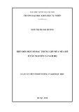
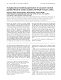
Phytanic acid is derived from phytol, the isoprenoid side
chain of chlorophyll. As chlorophyll-bound phytol cannot
be metabolized by humans, and free phytol is present only
in minimal quantities in food, the phytanic acid present in
the human body is mostly provided by external sources
(Fig. 1). Ruminants ingest large amounts of chlorophyll,
from which phytol is efficiently cleaved off by bacteria in the
gastrointestinal tract. Phytol is subsequently taken up and
converted to phytanic acid, which is deposited in fat tissues
and in milk, the major sources of phytanic acid for humans
[2].
Accumulation of phytanic acid is typically seen in Adult
Refsum Disease (ARD) and is due to a deficient degrada-
tion of this exogenous 3-methyl-branched fatty acid [2,3].
Elevated phytanic acid levels can also be seen in peroxisome
biogenesis disorders, in which a defective a-oxidation is only
one of the deficiencies present [4]. Degradation of phytanic
acid via x-oxidation, by which a carboxylic acid group is
introduced at the omega end, has also been described [5,6],
but appears to be quantitatively less important under
physiological conditions. Its importance increases when
phytanic acid levels in serum are elevated as is seen in ARD
[7].
The degradation of phytanic acid via a-oxidation is
presently proposed to evolve completely in peroxisomes,
some doubts remaining, however, concerning the first
(activation) and last (aldehyde dehydrogenation) enzymatic
steps.
Degradation of 3-methyl-branched fatty acids
The classic catabolic pathway by which fatty acids are
degraded is b-oxidation and a mitochondrial as well as a
peroxisomal b-oxidation pathway is known [8]. Very long
chain fatty acids, 2-methyl-branched fatty acids, the side
chains of bile acid intermediates and eicosanoids are mainly/
exclusively handled by the peroxisomal pathway, whereas
short and medium chain fatty acids are oxidized mainly in
mitochondria [8].
Phytanic acid and other 3-methyl-branched fatty acids
cannot undergo b-oxidation because the 3-methyl-group
prevents the formation of a 3-keto substituent in the
dehydrogenation step. Therefore, 3-methyl-branched fatty
acids first undergo a-oxidation. In the case of phytanic acid,
this results in the generation of 2-methyl-branched pristanic
acid (2,6,10,14-tetramethylpentadecanoic acid), which is
then shortened to 4,8-dimethylnonanoic acid via peroxi-
somal b-oxidation. The dimethyl fatty acid is then degraded
further via mitochondrial b-oxidation.
Peroxisomes, in which most or all steps of the a-oxidation
pathway evolve, are subcellular organelles involved in a
number of anabolic (e.g. plasmalogen synthesis) and
catabolic processes, including a-andb-oxidation [8].
Peroxisomal enzymes are synthesized on polyribosomes in
the cytosol and are post-translationally imported into the
peroxisome. Therefore, these enzymes contain a series of
conserved amino acids or so called peroxisome targeting
signals (PTSs) [9]. Two classes of these topogenic sequences
Correspondence to M. Casteels, Afdeling Farmacologie, Department
of Molecular Cell Biology, Katholieke Universiteit Leuven,
Campus Gasthuisberg, Herestraat 49, B 3000 Leuven, Belgium.
Fax: + 32 16 345699, Tel.: + 32 16 345816,
E-mail: minne.casteels@med.kuleuven.ac.be
Abbreviations: PAHX, phytanoyl-CoA hydroxylase; 2-HPCL,
2-hydroxyphytanoyl-CoA lyase; ARD, Adult Refsum Disease;
PTS, peroxisome targeting signal; TPP, thiamine pyrophosphate.
(Received 15 November 2002, revised 15 February 2003,
accepted 21 February 2003)
Eur. J. Biochem. 270, 1619–1627 (2003) FEBS 2003 doi:10.1046/j.1432-1033.2003.03534.x

have been described: PTS1, a carboxy-terminal tripeptide,
and PTS2, an amino-terminal nonapeptide [9]. A defect in
the PTS-receptors or other components of the import
machinery results in a generalized peroxisome biogenesis
disorder [4].
a-Oxidation of 3-methyl-branched fatty acids has already
been studied in the sixties and seventies, but only in the last
decade have most aspects of a-oxidation been unravelled [8].
For the study of this pathway both the natural substrate
phytanic acid, racemic at carbon 3, and the synthetic
(3-R,S)-methylhexadecanoic and (3-R,S)-methylheptadeca-
noic acids, have been used. It has been shown that the
synthetic 3-methyl-branched fatty acids are metabolized in
the same way as phytanic acid [10], and can validly be used
as substitutes for the latter substrate when studying
a-oxidation. A major breakthrough in a-oxidation research
was Poulos’ finding that in fibroblasts a-oxidation of
3-methyl-branched fatty acids generates not only CO
2
,as
was generally believed, but also formate [11]. Up till then
only CO
2
had been measured as an end product, and major
discrepancies existed between oxidation rates obtained in
intact cells (isolated hepatocytes, confluent fibroblasts),
permeabilized hepatocytes and broken cell systems (liver
homogenates, subcellular fractions) [8]. Subsequent meas-
urements of formate (plus formyl-CoA, see below) and CO
2
resolved the discrepancies between intact and permeabi-
lized/broken systems and allowed for the dissection of the
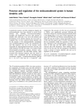
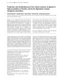
a-oxidation process. Our present knowledge of the enzy-
matic sequence is shown in Fig. 2.
In a first step the 3-methyl-branched fatty acid is activated
to the corresponding CoA-ester by an acyl-CoA synthetase
which is most probably present in the peroxisomal
membrane. It is not yet clear which synthetase is responsible
for the activation step: a nonspecific long chain fatty acyl-
CoA synthetase [12], a specific phytanoyl-CoA synthetase
[13] or a very long chain fatty acyl-CoA synthetase [14].
The second step is responsible for the iron dependence of
the pathway [15], which had been described by several
authors in the past but was regarded as doubtful concerning
its physiological relevance [16,17]. In this step the
3-methylacyl-CoA is hydroxylated in position 2 by a
dioxygenase, which is dependent on molecular O
2
, iron,
2-oxoglutarate, ascorbate, ATP/GTP and Mg
2+
[18–21].
This dioxygenase, named phytanoyl-CoA hydroxylase
(PAHX), contains a PTS2-signal and is present in the
peroxisomal matrix [22,23]. The product of the reaction
Fig. 1. Chemical structures of chlorophyll, phytol and phytanic acid
(3,7,11,15-tetramethylhexadecanoic acid).
Fig. 2. a-Oxidation of 3-methyl-branched fatty acids. The scheme
represents the a-oxidation pathway of phytanic acid. The numbers
indicate the enzymes catalysing the different steps: (1) acyl-CoA syn-
thetase; (2) phytanoyl-CoA hydroxylase (PAHX); (3) 2-hydroxy-
phytanoyl-CoA lyase (2-HPCL); (4) aldehyde dehydrogenase; and (5)
formyl-CoA hydrolase.
1620 M. Casteels et al. (Eur. J. Biochem. 270)FEBS 2003

catalysed by PAHX is a 2-hydroxy-3-methylacyl-CoA, or, if
phytanic acid is the substrate, 2-hydroxyphytanoyl-CoA.
The PAHX gene is located on chromosome 10 [22], and
mutations of this gene are probably the most frequent cause
of ARD [22–25]. Structure-function analysis of PAHX
further revealed that at least four different types of
mutations can cause loss of enzyme activity [25].
In the third step, 2-hydroxy-3-methylacyl-CoA is cleaved
in the peroxisomal matrix [26,27] by 2-hydroxyphytanoyl-
CoA lyase (2-HPCL), which uses thiamine pyrophosphate
(TPP) as cofactor [26]. Products of this reaction are formyl-
CoA [28] and a 2-methyl-branched fatty aldehyde (pristanal
when 2-hydroxyphytanoyl-CoA is cleaved) [29,30], both of
which had been identified before the discovery of the lyase
(see below).
The 2-methyl-branched fatty aldehyde is subsequently
dehydrogenated by an NAD
+
-dependent aldehyde dehy-
drogenase to a 2-methyl-branched fatty acid (pristanic acid
in the case of pristanal), which can be activated to the
corresponding acyl-CoA ester. This CoA-ester can then
enter the peroxisomal b-oxidation sequence. The 2-methyl
aldehyde dehydrogenase activity is located in the peroxi-
somal matrix according to Croes et al.[29]andinthe
endoplasmic reticulum (microsomes) according to Verho-
even et al. [30]. It remains at present unclear which aldehyde
dehydrogenase is involved. Measurements in Sjo
¨gren–
Larsson syndrome (SLS) fibroblasts, the microsomal alde-
hyde dehydrogenase of which is deficient, show only a 30%
decrease in dehydrogenation rates of pristanal [31,32] and
make an exclusive role of a microsomal aldehyde dehy-
drogenase unlikely.
The major part of formyl-CoA is enzymatically converted
to formate in peroxisomes [28]. It was shown previously [33]
that in rats, aminotriazole, known as an inhibitor of
catalase, had little effect on the conversion of
14
C-formate to
CO
2
(but decreased the rates of a-oxidation by 90%). In rat
formate is metabolized by two pathways: the catalase
pathway and the tetrahydrofolate pathway, important in
one carbon-metabolism [34]. The data on aminotriazole
indicate that at least in the rat the catalase pathway is of no
paramount importance, and suggest that the tetrahydro-
folate pathway is quantitatively more important for formate
metabolism [33]. We studied the conversion of
14
C-formate
to
14
CO
2
in rat and found it to be localized mainly in the
cytosolic fraction, and to be stimulated by NAD
+
[19]. No
further work on the fate of formate as a product of
a-oxidation has been published since. Nothing is known
on the export of formate from the peroxisome, but it is
supposed that formate, as well as other small organic acids
can leak from the peroxisomes [35].
Table 1 gives an overview of the presently known
characteristics of the four main enzymes of the a-oxidation
pathway.
Stereospecificity of the a-oxidation pathway
Phytol has two chiral centres, one at carbon 7 and one at
carbon 11, both of which are of the R-configuration [41].
Non-specific reduction of the double bond in phytol leads
to the production of two diastereoisomers: (3S,7R,11R)-
and (3R,7R,11R)-phytanic acid [42]. Phytanic acid
from all common sources is a mixture of these two
Table 1. Properties of the enzymatic steps/enzymes of the a-oxidation pathway. The table gives an overview of the present knowledge of some of the
properties of the enzymes involved in the initial degradation of 3-methyl-branched fatty acids in humans. See text for details.
Acyl-CoA
synthetase
Phytanoyl-CoA hydroxylase
(PAHX)
2-Hydroxyphytanoyl-CoA lyase
(2-HPCL)
Aldehyde
dehydrogenase
Accession number O14832 Q9UJ83
Gene mapping 10p15.1 [22] 3p25 [39]
Mass of subunit Unprocessed: 38 556/
mature: 35 436 Da
Monomer: 63 732 Da
Cofactors ATP, CoA, Mg
2+
O
2
,Fe
2+
, ascorbate, 2-oxoglutarate
[18,19]
TPP, Mg
2+
[26] NAD
+
[29,30]
ATP/GTP, Mg
2+
[21]
K
m
for CoA-ester 29.5 ± 1.7 lM
b
[36] 15 lM
d
[26]
Subcellular localization Peroxisomal
membrane [12–14]?
Peroxisomal matrix [19,20] Peroxisomal matrix [26,27] Peroxisomes
[29,32]?
Targeting PTS-2 [22,23] PTS-1 [26]
Stereochemistry Not stereospecific
a
3Rfi2S,3R;3Sfi2R,3S
c
[37,38] Not stereospecific [38] Unknown
e
Heterologous expression
systems
E. coli Mammalian cells,
S. cerevisiae [26,39]
Mutagenesis studies Yes [22,24,25] No
Structural information Yes [25] TPP binding domain [26,39]
a
As both phytanic acid and phytanoyl-CoA are racemic at position 3, it is supposed that the acyl-CoA synthetase is not stereospecific.
Whether the activation rates for the R- and S-isomers are different, as shown for the conversion of 2-methyl-branched fatty acids to the
corresponding acyl-CoA esters in human liver [40], is not known.
b
K
m
determined for phytanoyl-CoA with recombinant PAHX, in the
presence of equimolar concentrations of SCP-2.
c
Phytanoyl-CoA hydroxylase is not stereospecific, but the configuration of the methyl-
branch at position 3 determines the orientation of the hydroxy-group at position 2. Eventually, only (2R,3S) and (2S,3R) isomers are
formed.
d
K
m
determined for 2-hydroxy-3-methyl-C16-CoA with partially purified enzyme.
e
Although nothing is known about the stereo-
specificity of aldehyde dehydrogenases, it can be postulated from all different data concerning the stereochemistry of the a-oxidation
pathway that this last step of the reaction sequence is not stereospecific.
FEBS 2003 Alpha-oxidation and its TPP dependence (Eur. J. Biochem. 270) 1621

diastereoisomers and their ratios are variable and depend-
ent on sample origin. As the a-oxidation product of
racemic phytanic acid, pristanic acid, is racemic at
position 2, it seems obvious that both stereoisomers can
undergo a-oxidation without a previous isomerization at
the initial 3-methyl-branch. Croes et al.[38]provided
indeed evidence that isomerization of the 3-methyl-branch
during a-oxidation does not occur and that the configur-
ation of the methyl-branch is conserved throughout the
whole a-oxidation process. It was also demonstrated that
the configuration of the 3-methyl-branch does not influ-
ence the rate of a-oxidation, but determines the orienta-
tion of the 2-hydroxylation. This explains the formation
of only the (2S,3R)and(2R,3S) isomers of 2-hydroxy-3-
methylhexadecanoyl-CoA by purified peroxisomes, despite
the experimental finding that all four possible isomers
(although each to a different extent) can be metabolized
[38]. The data of Croes et al. confirm the earlier findings
of Tsai [37], who concluded that the introduction of the
hydroxy group at position 2 is stereospecific and deter-
mined by the configuration of the methyl group at
position 3. The stereochemistry of the a-oxidation path-
way is presented in Fig. 3.
The lack of stereospecificity of the a-oxidation pathway is
in contrast with the stereospecificity of both the peroxisomal
and mitochondrial b-oxidation systems. As a-oxidation of
phytanic acid results in both stereoisomers of pristanic acid,
the produced (2R,6R,10R) isomer has to undergo racemi-
zation at carbon 2 before b-oxidation can take place. In
addition, racemization at the other chiral centres is an
essential step for the further b-oxidation of the intermediate
a-methyl fatty acids [40].
2-HPCL: a thiamine dependent enzyme
2-HPCL identification
After the discovery by Poulos et al. [11] of formate as a
product of a-oxidation in fibroblasts, a finding which was
confirmed in isolated hepatocytes [33], Croes et al. found in
1997 that not formate (or CO
2
) was the primary end
product but formyl-CoA [28]. This finding led several
authors to propose a reaction mechanism in which the
other product would be a 2-methyl-branched aldehyde
(or pristanal in case phytanic acid is the substrate). Soon,
the formation of a 2-methyl-branched aldehyde, using
2-hydroxy-3-methylacyl-CoA or 2-hydroxyphytanoyl-CoA
as precursor, was demonstrated simultaneously by Croes
et al. [29] and Verhoeven et al.[30].
Foulon et al. used 2-hydroxy-3-methylhexadecanoyl-
CoA as substrate for studying the third reaction of the
a-oxidation pathway, and measured formate (together with
formyl-CoA, which is, partly enzymatically, converted to
formate) as the reaction product [26].
Subcellular fractionation studies in rat liver demonstra-
ted that the lyase activity colocalized with catalase in the
peroxisomal fraction [26]. Hence, isolation of the pre-
sumptive cleavage enzyme was started from the matrix
protein fraction of isolated rat liver peroxisomes. The
purified lyase was made up of four identical subunits of
63 kDa. Formyl-CoA and 2-methylpentadecanal (meas-
ured by GC-analysis) were identified as reaction products
when the enzyme (in the presence of thiamine pyrophos-
phate (TPP), see below) was incubated with 2-hydroxy-
3-methylhexadecanoyl-CoA as the substrate. Quantitative
measurements of both reaction products further confirmed
the stoichiometry of the cleavage step. Incubations in the
presence of NAD
+
(a cofactor for fatty aldehyde
dehydrogenation [43]) did not alter the amount of formate
(formyl-CoA) and 2-methyl-pentadecanal formed, and no
conversion of the aldehyde to a fatty acid could be
demonstrated indicating that this reaction is performed by
a separate enzyme. Hence, as the only activity of the
purified enzyme is the specific cleavage of a carbon-carbon
bond, it was called 2-hydroxyphytanoyl-CoA lyase or
2-HPCL [26].
An apparent Km of 15 l
M
for 2-hydroxy-3-methylhexa-
decanoyl-CoA was calculated. The pH optimum was
between 7.5 and 8.0 [26].
TPP-dependence of 2-HPCL
Originally, 2-HPCL had been purified in the absence of TPP
and the enzyme lost virtually all of its activity during
purification. The amino-acid sequences of tryptic peptides
from the purified and barely active 2-HPCL suggested that
the cleavage enzyme is related to a putative Caenorhabditis
elegans protein that displays homology to bacterial oxalyl-
CoA decarboxylases [44,45]. These enzymes, which have
hitherto only been described in bacteria, catalyse the TPP-
dependent decarboxylation of oxalyl-CoA to formyl-CoA
Fig. 3. Stereochemistry of the a-oxidation pathway. The scheme rep-
resents the a-oxidation pathway of (3R,3S)-methylhexadecanoic acid
and the stereochemical configuration of the intermediates involved.
The numbers indicate the enzymes catalysing the different steps: (1)
acyl-CoA synthetase; (2) phytanoyl-CoA hydroxylase (PAHX); (3)
2-hydroxyphytanoyl-CoA lyase (2-HPCL); (4) aldehyde dehydro-
genase; (5) formyl-CoA hydrolase; (6) acyl-CoA synthetase; and (7)
2-methylacyl-CoA racemase, responsible for the conversion of the
2R-methylacyl-CoA into the 2S-methylacyl-CoA, as only the S-isomer
can undergo b-oxidation.
1622 M. Casteels et al. (Eur. J. Biochem. 270)FEBS 2003

and CO
2
[44,45]. This homology suggested that also
2-HPCL might require TPP, an unexpected cofactor for
a-oxidation.
In the presence of 0.8 m
M
Mg
2+
, optimum activity for
the purified enzyme was reached at 20 l
M
TPP (K
m
for
TPP ¼8.43 l
M
). Only minor stimulation by TPP was
noted in a fresh liver homogenate (1.3 fold), and a gradually
more potent stimulation of the lyase activity was observed
as the enzyme became more purified. Hence, optimal lyase
measurements have to be performed in the presence of TPP
and MgCl
2
.
cDNA and amino-acid sequence
The cDNA sequence of the human lyase contains an open
reading frame of 1734 nucleotides encoding a polypeptide
with a calculated molecular mass of 63 732 Da. Similarly
to other TPP-dependent enzymes (e.g. bacterial oxalyl-
CoA decarboxylases), a TPP-binding consensus domain
could be identified in the C-terminal part of the lyase. The
corresponding peptide sequences of this domain in the
human, mouse and rat enzyme, comply exactly with
the TPP consensus domain of pyruvate decarboxylase of
Saccharomyces cerevisiae, acetolactate synthase of Escheri-
chia coli, oxalyl-CoA decarboxylase of Oxalobacter formi-
genes and the putative oxalyl-CoA decarboxylases of
Caenorhabditis elegans and S. cerevisiae [44,45] (Fig. 4
[46]).
Substrate specificity of 2-HPCL
Recombinant human protein, expressed in mammalian cells
or in a yeast system, clearly exhibited lyase activity, whereas
expression in a bacterial system did not result in a
functionally active enzyme [26].
Study of the substrate specificity of recombinant
human lyase revealed that the enzyme is not only active
towards 2-hydroxy-3-methylhexadecanoyl-CoA (the
analogue of 2-hydroxyphytanoyl-CoA), but also,
although to a minor extent, towards 2-hydroxyoctadeca-
noyl-CoA (± 12% of control activity) at equal substrate
concentration. The latter compound, however, as well as
2-hydroxyhexadecanoyl-CoA, effected a very strong inhi-
bition on the cleavage of 2-hydroxy-3-methylhexadeca-
noyl-CoA, most probably due to competition [39]. No
activity at all was seen with 2-hydroxy-3-methylhexadeca-
noic acid, 3-methylhexadecanoic acid or 3-methylhexa-
decanoyl-CoA, indicating that both a 2-hydroxy group
and a CoA-moiety, but not a 3-methyl-branch, are
necessary for lyase activity [39].
Identification of novel PTS
At first glance, the Hs 2-HPCL sequence did not contain a
C-terminal or N-terminal peroxisome targeting signal
(PTS). As the C. elegans orthologue ends in a putative
PTS1 (SKM) and as PRL, the C-terminal tripeptide of the
S. cerevisiae orthologue, had been shown to bind to the
human PTS1 import receptor [47], the C-terminal sequence
SNM, which is also conserved in the mouse counterpart,
was considered to have a targeting function. Transfection
studies with constructs coding for 2-HPCL fused to GFP
revealed that the fluorescence localized to peroxisomes in
fibroblasts from PEX5
+/–
miceandtothecytosolin
fibroblasts from PEX5
–/–
mice [26]. The latter mice lack the
PTS1 receptor (Pex5p) and do not import PTS1-containing
proteins into their peroxisomes [48]. As a GFP-construct
containing only the last 5 amino acids of 2-HPCL localized
to peroxisomes in fibroblasts from normal mice, we can
conclude that targeting information is present within this
pentapeptide and that SNM, preceded by a positive charge,
is a hitherto unrecognized PTS1 [26].
Reaction mechanism of 2-HPCL
A 2-hydroxy carboxyl compound (instead of a 2-keto
carboxyl compound) is a rather unusual substrate for
thiamine dependent decarboxylases. In all TPP-dependent
reactions described so far, catalysis involves activation of
the C2-H of the thiazole ring, followed by a nucleophilic
attack at the carbonyl carbon of the substrate [49]. By use of
nuclear magnetic resonance spectroscopy, it has been shown
that in the enzyme-bound state, the C2 proton of TPP is
undissociated, but that the protein component dramatically
accelerates the deprotonation, producing an intermediate
C2 carbanion with a short lifetime [50,51]. Most likely, the
formation of a carbanion is also required for the cleavage of
2-hydroxy-3-methylacyl-CoAs by 2-HPCL (Fig. 5). How-
ever, this carbanion will attack carbon 1 of the substrate,
which is highly reactive due to the nature of the thioester
bond. Ultimately this leads to the formation of formyl-CoA
and a 2-methyl-branched fatty aldehyde.
Fig. 4. Alignment of the cofactor-binding consensus domain in TPP-dependent enzymes. An alignment [26] is given of the cofactor-binding consensus
domain in several TPP-dependent enzymes (Sc PDC: S. cerevisiae pyruvate decarboxylase; Ec ALS: E. coli acetolactate synthase; Of OCD:
O. formigenes oxalyl-CoA decarboxylase) and in Hs 2-HPCL and its homologues in lower organisms (Ce OCD: C. elegans putative oxalyl-CoA
decarboxylase; Sc OCD: S. cerevisiae putative oxalyl-CoA decarboxylase). The TPP-binding consensus motif, here represented with 10 residues
upstream and downstream, is defined as G-D-G-x-(24–27)-N-N [46]. About 10 residues downstream of the G-D-G sequence, a negatively charged
amino acid is present (E or D), followed about 5 and 11 residues further by a generally conserved alanine and proline residue, respectively.
Immediately preceding the N-N sequence is a cluster of 6 or 7 largely hydrophobic side-chains.
FEBS 2003 Alpha-oxidation and its TPP dependence (Eur. J. Biochem. 270) 1623


![Bộ Thí Nghiệm Vi Điều Khiển: Nghiên Cứu và Ứng Dụng [A-Z]](https://cdn.tailieu.vn/images/document/thumbnail/2025/20250429/kexauxi8/135x160/10301767836127.jpg)
![Nghiên Cứu TikTok: Tác Động và Hành Vi Giới Trẻ [Mới Nhất]](https://cdn.tailieu.vn/images/document/thumbnail/2025/20250429/kexauxi8/135x160/24371767836128.jpg)


