Properties of group I allergens from grass pollen and their relation
to cathepsin B, a member of the C1 family of cysteine proteinases
Kay Grobe
1
, Marco Po¨ ppelmann
2
, Wolf-Meinhard Becker
2
and Arnd Petersen
2
1
University of California San Diego, La Jolla, USA;
2
Forschungszentrum Borstel, Borstel, Germany
Expansins are a family of proteins that catalyze pH-
dependent long-term extension of isolated plant cell walls.
They are divided into two groups, aand b, the latter con-
sisting of the grass group I pollen allergens and their veget-
ative homologs. Expansins are suggested to mediate plant
cell growth by interfering with either structural proteins or
the polysaccharide network in the cell wall.
Our group reported papain-like properties of b-expansin of
Timothy grass (Phleum pratense) pollen, Phl p 1, and sug-
gested that cleavage of cell wall structural proteins may be
the underlying mechanism of expansin-mediated wall
extension. Here, we report additional data showing that
b-expansins resemble ancient and modern cathepsin B,
which is a member of the papain (C1) family of cysteine
proteinases. Using the Pichia pastoris expression system, we
show that cleavage of inhibitory prosequences from the
recombinant allergen is facilitated by its N-glycosylation and
that the truncated, activated allergen shows proteolytic
activity, resulting in very low stability of the protein. We
also show that deglycosylated, full-length allergen is not
activated efficiently and therefore is relatively stable. Motif
and homology search tools detected significant similarity
between b-expansins and cathepsins of modern animals as
well as the archezoa Giardia lamblia, confirming the presence
of inhibitory prosequences, active site and other functional
amino-acid residues, as well as a conserved location of these
features within these molecules. Lastly, we demonstrate by
site-directed mutagenesis that the conserved His104 residue
is involved in the catalytic activity of b-expansins. These
results indicate a common origin of cathepsin B and
b-expansins, especially if taken together with their previously
known biochemical properties.
Keywords: cathepsin B; cell wall; expansin; group I allergen;
proteinase.
Pollen triggers allergic reactions such as hayfever and
seasonal asthma, which affect up to 25% of adults in
industrialized countries. Of the diverse allergens of grass
pollen, group I allergens are the major components [1] to
which most patients possess specific IgE antibodies. They
are glycoproteins of about 30 kDa with a carbohydrate
content of 5% and are exclusively expressed in pollen of
all grasses [2,3]. Grass group I allergens constitute the
b-expansin subfamily of expansins [4]. Besides functioning
as mediators of acid-induced cell wall loosening in plants,
expansins are also essential for fruit ripening [5–8], fertiliza-
tion [9] and differentiation [10,11]. However, the mechanism
by which they mediate plant cell wall growth is highly
controversial. Three main hypotheses have been put
forward to explain their wall-loosening properties.
Several reports have suggested that expansins may
interfere with hydrogen bonds between cellulose and
hemicellulose microfibrils by a unique and novel mechan-
ism, reducing the rigidity of the cell wall [12]. This was
supported by experiments showing that a-expansins asso-
ciate with hemicellulose-coated cellulose microfibrils in vitro
[13]. Expansins were therefore suggested to possess a
C-terminal cellulose-binding domain (CBD) resembling
bacterial CBDs, based on the spacing between highly
conserved Trp (W) residues. They were also reported to be
able to induce loosening of cellulosic paper [14]. On the basis
of these findings, expansins were suggested to bind cellulose
fibrils with their C-terminal CBDs, allowing interference
with hydrogen bonds between wall polysaccharides via their
N-terminal domain. The resulting weakening of the poly-
saccharide network was suggested to subsequently allow
turgor-driven extension (relaxation) of the structure.
Another model indicates possible hydrolysis of polysac-
charides, based on a 30% sequence similarity within a
restricted region between expansins and a small (F45) family
of fungal endoglucanases. However, hydrolytic activity (exo
and endo type) of expansins on polysaccharides has never
been detected, and F45 hydrolases fail to stimulate plant cell
wall extension [15,16]. Transglycosidase activity, another
proposed mechanism, has also not been established.
A summary of these models was recently published [17].
The third hypothesis proposed that expansins possess C1
(papain) proteinase family-related proteolytic activity,
mediating plant cell wall loosening by cleavage of structural
wall proteins, namely the extensins (hydroxyproline-rich
glycoproteins) and associated proteins [18]. This concept
requires a fundamental revision of the model of plant cell
wall organization and growth. In accordance with this
hypothesis, several potent allergens have been identified as
Correspondence to K. Grobe, UCSD Cancer Center, University of
California San Diego, 9500 Gilman Drive, M/C 0687, La Jolla, CA
92093-0687, USA. Fax: + 1 858 534 5611, Tel.: + 1 858 822 1102,
E-mail: kgrobe@ucsd.edu
Abbreviations: Phl p 1, grass group I allergen derived from Phleum
pratense; Hol l 1, grass group I allergen derived from Holcus lanatus;
CBD, cellulose-binding domain.
Enzymes: cathepsin B (EC 3.4.22.1); papain (EC 3.4.22.2); bromelain
(EC 3.4.22.32).
(Received 31 October 2001, revised 18 February 2002, accepted 25
February 2002)
Eur. J. Biochem. 269, 2083–2092 (2002) FEBS 2002 doi:10.1046/j.1432-1033.2002.02856.x

proteinases, and their function suggested to contribute to
their allergenicity. Thus, a proteinase function of group I
allergens could explain the high prevalence of allergic
individuals sensitized to these molecules. This model of
expansins acting as proteinases was based on the finding
that the recombinant b-expansin/allergen of Timothy grass,
P. pratense, expressed in the yeast Pichia pastoris, catalyzed
the degradation of a synthetic substrate containing a
papain-cleavage site, as well as other proteins. Moreover,
a protein with strong proteolytic activity was coeluted with
the recombinant allergen after affinity purification using the
mAb IG12 [18]. The natural allergen Phl p 1 was also found
to be capable of degrading a synthetic substrate at a papain-
cleavage site after incubation under acidic and reducing
conditions, which are known to activate C1 proteinases. At
that time, limited sequence similarity to motifs surrounding
the active-site residues of papain was also established.
However, the proposed putative proteinase identity of
expansins seemed to be at odds with reports in the literature,
e.g. that expansins loosened cellulosic paper [14] and that
proteinases did not mediate plant cell wall extension in vitro
[19,20].
EXPERIMENTAL PROCEDURES
Site-directed mutagenesis and subcloning
Phl p 1 cDNA (GeneBank/EMBL accession number
Z27090) was ligated in pBluescript (Stratagene, La Jolla,
CA, USA). Elimination of the putative N-glycosylation site
NIT to QIT in position 9 of the mature protein product was
performed by PCR with modified sense primer Phl p 1 Q
(5¢-ATCCCCAAGGTCCCCCCCGGCCCGCAGATC
ACG-3¢) Here, AAC coding for Asn (N) in the wild-type
sequence Phl p 1 N (5¢-ATCCCCAAGGTCCCCCCCGG
CCCGAACATCACG-3¢) was changed into CAG coding
for Gln (Q). PCR in combination with antisense primer
Phl p 1 rev (5¢-TGGTGATCTTCTCGAGTCAAAATTG
AACTT-3¢), containing a XhoIsite,wasperformedusing
Pfu polymerase (Stratagene) under the following conditions:
Hotstartfor5minat95C; followed by 20 cycles
consisting of 95 Cfor30s,70Cfor1minand72C
for 2 min; and terminated by an extension step for 5 min at
72 C. The reaction mixture consisted of 10 ng template
DNA, 0.5 m
M
dNTPs and 1 l
M
each primer in a total
volume of 20 lL. The PCR products were purified using the
PCR Purification Kit (Qiagen, Hilden, Germany), sub-
cloned into EcoRV-digested pBluescript, and sequenced.
Inserts coding for rPhl p 1 N and Q were then separated
using EcoRV and XhoI; the latter restriction site was then
blunted with Pfu polymerase. This was followed by ligation
into SnaBI-digested vector pPIC9 (P. pastoris Expression
Kit; Invitrogen, San Diego, CA, USA), directly after the
a-mating factor leader sequence, which mediates export of
rPhl p 1 into the medium. Correct orientation of the
constructs was confirmed by restriction analysis with
subsequent sequencing and resulted in pPIC9 Phl p 1 N
and Q, which were used for transformation after lineariza-
tion with BglII. Mutagenesis of His104 to Val was
performed by PCR using the primer phlp1s (5¢-ACC
CGGGAGGAGGAATCCCCAAGGTCCCCCCCG-3¢)
with phlp1-Has (5¢-TACGTACGCGGCGATGGGCTCC
TCG-3¢), and phlp1as2 (5¢-AGAATTCTCAGTCCTT
GGCCTCGCCCTTG-3¢) with phlp1-Hs (5¢-TACGTAT
TCGACCTCTCCGGCATCGC-3¢).Thewild-typecontrol
was produced by using the primers phl p1s with phlp1as2.
PCR products were obtained as described above,
TA-cloned (pGEM, Promega, Madison, WI, USA), and
sequenced. To construct the mutated form, fragments were
released using the restriction enzymes SmaIandSnaBI or
EcoRI and SnaBI, respectively, and both ligated in pBS,
which had previously been linearized using the restriction
enzymes EcoRI and SmaI. After transfection, positive
clones were sequenced. All restriction enzymes were
obtained from New England Biolabs, Beverly, MA, USA.
Pichia
-transformation, identification of transformants,
and expression
Transformation of P. pastoris strains GS115 and PEP4-
(thus proteinase A)-deficient SMD1168 (Invitrogen) was
performed by electroporation (Gene Pulser, Bio-Rad,
Hercules, CA, USA) at 1.5 kV, 200 Wand 25 lFwith
5lg linearized pPIC9 Phl p 1 per transformation, using
1-mm cuvettes (Bio-Rad). Transformants were identified by
Mut
s
phenotype (methanol utilization slow) and PCR with
Phl p 1-specific primers. Cells were grown in BMDY
(buffered minimal glucose + yeast extract; 2% bactotryp-
tone, 1% yeast extract, 1.3% yeast nitrogen base with
ammonium sulfate, 1% glucose, 0.00004% biotin in 0.1
M
potassium phosphate buffer, pH 6.0) for 2 days, transferred
in BMGY [buffered minimal glycerol (1%) + yeast extract]
for 1 day and subsequently induced in BMMY Mod
[buffered minimal methanol (0.5%) + yeast extract;
10 gÆL
)1
milk powder, 1 gÆL
)1
cysteine, 0.5% glycerol, in
0.1
M
potassium phosphate buffer, pH 5.0] at a cell density
of 10 D
600
units for 1 day, all at 30 Candshakingat
150 r.p.m. in baffled flasks. Expression and export of
rPhl p 1 was confirmed by dot-blotting of culture superna-
tant on to nitrocellulose membranes and detection with
Phl p 1-specific mAbs IG12 [2], Bo14 and HB7. Cells were
centrifuged at 1500 gfor 10 min. The supernatant was
collected, concentrated 20 times using Amicon concentra-
tors (10-kDa membrane filters; Amicon, Beverly, MA,
USA), and stored at )20 C. The supernatant was washed
twice with 0.1
M
potassium phosphate buffer at pH 5.0.
Baculovirus expression of rPhl p 1 and rPhl p 1*His104
Sequences coding for rPhl p 1 as well as rPhl p 1*His were
released from pBS or pGEM and ligated into the expression
vector pAcSecG2T (Pharmingen, San Diego, Ca, USA)
using the restriction endonuclease sites SmaIandEcoRI.
Recombinant virus was produced and amplified according
to the manufacturer’s instructions (Pharmingen). Briefly,
recombinant virus was produced by cotransfection of Sf9
cells with linearized BaculoGold virus and pAcSecG2T-
Phlp1 or pAcSecG2T-Phlp1*His. AcNPV wild-type virus
was used as a control. Pure virus clones were isolated by
plaque purification. Three clones each were tested for levels
of expression, which was confirmed to be identical among a
given construct. Virus was amplified until a titer of 10
9
mL
)1
was achieved. An infectious dose of 10 virus particles per cell
(multiplicity of infection ¼10) was used for infection of
cells (1 ·10
7
Sf9 cells in a 10-cm dish). Expressing cells were
either lysed 3 days after infection directly in SDS buffer, or
2084 K. Grobe et al.(Eur. J. Biochem. 269)FEBS 2002

recombinant GST fusion protein was purified from the
medium using glutathione/agarose (Sigma, St Louis, MO,
USA),elutedin50m
M
acetic acid/sodium acetate buffer,
pH 6.0, containing 5 m
M
GSH (Sigma), and processed as
described below. Recombinant protein was detected after
Western blotting using a monoclonal antibody to GST
(Pharmingen).
SDS/PAGE and Western-blot analysis
Proteins were separated by discontinuous SDS/PAGE
(T ¼15%, C ¼4%) and transferred to nitrocellulose
membrane by semidry blotting [21]. Immunostaining was
performed with mAbs IG12, Bo14, and HB7, binding to the
peptide epitopes K(48)PPFS(52) (unpublished result), a
C-terminal peptide and an N-terminal peptide, respectively
(A. Petersen, personal communication). Subsequently alka-
line phosphatase-conjugated goat anti-(mouse IgG and
IgM) Ig (Dianova, Hamburg, Germany) was added before
development (Nitro Blue tetrazolium/5-bromo-4-chloroin-
dol-2-yl phosphate). Polyacrylamide gels were stained with
Coomassie Brilliant Blue R250 [21]. For dot-blots, probes
were applied directly to nitrocellulose and developed.
Zymograms
Zymograms were run as for SDS/PAGE, with 1% evap-
orated milk powder copolymerized in the resolving gel [22].
After electrophoresis, the gels were incubated in buffer
containing 0.1
M
glycine, 10 m
M
Ca
2+
,5m
M
dithiothreitol
and 10 m
M
cysteine, pH 3.6, for 16 h, followed by staining
with Coomassie Blue. Protein probes (rPhl p 1 Q and
rPhl p 1 N) were incubated in SDS sample buffer under
nonreducing conditions at 65 C for 10 min, before being
loaded on to the Zymogel.
Preparative isoelectric purification of allergen
Concentrated P. pastoris expression supernatant was cen-
trifuged at 3500 gfor 30 min, filtered through a 0.2-lm
filter, and dialyzed overnight against double-distilled water
at 4 C. A preparative Rotophor cell (Bio-Rad) was
assembled according to the manufacturer’s instructions,
and precooled to 4 C. Ampholyte (pH 2–11; Serva,
Heidelberg, Germany) was added to 60 mL dialyzed
expression supernatant to a 2% final concentration, and
separation was achieved at 12 W constant power for 5 h at
4C. The fractions were collected, and the respective pH
values determined; the fractions were stored at )20 C until
analyzed.
Deglycosylation of Phl p 1 N with N-glycosidase A
Deglycosylation of proteins in the Phl p 1 N expression super-
natant was achieved using N-glycosidase A (Boehringer-
Mannheim, Mannheim, Germany). Twenty microliters
deglycosylation buffer (100 m
M
citrate/sodium dihydrogen-
phosphate buffer, pH 5.0, 1 m
M
dithiothreitol) and 0.3 mU
N-glycosidase A were added to 10 lL expression superna-
tant (25 lg total protein) and incubated at 37 Covernight.
Buffer alone served as the negative control. Deglycosylated
and control supernatant was subsequently analyzed in
zymograms.
Alignments and computer analysis
Sequence data were analyzed with
PCGENE
software (Intel-
ligenetics, Geel, Belgium). Alignments to conserved
sequences of cysteine proteases and among Phl p 1 and
Hol l 1 were performed using
WU
-
BLAST
p2 (PAM270
matrix), modified manually and displayed using
CLUSTALW
and
SEQVU
software. Motifs were analyzed by the
IMPALA
BLOCKS
search tool using the
BLOSUM
62 matrix. The
percentage of identical amino acids between each pair of
proteins was calculated by setting the number of compar-
able (e.g. within the same position) amino acids at 100%.
RESULTS
Expansins show significant sequence similarities
to cysteine proteinases, especially cathepsin B
Analysis of the amino-acid sequence of Phl p 1 for con-
served, functional motifs using the
IMPALA BLOCKS
search
tool resulted in the following order of hits: (1) major pollen
allergen Lol p 1 signature (e
)103
); (2) expansin signature
(1e
)13
); (3) allergen pollen CIM1/Hol l 1 signature (0.009);
(4) eukaryotic thiol (cysteine) proteases active-site signature
IPB000169 (1.6). Moreover, the
WU
-
BLAST
p2 program,
employing the PAM270 matrix which allows detection of
distantly related proteins, computed similarities between
expansins and cathepsin (Q10834, 1.9e
)7
)aswellasother
cysteine proteinases (Q40261, 4.2e
)6
). Additional cathep-
sin B-like cysteine proteinases as well as cysteine proteinases
from Giardia lamblia could also be detected [23]. From these
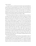
findings, an alignment of Phl p 1 with Gallus gallus and
G. lamblia cysteine proteinases was generated (Fig. 1). The
identity between Phl p 1 and CP2 of Giardia within com-
parable regions was 21%, and the combined identity and
similarity amounted to 34%. The identity between Hol l 1
and CP2 was 22%, between Phl p 1 and CP1 as well as
CP3 19%, and between Phl p 1 and CatB 15%. Moreover,
the putative active-site amino acid Cys72 of Phl p 1 and
Hol l 1 is very similarly positioned if compared with the
Giardia proteinases 1 (residue 71), 2 (residue 67) or 3 (residue
66). The catalytically essential Trp residues are also similarly
located within the C-terminal region. Another striking
feature is the well-conserved relative location of Cys41, 57,
69, 72, 83 and 139 when compared with the proteinases of
G. lamblia and G. gallus. All Cys and Trp residues are
absolutely conserved in a-expansins and b-expansins as well
as in the C1 cysteine proteinases. Other highly conserved
amino acids of cathepsin B-like proteinases are also well
conserved in most b-expansins, notably the Pro2 residues and
the Glu216 residue. However, the amino acids His158 and
Asn193 (C1 numbering; cathepsin B: His260 and Asn280) of
the catalytic triad are not present in a comparable position in
C1 proteinases and group I allergens. Subsequently, func-
tional tests on recombinant (r) Phl p 1 were refined to further
explore the biochemical function of group I allergens.
Expression of glycosylated and nonglycosylated
rPhl p 1 reveals differing stability of these allergens
The expression of rPhl p 1 in the yeast P. pastoris was
attempted to obtain a post-translationally modified allergen
in a natural conformation. In addition to the wild-type
FEBS 2002 Proteolytic properties of grass group I allergens (Eur. J. Biochem. 269) 2085
sequence rPhl p 1 N, which contains an N-glycosylation
site in position 9, another recombinant allergen rPhl p 1 Q
lacking this site was produced by site-directed mutagenesis,
the N-glycosylation site NIT being changed to QIT in the
mutant protein. This allowed absolute discrimination of the
biochemical characteristics between glycosylated and non-
glycosylated allergens compared with other methods, such
as enzymatic deglycosylation or expression in the presence
of tunicamycin. Both proteins were produced by protein-
ase A-deficient P. pastoris SMD1168 cells and secreted into
the medium. The identity of the proteins was confirmed by
Western blotting, using grass group I-specific monoclonal
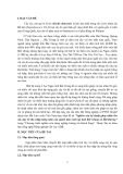
antibodies or sera from patients. Figure 2A shows a
Western blot of rPhl p 1 N, rPhl p 1 Q and the albumin-
expressing control as detected with mAb IG12. The
expressions were performed in a protein-enriched medium
for a limited time (< 24 h). The hyperglycosylated (15%
carbohydrate content) rPhl p 1 N has a size of about
40 kDa, whereas the nonglycosylated form, Phl p 1 Q, has
a size of about 33 kDa. The identity of the respective
N-termini was determined by N-terminal sequencing,
resulting in the sequence Y-I-P-K-V, confirming the correct
processing of the yeast (a-mating factor) signal sequence for
protein export. The additional tyrosine resulted from the
cloning site.
Induction of expression of rPhl p 1 in protein-free
medium, even for a short time (< 24 h), consistently led
to heavy degradation and a low yield of the recombinant
proteins, which was not seen in albumin-expressing
controls. In particular, rPhl p 1 N displayed very low
stability (data not shown). By using a modified, protein-
enriched medium and a short expression time (< 24 h)
expression of nondegraded allergen could be achieved
(Fig. 2A). However, expression over prolonged periods of
time, even in protein-rich medium, did not allow expression
of intact allergens. Elimination of the protecting proteins
during purification also led to degradation of the allergens,
predominantly at low pH. As rPhl p 1 N was consistently
much less stable than rPhl p 1 Q during expression and
purification, and rPhl p 1 N-containing supernatant
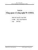
Fig. 1. Alignment of Phl p 1 (GenBank
Z27090), Hol l 1 (Z68893), G. lamblia
cathepsins 1–3 (U83275, U83276 and U83277,
respectively) and G. gallus cathepsin B
(U18083). Areas of identity are boxed, and
areas of similarity are shaded. Binding
epitopes for mAb HB7 (N-terminal), IG12
(central) and Bo14 (C-terminal) are marked.
Important conserved cysteine residues and
other essential amino acids are numbered.
Areas of high similarity exist around Cys69
and Cys72 and around Trp186, Trp193 and
Trp197. The positions of Cys41, 57, 69 and 72
are especially conserved between b-expansins
and cathepsin B. The His104 residue, which is
absolutely conserved in all expansins, is also
present in CP2 of G. lamblia.
Fig. 2. Comparison of the expression and enzymatic activity of recom-
binant Phl p 1 N and Q. Sizes in kDa are indicated. (A) P. pastoris
expression of Phl p 1 N (N) and Phl p 1 Q (Q), as well as albumin (c)
as detected by Western blotting and IG12 binding. IG12 specifically
detected the recombinant allergens, but not any yeast proteins. (B)
Activity of Phl p 1 N (N) or Phl p 1 Q (Q) after prolonged expression
in zymograms. The glycosylated allergen Phl p 1 N shows a much
more pronounced proteolytic activity than the nonglycosylated aller-
gen. (C) Effect of enzymatic deglycosylation on rPhl p 1 N activity in
zymograms. Expression supernatant was applied in lane 1. Expression
supernatant in deglycosylation buffer is applied in lane 2. Addition of
N-glycosidase A leads to abolished proteolytic activity of the allergen
in lane 3. Lane 4, albumin-expressing control.
2086 K. Grobe et al.(Eur. J. Biochem. 269)FEBS 2002
showed much more pronounced proteolytic activity in
zymograms (Fig. 2B), we investigated whether enzymatic
deglycosylation of protein in rPhl p 1 N-containing super-
natant after brief expression would lead to decreased
(rPhl p 1 Q-like) enzymatic activity. As shown in Fig. 2C,
deglycosylation of the allergen by N-glycosidase A indeed
resulted in reduced proteolytic activity in zymograms
compared with rPhl p 1 in buffer without N-glycosidase A.
This result led us to investigate the behavior of full-length
glycosylated vs. nonglycosylated recombinant allergens at
various pH values by preparative isoelectric focusing.
Protein-rich BMMY Mod expressions of intact
rPhl p 1 N and rPhl p 1 Q (as judged by Western blotting,
Fig. 2A) were subjected to isoelectric focusing, concentra-
ting the allergen according to the isoelectric point (pI) of the
molecule. A pH gradient from 2 to 11 was established for
the characterization of Phl p 1. After completion of the run,
the individual fractions were collected and their pH values
determined. Proteins in the respective fractions were subse-
quently analyzed by SDS/PAGE followed by Western
blotting, using mAb IG12 and Bo14 for detection of the
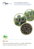
allergen. As can be seen in Fig. 3B,D, rPhl p 1 Q showed
the appropriate size of 33 kDa and was detected by both
antibodies close to the theoretical pI of about 8.0. No
degradation products of the allergen could be detected by
either antibody. However, the expression supernatant
containing rPhl p 1 N showed strong degradation of
the full-length allergen and accumulation of a truncated
15-kDa fragment at about pH 4.5 (Fig. 3A). This sharp
band lacked the N-terminal peptide, as the N-glycosylated
Asn residue was located in position 9 of the molecule, and
glycosylation would have resulted in a fuzzy band (Fig. 1A).
It also lacked the C-terminal peptide normally detected by
mAb Bo14. It is notable that degradation of the glycosyl-
ated form yielded only a limited number of defined
fragments but no smear. Thus, a specific endoproteinase,
and not a nonspecific digestive enzyme or exoproteinase,
probably produced the observed fragments of rPhl p 1 N.
The detection of the respective antibody-binding sites
(HB7: N-terminal; IG12: central; Bo14: C-terminal, Fig. 1)
on dot-blots of various allergen expressions in various
systems confirms that N-terminal and C-terminal peptides
were cleaved off the active allergen Phl p 1 N (Fig. 4). The
allergens were expressed in Pichia over a prolonged period
of time (5 days) in protein-rich medium, concentrated,
washed over 10-kDa membranes to eliminate short peptides
and tested in zymograms (Fig. 2B). Also, a natural allergen
isolated from pollen, Escherichia coli-expressed allergen as
well as Pichia-expressed rPhl p 1 Q were tested in zymo-
grams and turned out to be inactive or weakly active in the
case of rPhl p 1 Q (Fig. 2B). They all possessed binding
sites for antibodies HB7, IG12 and Bo14. However, the
proteolytically active Phl p 1 N was not bound by the
mAbs HB7 and Bo14, indicating truncations on both sides
of the allergen. Supernatant of albumin-expressing P. pas-
toris showed no cross-reactivity with any allergen-specific
monoclonal antibody. It was further tested whether the
truncated IG12-binding forms still possessed proteolytic
activity after affinity purification. IG12 affinity purification
of supernatant containing the truncated, active allergen as
shown in Fig. 4 was thus performed. This led to strong
proteolytic activity of the eluate, whereas supernatants of
albumin-expressing Pichia cells did not show any proteolytic
activity [18]. Even preincubation of the supernatant con-
taining truncated rPhl p 1 N with 0.5% SDS, which
strongly interferes with protein–protein interactions and
thus further reduces the possibility of coelution of another
proteinase, allowed IG12 affinity purification of a proteo-
lytically active allergen (data not shown).
Site-directed mutagenesis was then conducted in order to
identify the catalytic His residue of the C1 catalytic triad.
Analysis of hydrophobicity plots of the Phl p 1 amino-acid
sequence (data not shown) indicated that His104, which is
the only histidine residue conserved in all aand b-expansins,
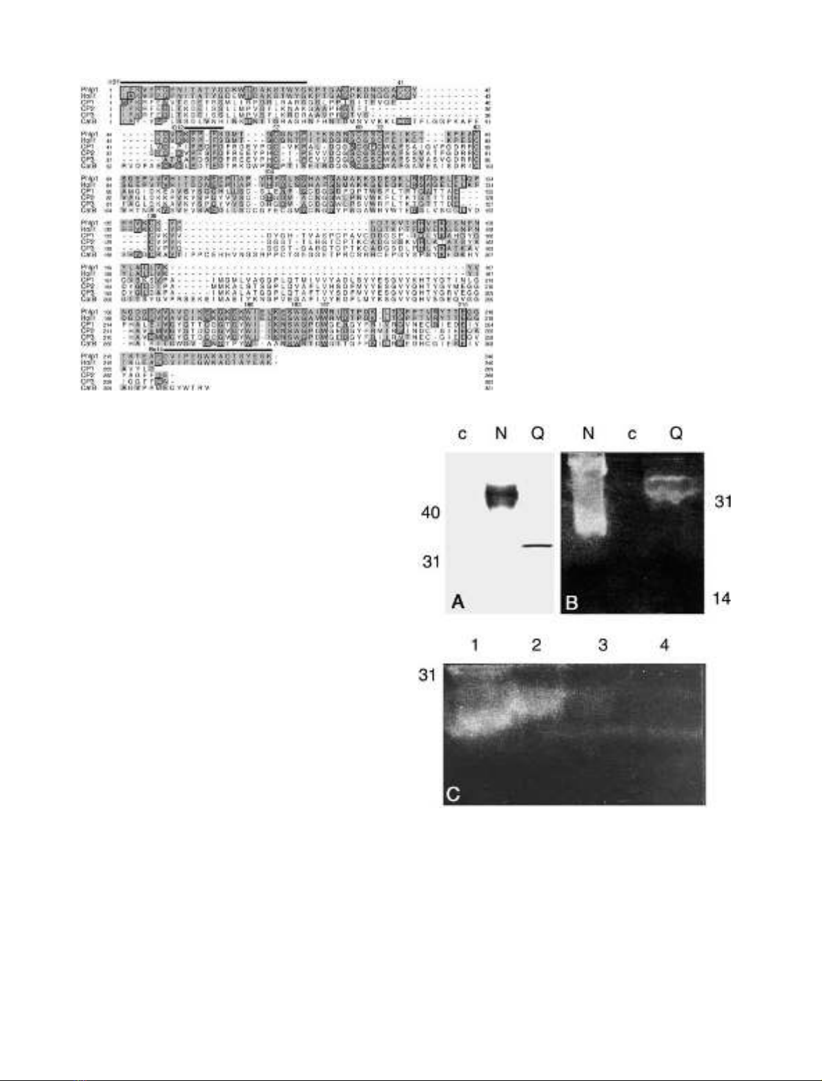
Fig. 3. Isoelectric focusing of rPhl p 1 N and rPhl p 1 Q, as detected
by mAb IG12. The pH values of the respective fractions and the size of
the protein markers used are indicated. (A) The full-length allergen
Phl p 1 N mostly disintegrates. Notably, a 15-kDa fragment can be
seen in the pH 4.5 fraction (arrow). Further degradation products can
be seen in the more basic fractions. (B) The allergen Phl p 1 Q can be
detected at about pH 8.0, which is the pI computed for Phl p 1 and
does not show any degradation products (arrow). (C) and (D)
Isoelectric focusing of rPhl p 1 N and rPhl p 1 Q, as detected with
mAb Bo14. As can be seen, only Phl p 1 Q was detected with this
antibody, indicating an intact allergen (arrow). None of the Phl p 1 N
fragments could be detected with mAb Bo14, demonstrating lack of a
C-terminal peptide.
FEBS 2002 Proteolytic properties of grass group I allergens (Eur. J. Biochem. 269) 2087