Open Access
Available online http://ccforum.com/content/12/2/R43
Page 1 of 8
(page number not for citation purposes)
Vol 12 No 2
Research
Danaparoid sodium inhibits systemic inflammation and prevents
endotoxin-induced acute lung injury in rats
Satoshi Hagiwara, Hideo Iwasaka, Seigo Hidaka, Sohei Hishiyama and Takayuki Noguchi
Department of Brain and Nerve Science, Anesthesiology, Oita University Faculty of Medicine, Oita, Japan
Corresponding author: Satoshi Hagiwara, saku@med.oita-u.ac.jp
Received: 27 Nov 2007 Revisions requested: 16 Jan 2008 Revisions received: 5 Feb 2008 Accepted: 2 Apr 2008 Published: 2 Apr 2008
Critical Care 2008, 12:R43 (doi:10.1186/cc6851)
This article is online at: http://ccforum.com/content/12/2/R43
© 2008 Hagiwara et al.; licensee BioMed Central Ltd.
This is an open access article distributed under the terms of the Creative Commons Attribution License (http://creativecommons.org/licenses/by/2.0),
which permits unrestricted use, distribution, and reproduction in any medium, provided the original work is properly cited.
Abstract
Introduction Systemic inflammatory mediators, including high
mobility group box 1 (HMGB1), play an important role in the
development of sepsis. Anticoagulants, such as danaparoid
sodium (DA), may be able to inhibit sepsis-induced
inflammation, but the mechanism of action is not well
understood. We hypothesised that DA would act as an inhibitor
of systemic inflammation and prevent endotoxin-induced acute
lung injury in a rat model.
Methods We used male Wistar rats. Animals in the intervention
arm received a bolus of 50 U/kg of DA or saline injected into the
tail vein after lipopolysaccharide (LPS) administration. We
measured cytokine (tumour necrosis factor (TNF)α, interleukin
(IL)-6 and IL-10) and HMGB1 levels in serum and lung tissue at
regular intervals for 12 h following LPS injection. The mouse
macrophage cell line RAW 264.7 was assessed following
stimulation with LPS alone or concurrently with DA with
identification of HMGB1 and other cytokines in the supernatant.
Results Survival was significantly higher and lung
histopathology significantly improved among the DA (50 U/kg)
animals compared to the control rats. The serum and lung
HMGB1 levels were lower over time among DA-treated animals.
In the in vitro study, administration of DA was associated with
decreased production of HMGB1. In the cell signalling studies,
DA administration inhibited the phosphorylation of IκB.
Conclusion DA decreases cytokine and HMGB1 levels during
LPS-induced inflammation. As a result, DA ameliorated lung
pathology and reduces mortality in endotoxin-induced systemic
inflammation in a rat model. This effect may be mediated through
the inhibition of cytokines and HMGB1.
Introduction
Despite extensive investigation of strategies for treating acute
lung injury (ALI), the overall mortality still remains high at
approximately 30 to 50% [1]. One of the mechanisms of sep-
sis-induced acute lung injury involves bacterial endotoxin
release into the circulation that activates interconnected
inflammatory cascades in the lung, ultimately leading to lung
damage [2,3]. The production of inflammatory mediators plays
an important role in the pathophysiology of inflammation in
lung injury.
High mobility group box 1 (HMGB1) protein is an intranuclear
protein that was originally identified as a DNA-binding protein,
[4], but is also a late-phase mediator in the pathogenesis of
sepsis [5]. HMGB1 acts as a pro-coagulant [6], thereby
enhancing the inflammatory response in septic shock [7,8].
The timing of its release and action is typically later than other
cytokines, such as TNFα and IL-1β [5]. Inhibitors of HMGB1
might therefore be beneficial in the treatment of various inflam-
matory diseases.
The role of clotting factors as inflammatory mediators has
attracted close attention. Initiation of the coagulation cascade
and the subsequent production of proinflammatory cytokines
(particularly in response to factor Xa (FXa)) are central to the
pathogenesis of sepsis [9,10]. Danaparoid sodium (DA) is a
low molecular weight heparinoid consisting of heparan sulfate,
dermatan sulfate and chondroitin sulfate that has both antico-
agulant and anti-inflammatory effects. DA inhibits of FXa and
factor IIa (FIIa) at ratios greater than heparin, while enacting
minimal effects on platelet function [11-13]. Anti-inflammatory
and anticoagulant agents have thus become a focus of new
treatments for sepsis [14,15].
We hypothesised that DA would act as an inhibitor of systemic
inflammation and prevent acute lung injury in a rat model. To
ALI = acute lung injury; ARDS = acute respiratory distress syndrome; DA = danaparoid sodium; FIIa = factor IIa; FXa = factor Xa; HMGB1 = high
mobility group box 1; IKK = IkB kinase; LPS = lipopolysaccharide; NF-kB = nuclear factor kB.

Critical Care Vol 12 No 2 Hagiwara et al.
Page 2 of 8
(page number not for citation purposes)
test this hypothesis, we investigated the impact of DA admin-
istration on serum and lung levels of HMGB1, serum cytokine
levels and on lung histopathology in rats with lipopolysaccha-
ride (LPS)-induced systemic inflammation. To further elucidate
the mechanism of action of these effects, we assessed the
impact of DA on HMGB1 and cytokine secretion by
RAW264.7 cells.
Materials and methods
In vivo study
Materials
Danaparoid sodium was purchased from Organon Co. Ltd.
(CC, Oss, The Netherlands). Lipopolysaccharide (LPS,
O127:B8) was obtained from Sigma (St Louis, MO, USA).
Antibodies to rabbit polyclonal IgE anti-HMGB1 were pur-
chased from Becton Dickinson and Company (Franklin Lakes,
NJ, USA). Antibodies to β-actin were obtained from Abcam
PLC (Cambridge, UK).
Treatment protocol
The study was approved by the Ethical Committee of Animal
Research at the College of Medicine, Oita University, Oita,
Japan. Male Wistar rats weighing 250 to 300 g (Kyudou,
Saga, Japan) were used. Anaesthesia was induced by 4%
sevoflurane. The animals were randomly assigned to one of
three groups: (1) untreated LPS group: rats received a bolus
of a 0.9% NaCl solution (1.0 ml/kg) and LPS (7.5 mg/kg) into
the tail vein; (2) DA-treated LPS group: rats received a bolus
of DA (50 U/kg), and LPS (7.5 mg/kg) into the tail vein; (3)
Negative control group: rats received a bolus of 0.9% NaCl
solution (1.0 ml/kg) into the tail vein. Before and after surgery,
animals had unlimited access to food and water.
Histological analysis
A pathologist blind to group assignment analysed the samples
and determined levels of lung injury according to Murakami's
technique [16]. Briefly, 24 areas in the lung parenchyma were
graded on a scale of 0 to 4 (0, absent and appears normal; 1,
light; 2, moderate; 3, strong; 4, intense) for congestion,
oedema, infiltration of inflammatory cells, and haemorrhaging.
Measurements of cytokine and HMGB1 secretion
HMGB1, Il-6 and TNFα levels were determined using a com-
mercial enzyme-linked immunosorbent assay kit. HMGB1 was
from Shino-Test Corporation, Tokyo, Japan; IL-6, IL-10 and
TNFα were from R&D Systems Inc, Minneapolis, MN, USA.
Western blotting
Proteins were subjected to SDS-PAGE, and then transferred
to polyvinylidene difluoride (PVDF) membranes (Millipore,
Bedford, MA, USA). The membranes were incubated with pri-
mary antibody (1:1,000 dilution). After incubation with sec-
ondary antibody, blots were developed using an enhanced
chemiluminescence detection kit (Amersham, Buckingham-
shire, UK) and exposed on Hyperfilm ECL (Amersham). We
used the NIH ImageJ software (National Institutes of Health,
Bethesda, MD, USA) to quantitate protein band
concentrations.
Cell culture study
The murine macrophage cell line, RAW264.7, was maintained
in RPMI 1640 medium containing 5% heat-inactivated foetal
bovine calf serum and antibiotics at 37°C under 5% CO2. The
medium was removed and replaced with RPMI 1640 contain-
ing 5% fetal bovine serum (FBS) for most experiments, or
Opti-MEM (Sigma) for experiments designed to measure
HMGB1 in conditioned media.
Nuclear factor (NF)-κB binding assay
The DNA binding activity of NF-κB (p50/p65) was determined
using an ELISA-based non-radioactive NF-κB p50/p65 tran-
scription factor assay kit (Chemicon, Temecula, CA).
Statistical analysis
For descriptive purposes, all continuous data were presented
as mean ± SD. The data were analysed by Mann-Whitney U
test for comparison between two independent groups. A p
value of less than 0.05 was considered to be statistically sig-
nificant. Survival data were analysed with the Kaplan-Meier
program included in the Prism 4.0 software package (San
Diego, CA, USA). p Values less than 0.05 were considered
statistically significant.
Results
In vivo study
Mortality
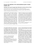
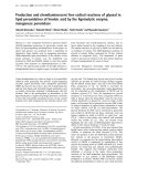
A total of 40% of the rats in the untreated LPS group died
within 12 h, and an additional 30% died within 24 h, while all
rats in the DA-treated LPS group (50 U/kg) survived (Figure
1). In addition, only 20% of rats treated with 1 U/kg DA and
50% of rats treated with 10 U/kg DA survived for 24 h, sug-
gesting a dose-dependent effect of DA on the survival rate of
LPS-treated rats (data not shown). All of the saline-treated
control animals survived for 7 days. Kaplan-Meier analysis
revealed a significantly shorter time-to-death among the
untreated LPS group compared to the DA (50 U/kg)-treated
LPS group (p < 0.05).
Effect of DA on lung tissue specimens
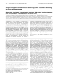
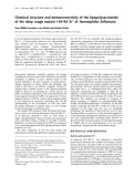
In the negative control group, no histological alterations were
observed (Figure 2a,d,g). Among the LPS group with sepsis,
the microscopic changes in the lung tissue specimens
observed 12 h after LPS administration showed oedema-like
formation, and interstitial infiltration by neutrophils (Figure
2b,e,h). The interstitial oedema and inflammatory cell infiltra-
tion were markedly reduced in the DA-treated group; DA treat-
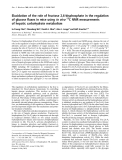
ment reduced each of these parameters. All of the scores
were significantly lower in the DA (50 U/kg) group than in the
LPS group (p < 0.05) (Figure 3).

Available online http://ccforum.com/content/12/2/R43
Page 3 of 8
(page number not for citation purposes)
Effects of DA on the serum levels of IL-6, TNF
α
, IL-10 and
HMGB1
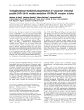
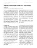
Prior to LPS administration, IL-6, TNFα, IL-10 and HMGB1 in
the serum were below levels detectable by the assays. Subse-
quent to LPS infusion, serum levels of IL-6 increased, with a
peak value observed at 3 h in both groups. Treatment with DA
following LPS administration led to a significantly decreased
concentration of IL-6 at all assay times (p < 0.05) (Figure 4a).
Likewise, serum levels of TNFα peaked 3 h post-LPS-infusion,
with the DA treatment group showing significantly decreased
levels at this time point (p < 0.05). During the investigation
period, TNFα levels of DA-treated LPS group were lower than
the LPS group at all assay times (Figure 4b). Serum levels of
HMGB1 increased over time following LPS infusion. This
increase was less prominent in DA-treated rats compared to
the untreated ones. At 6, 9 and 12 h following LPS administra-
tion, HMGB1 was significantly lower in the DA-treated LPS
group compared to the untreated LPS group (p < 0.05) (Fig-
ure 4c). By contrast, serum levels of IL-10 peaked 3 h post-
LPS-infusion, with the DA-treatment group showing increased
levels at all assay times. At 6, 9 and 12 h following LPS admin-
istration, IL-10 was significantly higher in the DA-treated LPS
group compared to the untreated LPS group (p < 0.05) (Fig-
ure 4d).
Figure 1
Effect of danaparoid sodium (DA) on the survival rate of lipopolysac-charide (LPS)-treated ratsEffect of danaparoid sodium (DA) on the survival rate of lipopolysac-
charide (LPS)-treated rats. The survival rate of animals treated with a
bolus of LPS (7.5 mg/kg) into the tail vein (LPS group, n = 10) is repre-
sented by black squares. The survival rate of animals that received DA
(50 U/kg) in addition to the intravenous injection of LPS (7.5 mg/kg)
into the tail vein (DA treated LPS groups, n = 10) is represented by
black circles.
Figure 2
Effects of danaparoid sodium (DA) on lung histopathology in lipopolysaccharide (LPS)-administered ratsEffects of danaparoid sodium (DA) on lung histopathology in lipopolysaccharide (LPS)-administered rats. Rats were intravenously infused with either
saline (control group), 7.5 mg/kg LPS (LPS group), or 7.5 mg/kg LPS with 50 U/kg DA (DA+LPS group). Lung tissue specimens were obtained
from the negative control (a) magnification ×40, (d) magnification ×100, (g) magnification ×400); LPS (b) magnification ×40, (e) magnification
×100, (h) magnification ×400; and DA+LPS (c) magnification ×40, (f) magnification ×100, (i) magnification ×400 groups, respectively. Haematox-
ylin and eosin staining was used.

Critical Care Vol 12 No 2 Hagiwara et al.
Page 4 of 8
(page number not for citation purposes)
Effect of DA on the HMGB1 levels in the lung
HMGB1 expression in lung tissue increased following LPS
injection. This increase was less pronounced among DA-
treated rats compared to the untreated LPS group (Figure
5a,b). In an immunohistochemical analysis, cells expressing
HMGB1 increased following LPS administration (Additional
file 1a). By contrast, the percentage of cells expressing
HMGB1 decreased dramatically in the LPS-administered rats
treated with DA (Additional file 1b).
In vitro study
Effect of DA on the culture supernatant and cell protein of
HMGB1
The secretion of HMGB1 was measured in the culture super-
natant at 20 h after the administration of LPS. The HMGB1
level of the culture supernatant increased after the administra-
tion of LPS, but the secretion of HMGB1 was inhibited by the
administration of DA. In addition, the inhibition of HMGB1 by
DA was minimal at a dose of 1 U/ml, was intermediate at a
dose of 15 U/ml, and was maximal at a dose of 50 U/ml (Figure
6). We therefore used a concentration of 50 U/ml DA for sub-
sequent experiments.
Effect of DA on the culture supernatant of cytokines
The TNFα level in the culture supernatant increased 3 h follow-
ing the administration of LPS. The administration of DA signif-
icantly inhibited the secretion of TNFα. The IL-6 level in the
culture supernatant also increased after the administration of
LPS. The administration of DA was thus found to significantly
inhibit the secretion of IL-6 in a manner similar to TNFα (Figure
7).
DA inhibits the IKK pathway and modulates NF-
κ
B
Since the NF-κB pathway plays a critical role in the secretion
of cytokines, we measured the quantity of p50 and p65 in the
nucleus. Treatment with LPS led to a robust activation of the
NF-κB transcription factor p50/p65. This activation was par-
tially blocked by DA (Figure 8).
We subsequently examined the IκB kinase (IKK) system as
another activation agent of NF-κB. Treatment with LPS
resulted in the degradation of IκB alpha and this degradation
was inhibited by DA (Additional file 2). In addition, the phos-
phorylation of p-IκB alpha in RAW264.7 cells increased after
LPS administration, and was also inhibited by DA (Additional
file 2).
Discussion
This is the first study to demonstrate the anti-inflammatory
actions of DA in a rat model of endotoxin-induced lung injury.
Acute inflammatory events, such as those that occur in ALI,
lead to dysregulation of the coagulation cascade. Indeed, ALI
is characterised by profound alterations in both systemic and
intra-alveolar coagulation and fibrinolysis [17]. Activation of
coagulation with resultant fibrin deposition also has proinflam-
matory consequences, serving to further amplify the inflamma-
tory cascade [18]. Lung damage may result not only from the
release of inflammatory mediators, but also from coagulation.
These results suggest that coagulation and inflammation are
related and therefore, anticoagulant therapy, such as treat-
ment with DA, will benefit patients with ALI.
In this study, we demonstrated that treatment with the antico-
agulant DA significantly improved acute lung injury and mortal-
ity in a rat model. Acute lung injury is characterised by non-
cardiogenic oedema, pulmonary inflammation and severe sys-
temic hypoxemia. Many sequelae associated with ALI result
from excessive production of cytokine mediators (such as
TNFα and IL-6) by activated monocytes [19]. In addition, stud-
ies have shown that HMGB1 is an important late mediator of
inflammation and acute lung injury in sepsis [20-22]. This
study adds to the previous findings by suggesting that DA may
prevent LPS-induced lung injury by inhibiting cytokine and
HMGB1 secretion.
We demonstrated that IL-10 increased following the adminis-
tration of DA during endotoxin-induced systemic inflammation.
A previous study showed that IL-10 inhibited the action of
inflammatory cytokines [23] and had profound negative effects
on macrophage activation [24]. In particular, IL-10 was closely
related to the secretion of TNFα [25]. IL-10 has been identified
in the lungs of patients with ARDS, where it was correlated
with improved survival [26]. Based on our results, the inhibition
of cytokines and prevention of lung injury might be related to
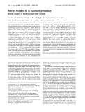
Figure 3
Effects of danaparoid sodium (DA) on lung histopathology score in lipopolysaccharide (LPS)-administered ratsEffects of danaparoid sodium (DA) on lung histopathology score in
lipopolysaccharide (LPS)-administered rats. The histological changes
identified included congestion, oedema, inflammation, and haemorrhag-
ing 12 h after the administration of LPS. White bars represent the non-
injected control animals, black bars represent the animals injected with
LPS, and slashed bars represent animals injected with DA and LPS.
The data are expressed as the mean ± SD. *Denotes a significant dif-
ference compared with the LPS group (p < 0.05).

Available online http://ccforum.com/content/12/2/R43
Page 5 of 8
(page number not for citation purposes)
increased serum levels of IL-10 resulting from administration
of DA at LPS-induced systemic inflammation.
NF-κB-dependent genes are related to the development of
septic shock and to septic lethality. Studies using an LPS
model of septic shock have consistently demonstrated that
blocking the NF-κB pathway improves outcome [27,28]. Fol-
lowing LPS stimulation, NF-κB is phosphorylated and coordi-
nates the induction of several genes encoding the production
and secretion of pro-inflammatory cytokines [29]. Therefore,
inhibiting NF-κB activation is crucial for treating inflammation.
Here, we showed that DA inhibits LPS-induced NF-κB activa-
tion, and may in turn inhibit the secretion of inflammatory medi-
ators and improve survival rate.
Recent studies have demonstrated that coagulation, particu-
larly the generation of thrombin, FXa, and the tissue factor-
factor VIIa complex, is related to acute inflammatory
responses [30]. Indeed, Riewald M et al., reported that FXa
activates NF-κB [31]. DA is a strong inhibitor of FXa. Binding
of DA to AT III leads to an accelerated inhibition of FXa,
resulting in the antithrombotic effect of DA. [32]. These
results suggest that the inhibitory effects of DA on NF-κB
may be partially due to inhibition of FXa. Further studies are
needed to clarify the signalling mechanisms that mediate the
beneficial anti-inflammatory effects of DA.
Recent studies have elucidated how LPS is recognised by
monocytes and macrophages of the innate immune system.
LPS stimulation of murine macrophages activates several
intracellular signalling pathways, including the IκB kinase
(IKK)-NF-κB pathway [33,34]. We used a murine macro-
phage cell line to show that DA suppresses the activation of
NF-κB by preventing the phosphorylation of IκB. Accord-
ingly, the inhibition of IκB phosphorylation following DA
administration in sepsis may lead to the inhibition of NF-κB
activation. As a limitation of this study, the mechanisms that
mediate these effects of DA in the LPS-induced systemic
inflammatory model are not understood, and we need to fur-
ther investigate the mechanisms of DA on the inhibition of
NF-κB activation.
Materials and methods
Antibodies to phosphorylated IkB and IkB-alpha were
obtained from Cell Signaling Technology (Beverly, MA).
Immunohistochemical analysis
Immunohistochemistry was performed after blocking endog-
enous peroxidase activity. Blocked sections were incubated
with anti-HMGB1 polyclonal antibody (1:1000 dilution). Pri-
mary antibody binding was visualized with horseradish perox-
idase conjugate and diaminobenzidine.
Western blotting
Proteins were subjected to sodium dodecyl sulfate-polyacryla-
mide gel electrophoresis (SDS-PAGE), and then transferred
to polyvinylidene difluoride membranes (Millipore, Bedford.
MA.). The membranes were incubated with primary antibody
Figure 4
Temporal changes in the tumour necrosis factor (TNF)α, interleukin (IL)-6, IL-10, and high mobility group box 1 (HMGB1) serum concentrations fol-lowing LPS administrationTemporal changes in the tumour necrosis factor (TNF)α, interleukin (IL)-6, IL-10, and high mobility group box 1 (HMGB1) serum concentrations fol-
lowing LPS administration. The IL-6 (a), TNFα (b), HMGB1 (c) and IL-10 (d) serum concentrations at the indicated times are shown for the lipopol-
ysaccharide (LPS) (n = 6; squares) and danaparoid sodium (DA)-treated (n = 6; circles) groups. All data are expressed as mean ± SD. *Denotes a
significant difference compared with the LPS group (p < 0.05).