607
ALI = acute lung injury; ARDS = acute respiratory distress syndrome; ARDSexp = extrapulmonary ARDS; ARDSp = pulmonary ARDS; CCW =
chest wall compliance; CHF = congestive heart failure; CL= lung compliance; COPD = chronic obstructive pulmonary disease; CPAP = continu-
ous positive airway pressure; ESPVR = end-systolic pressure-volume relationship; FRC = functional residual capacity; IAP = intra-abdominal pres-
sure; ITP = intrathoracic pressure; LV = left ventricular; PaCO2 = arterial carbon dioxide partial pressure; Palv = avleolar pressure; Paw = airway
pressure; PCRIT = critical closing pressure; PEEP = positive end-expiratory pressure; Pes = esophageal pressure; Pms = mean systemic pressure;
Ppc = pericardial pressure; Ppl = pleural pressure; PVR = pulmonary vascular resistance; RAP = right atrial pressure; RV = right ventricular; SV =
stroke volume.
Available online http://ccforum.com/content/9/6/607
Abstract
In patients with acute lung injury, high levels of positive end-
expiratory pressure (PEEP) may be necessary to maintain or
restore oxygenation, despite the fact that ‘aggressive’ mechanical
ventilation can markedly affect cardiac function in a complex and
often unpredictable fashion. As heart rate usually does not change
with PEEP, the entire fall in cardiac output is a consequence of a
reduction in left ventricular stroke volume (SV). PEEP-induced
changes in cardiac output are analyzed, therefore, in terms of
changes in SV and its determinants (preload, afterload, contractility
and ventricular compliance). Mechanical ventilation with PEEP, like
any other active or passive ventilatory maneuver, primarily affects
cardiac function by changing lung volume and intrathoracic
pressure. In order to describe the direct cardiocirculatory
consequences of respiratory failure necessitating mechanical
ventilation and PEEP, this review will focus on the effects of
changes in lung volume, factors controlling venous return, the
diastolic interactions between the ventricles and the effects of
intrathoracic pressure on cardiac function, specifically left
ventricular function. Finally, the hemodynamic consequences of
PEEP in patients with heart failure, chronic obstructive pulmonary
disease and acute respiratory distress syndrome are discussed.
Introduction
Cyclic opening and closing of atelectatic alveoli and distal small
airways with tidal breathing is known to be a basic mechanism
leading to ventilator-induced lung injury [1]. To prevent alveolar
cycling and derecruitment in acute lung injury, high levels of
positive end-expiratory pressure (PEEP) have been found
necessary to counterbalance the increased lung mass resulting
from edema, inflammation and infiltrations and to maintain
normal functional residual capacity (FRC) [2]. Therefore,
application of high levels of PEEP is often recommended [3],
despite the fact that ‘aggressive’ mechanical ventilation using
high levels of PEEP to maintain or restore oxygenation during
acute lung injury can markedly affect cardiac function in a
complex and often unpredictable fashion. Likewise, this notion
holds true for intrinsic PEEP caused by ventilation with high
respiratory rates resulting in dynamic hyperinflation. Except from
the failing ventricle, PEEP usually decreases cardiac output, a
well known fact since the classic studies of Cournand et al. [4],
in which the effects of positive-pressure ventilation were
measured. They concluded that positive-pressure ventilation
restricted the filling of the right ventricle because the elevated
intrathoracic pressure (ITP) restricted venous flow into the
thorax and, thereby, reduced cardiac output. This formulation of
intrathoracic responses to positive-pressure ventilation still is the
basis of our present day understanding of the cardiopulmonary
interactions induced by PEEP, although precise responses to
PEEP have not been simple to prove, and the intrathoracic
responses appear multiple and complex.
As heart rate usually does not change with PEEP [5], the
entire fall in cardiac output is a consequence of a reduction in
left ventricular (LV) stroke volume (SV). Therefore, the
discussion on PEEP-induced changes in cardiac output can
be confined to analyzing changes in SV and its determinants:
preload, afterload, contractility and ventricular compliance.
Before considering how PEEP affects the determinants of
SV, it has to be emphasized that ventilation with PEEP, like
any other active or passive ventilatory maneuver, primarily
affects cardiac function by changing lung volume and ITP [6].
To understand the direct cardiocirculatory consequences of
respiratory failure, one must, therefore, understand the effects
of changes in lung volume, factors controlling venous return,
the diastolic interactions between the ventricles and the
effects of ITP on cardiac function, specifically LV function.
Review
Clinical review: Positive end-expiratory pressure and cardiac output
Thomas Luecke1and Paolo Pelosi2
1Section Head, Critical Care, Department of Anesthesiology and Critical Care Medicine, University Hospital of Mannheim, Germany
2Associate Professor in Anaesthesia and Intensive Care, Dipartimento di Scienze Cliniche e Biologiche, Università degli Studi dell’Insubria, Varese, Italy
Corresponding author: Thomas Luecke, thomas.luecke@anaes.ma.uni-heidelberg.de
Published online: 18 October 2005 Critical Care 2005, 9:607-621 (DOI 10.1186/cc3877)
This article is online at http://ccforum.com/content/9/6/607
© 2005 BioMed Central Ltd

608
Critical Care December 2005 Vol 9 No 6 Luecke and Pelosi
This review will attempt to integrate basic mechanisms into
the global mechanisms of PEEP, and relate these concepts
to patient care. Analysis will focus on the relationships
between lung volume and ITP and using these relationships
to assess specifically the four primary components of the
circulatory system that are affected by ventilation (systemic
venous return, right ventricular (RV) output, LV filling, and LV
output) [7]. Subsequent analysis will be confined to
controlled mechanical ventilation and it needs to be
emphasized that hemodynamic effects during assisted
spontaneous ventilation, compared to controlled ventilation,
may be substantially different due to the difference in ITP.
Relationship between airway pressure,
intrathoracic pressure and lung volume
A lot of confusion exists, both in the literature and at the
bedside, in understanding and applying the concept of ITP
during mechanical ventilation. As outlined by Scharf [8], it
must be clear that the term ‘intrathoracic pressure’ does not
per se specify a pressure. Rather, one must ask, “which
intrathoracic pressure, esophageal, pleural, cardiac fossa, or
cardiac surface?” To make things even worse, it is common
practice to equate changes in airway pressure (Paw) with
changes in both ITP and lung volume.
Although positive-pressure ventilation increases lung volume
only by increasing Paw, the degree to which both ITP (being
esophageal, pleural or pericardial) and lung volume increase
will be a function of airway resistance as well as lung and
chest wall compliance.
Lateral chest wall pleural pressure (Ppl) and pericardial
pressure (Ppc) increase similarly in normal and acute lung
injury states for a constant tidal volume despite widely varying
lung compliance and a greater mean and plateau Paw during
the acute lung injury condition [9,10]. The primary
determinant of the increase in Ppl and Ppc during positive-
pressure ventilation is lung volume change [11]. During
sustained increases in lung volume, the increase in Ppl is
greater than the increase in Ppc. Thus, estimating Ppc by
measuring Ppl on any surface within the thorax may still
underestimate actual Ppc, which is LV surrounding pressure
[10]. Changes in Ppl induced by positive-pressure ventilation
are not the same in all regions of the thorax; Ppl at the
diaphragm increases least, and juxtacardiac Ppl increases
most [12]. These differences are in addition to the normally
described hydrostatic pressure gradient in the pleural space
from the posterior to anterior surface. As lung injury is often
non-homogeneous, large increases in Paw are often seen
during mechanical ventilation in such patients even when the
absolute tidal volume is low. This increased Paw should over-
distend these aerated lung units [13]. However, two separate
studies have demonstrated that, despite this non-
homogeneous alveolar distention, if tidal volume is kept
constant, the Ppl will increase equally, independent of the
mechanical properties of the lung [9,14]. Thus, if tidal volume
is kept constant, changes in peak and mean Paw will reflect
changes in the mechanical properties of the lungs and patient
cooperation, but will not reflect changes in Ppl nor alter
global dynamics of the cardiovascular system [10]. As
demonstrated by Pinsky and coworkers [15] in postoperative
patients, however, the percentage of Paw that will be
transmitted to the pericardial surface is not constant from one
subject to the next as PEEP is increased. Furthermore, the
degree to which Ppc will increase relative to Ppl is a function
of prior pericardial constraint [10].
Bearing in mind that the heart is a pressure chamber within a
pressure chamber (i.e. the thorax), the question of how much
of externally applied Paw (or PEEP) is actually transmitted to
the intrathoracic structures is of pivotal importance, especially
if one tries to measure and interpret filling pressures of the
heart in order to define its loading conditions. In addition, as
the heart is a pressure chamber within the pericardium, it is
also pericardial pressure applied over the surface of the atria
and ventricles that affect transmission of pressure to the
intracardial chambers, varying both with respiratory and
cardiac cycles and producing different surface pressures
over the four cardiac chambers during these cycles. The
catheter (central venous or pulmonary artery) measures an
intravascular pressure, relative to atmosphere. The
interpretation of hemodynamic data during positive-pressure
ventilation, however, requires thinking in terms of transmural
pressures, which is the pressure difference acting across the
wall of a vessel or cardiac chamber (i.e. inside minus outside
pressure). As neither the Ppc, which is the outside pressure
for the right and left ventricle, nor the Ppl are directly
accessible in clinical practice, the esophageal pressure (Pes)
is commonly used as the outside pressure. Thus, transmural
LV pressure would clinically be measured as LV intracavitary
pressure minus Pes, assuming that Pes represents cardiac
surface pressure.
While this is a common assumption, there are potential
pitfalls with that approach: Ppc may not increase as much as
juxtacardiac Ppl during positive-pressure ventilation,
especially in heart failure states. Presumably, as total cardiac
volume decreases with the application of positive Paw, its
venous return decreases and/or left ventricular ejection
increases [10]. Under these common conditions, if pericardial
restraint was limiting cardiac filling (i.e. Ppc exceeds
juxtacardiac Ppl), the pericardium will become less of a
limiting membrane [16]. Ppc is the surrounding pressure for
ventricular distention. Thus, estimates of Ppc made by using
Ppl (Pes) measurements may overestimate surrounding
pressure as Ppl is increasing.
In summary, one is faced with two important limitations
rendering the assessment of PEEP-induced changes in
cardiac function difficult. First, true transmural ventricular
filling pressures are not available and surrogate estimates
using Pes have to be used instead. Second, predicting how

609
much Paw is transmitted to the pericardial space is difficult at
best. According to O’Quin and Marini [17], one can estimate
how changes in avleolar pressure (∆Palv) translate into
changes in ITP (∆ITP), assuming that the compliances of the
lung (CL) and chest wall (CCW) are in series and
homogeneous:
∆ITP/∆Palv = 1/(1+ CCW/CL)
CCW/CLis not generally known with precision, and the validity
of the underlying assumptions is rather approximate.
Nevertheless, the above relationship is helpful for making
rough predictions. In most healthy subjects, CLis nearly the
same as CCW during normal tidal volume (0.2 L/cmH2O). In
this situation, ∆ITP/∆Palv = ½ or half of the applied PEEP
would be expected to be transmitted to ITP. Whereas a
popular rule of thumb is to subtract half of the applied PEEP
from hemodynamic measurements, this rule is helpful only
when the patient’s chest wall and lung compliance are normal
[18]. A decrease in lung compliance has been shown to
decrease the transmission of Paw to intrathoracic structures
(commonly measured as Ppl) [19,20], while these findings
have been challenged by O’Quin and Marini [17], who
measured juxtacardiac Ppl and found that the fractional
change of Ppl versus Paw was only slightly decreased after
acute lung injury in a canine model. These results were
confirmed by Scharf and Ingram [14] and Romand et al. [9],
who showed that the primary determinant of change in Ppl (or
ITP) during positive-pressure breathing is the amount of lung
inflation, not a specific change in compliance. Thus, the
PEEP-induced change in total intrathoracic volume, which
actually has to be considered in the diseased lung, when total
volume can be increased due to extensive edema even if
aerated lung volume is actually decreased, ultimately
determines the changes in ITP and the concomitant
hemodynamic effects.
In summary, it is extremely difficult to predict the amount to
which increases in Paw, either induced by PEEP or positive-
pressure ventilation, will increase ITP in an individual patient
with acute lung injury. Pes may serve as a reasonable
estimate for Ppl and Ppc, but is one step removed from these
values and may underestimate increases in either Ppl or Ppc
when lung volumes also increase [10]. Nevertheless, when
trying to understand the hemodynamic effects of PEEP in an
individual patient, the most important question to keep in
mind is: to what extend will PEEP change total lung volume
and ITP and how will these changes ultimately affect LV
preload, contractility and afterload?
Effects of PEEP
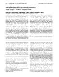
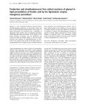
As proposed by Pinsky [6], all hemodynamic effects of
positive-pressure ventilation and PEEP can simply be
grouped into processes that, by changing lung volume and
ITP, affect left ventricular preload, afterload and contractility
(Fig. 1).
Left ventricular preload
The effects of PEEP on LV preload are dependent on
changes in systemic venous return, RV output and LV filling.
Due to the complexity of these changes, the single factors
will be discussed separately.
PEEP and the determinants of systemic venous return
Determinants of venous return
In steady state, cardiac output must equal the return of blood
to the heart. This in turn is determined by the mechanical
characteristics of the circuit, which is called circuit function.
This includes stressed vascular volume, venous compliance,
resistance to venous return and the outflow pressure for the
circuit, which is right atrial pressure (RAP). RAP is controlled
by cardiac function and the interaction of cardiac function
and circuit function determine cardiac output [21]. An
important concept for the understanding of venous return is
that of stressed and unstressed volume. The venous system,
like any other elastic structure, will fill with a certain volume,
called the ‘unstressed’ volume, without changing the
pressure or causing distention of the structures. Unstressed
volume represents as much as 25% of total blood volume
and constitutes a significant reservoir for internally recruiting
volume into the system. The difference between the total
volume in the system and the unstressed volume is the
relevant volume for causing pressure in the filling chamber,
the stressed volume [8]. The equivalent pressure in the veins
and venules to the hydrostatic pressure filling the system is
called mean systemic pressure (Pms). It is determined by the
volume filling the veins and the compliance of the veins. The
term that is used for describing the relationship of the total
volume for a given pressure is ‘capacitance’ and takes into
account both stressed and unstressed volume. This is not to
be confused with the term compliance, which is the change
in volume for the change in pressure [21]. In summary, the
determinants of venous return are the stressed volume (i.e.
the difference between total volume and unstressed volume),
venous compliance, resistance to venous return, and RAP.
Venous return is maximal when RAP equals zero. An increase
in venous return comes from an increase in stressed volume,
decrease in venous compliance, decrease in resistance to
venous return and a decrease in RAP. Vascular capacitance
is determined by the tone in the walls of the small venules
and veins. Contraction of smooth muscles in these vessels
due to neurosympathetic activation or exogenous catechol-
amines can decrease venous capacitance by converting
unstressed volume into stressed volume, thus raising mean
systemic pressure [21].
The sensitivity of systemic venous return to respiratory-
induced changes has been described in the classic
experiments by Guyton and colleagues [22,23]. The basic
principle is that systemic venous return is the major
determinant of circulation and is equal to left ventricular
output under steady state conditions [7,24,25]. Guyton et al.
[23] demonstrated that RAP represents the outflow pressure
Available online http://ccforum.com/content/9/6/607

610
(backpressure) for venous return. The relationship between
RAP and venous return is displayed by the venous return
curve. The pressure gradient driving blood from the periphery
to the right atrium can be defined as the difference between
the pressures in the upstream reservoirs, the Pms relative to
RAP. Pms, defined as the RAP at the point of zero flow, is a
function of blood volume, peripheral vasomotor tone and the
distribution of blood within the vasculature [26]. As RAP
increases, venous return decreases until RAP equals Pms. As
RAP decreases, venous return increases until the point of
flow limitation. The slope of the venous return curve is equal
to 1/resitance to venous return. The relationship between
right atrial end-diastolic pressure (representing preload) and
cardiac output is the familiar Frank-Starling relationship [8].
The superimposition of the venous return curve and the
Frank-Starling curve on the same set of axes was the creative
insight of Guyton [22] and provided an immensely useful
conceptual framework for studying cardiovascular control
[27]. Because, in steady state, cardiac output must equal
venous return, the point at which the two systems exist in
equilibrium is represented by the point of intersection of the
cardiac function (Frank-Starling) and venous return curves
[8]. Thus, for any given set of cardiac function and venous
return curves there exists only one combination of RAP and
cardiac output (= venous return) at which steady-state
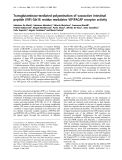
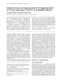
conditions apply (Fig. 2, point A).
Effect of PEEP on venous return
As the right atrium is a highly compliant structure, RAP would
reflect variations in ITP. Any increase in PEEP, by increasing
lung volume, and thus ITP, is expected to decrease venous
return by decreasing the pressure gradient in a manner
demonstrated in Fig. 2. The cardiac function curve is dis-
placed rightward by the amount by which ITP is increased,
thus maintaining the same transmural pressure-cardiac output
relationships. Postulating that Pms does not change with
PEEP, this would move the intersection of the cardiac
function and the venous return curves ‘downward’ on the
venous return curve (Fig. 2a, point B) [8]. As a result, the
gradient for venous return decreases, decelerating venous
blood flow [28], decreasing RV filling and, consequently,
decreasing RV SV [28-32].
However, as suggested by Scharf et al. [33] and later
demonstrated in experimental studies [34,35], PEEP also
increases Pms, thus preserving the gradient for venous
return. Jellinek and coworkers [36] confirmed that positive
Paw equally increased RAP and Pms in patients during
general anesthesia for implantation of defibrillator devices.
This increase in Pms, which may be due to an increase in
stressed volume or sympathoadrenal stimulation, could buffer
the PEEP-induced decrease in venous return and shift the
equilibrium point towards higher values of cardiac output
(Fig. 2a, point C). In addition to the effects of increased ITP, it
should be emphasized, however, that the actual compliance
of the right atrium is substantially defined by the pericardium.
As demonstrated by Tyberg and coworkers [37], as volume is
increased, the compliance of the entire right atrium is
constrained by the pericardium, thus markedly decreasing the
effective compliance of the right atrium. Tyberg and
colleagues’ work suggests that RAPs relative to atmosphere
as low as 5 mmHg are beginning to reflect pericardial
Critical Care December 2005 Vol 9 No 6 Luecke and Pelosi
Figure 1
Schematic representation of potential cardiopulmonary interactions with changes in intrathoracic pressure (ITP) and lung volume (redrawn with
permission from [137]). To obtain a more focused view of these numerous interactions, one can simply group all hemodynamic effects of ventilation
into processes that, by changing lung volume and ITP, affect left ventricular (LV) preload, contractility and afterload [6]. RV, right ventricular.

611
constraint and that pressures exceeding 10 to 12 mmHg are
dominated by pericardial constraint.
Tyberg et al. [38] also measured RV filling pressure defined
as RAP minus Ppc in patients undergoing elective cardiac
surgery. They demonstrated that RV filling pressure was
insignificantly altered by acute volume loading. While RAP
increased with volume loading, however, Ppc also increased
so that RV filling pressures remained unchanged. Thus, under
normal conditions, RV diastolic compliance is greater than
pericardial compliance. With RV filling, right heart sarcomere
length probably remains constant, and conformational
changes in the RV more than wall stretch are responsible for
RV enlargement [16]. Another study in postoperative surgical
patients [39] showed that when the RV end diastolic volume
was reduced by application of PEEP, both RAP and Ppc
increased, but RV filling pressure remained constant. Thus
changes in RAP do not follow changes in RV end diastolic
volume. The exact quantification of these mechanical heart-
pericardium-lung interactions is difficult in clinical practice,
however.
Whereas the pressure gradient for venous return (Pms-RAP)
was not altered by PEEP in the studies cited above [34-36],
venous return and cardiac output invariably fell, indicating an
increase in resistance of the venous conduits. According to
Fessler et al. [34], PEEP may either: decrease the caliber of
the conducting veins by constriction or compression,
resulting in reduced flow at the same driving pressure
through an increase in ohmic resistance (e.g. by abdominal
pressurisation); or increase the pressure around a portion of
the veins in excess of RAP.
If RAP were below a critical closing pressure (PCRIT) of the
veins, a condition termed a ‘vascular waterfall’ is said to exist.
This term was first applied to blood flow through the
pulmonary circulation when alveolar pressure exceeded left
atrial pressure [40]. Under these circumstances, the effective
downstream pressure for venous return is PCRIT, not RAP. If
PEEP were to elevate PCRIT in some parts of the circulation in
excess of RAP, then the effective pressure gradient for
venous flow from those regions could fall despite an
unaltered (Pms-RAP) difference [41], flow limitation at PEEP
would occur at higher pressures compared to ZEEP and the
ability of an increased Pms to buffer the PEEP-induced
decrease in venous return would be markedly less (Fig. 2b,
point B). In fact, Fessler and coworkers [42] demonstrated a
PEEP-induced vascular collapse at the inferior vena cava in
canine studies, consistent with a vascular waterfall [43] or
zone 2 condition [44], causing the back pressure to venous
return to be located upstream of the right atrium. With PEEP,
the vessels collapsed at higher pressure than normal, that is,
there was an increase in PCRIT of these veins, caused by
direct mechanical compression by the inflating lungs and/or
mechanical compression of intra-abdominal contents, especially
the liver [8,44,45]. The compression of the lung and liver of
course will have multiple effects, not only changing the time
constant (resistance × compliance) for enhancing venous
return, but also increasing the resistance and back pressure
to blood entering from the portal side into the liver and from
the right ventricle into the lung. Therefore, increased pressure
within the system can have the venous bed simultaneously
change its compliance and resistance, resulting in both a
discharging capacitator, and resistive changes that will have
Available online http://ccforum.com/content/9/6/607
Figure 2
Effects of positive end-expiratory pressure (PEEP) on venous return
and cardiac output. (a) Theoretical effects of PEEP on venous return
(VR) and cardiac output (CO). PEEP causes an increase in
intrathoracic pressure (ITP) and a right shift in the cardiac function
curve. If there were no change in the VR curve, then CO and VR would
decrease (from point A to point B). However, if there is a
compensatory increase in mean systemic pressure (from Pms1 to
Pms2), then the system will exist in equilibrium at point C, at which VR
and CO would be maintained compared to zero end-expiratory
pressure (ZEEP) conditions. Pms can increase either by an increase in
stressed volume or sympathoadrenal stimulation. (b) Another possible
scheme for the changes in VR with PEEP. If there is an increase in the
pressure at which flow limitation occurs, then the ability of an increase
in Pms to buffer PEEP-induced decreases in VR is markedly less. FL1,
flow limiting point at ZEEP; FL2, flow limiting point at PEEP. Modified
from [8], with permission.