Duy Hang Nguyen, Duc Van Khuat, Thang Huu Nguyen, Tuan Anh Tran
Abstract – The process of neural stem cell (NSC)
differentiation into neurons is crucial for the development
of potential cell-centered treatments for central nervous
system disorders. However, predicting, identifying, and
anticipating this differentiation is complex. In this study,
we propose the implementation of a convolutional neural
network model for the predictable recognition of NSC fate,
utilizing single-cell brightfield images. The results
demonstrate the model’s effectiveness in predicting NSC
differentiation into astrocytes, neurons, and
oligodendrocytes, achieving an accuracy rate of 91.27%,
93.69%, and 93.06%, respectively. Moreover, our
proposed model effectively distinguishes between various
cell types even within the initial day of culture.
Keywords—Neural stem cell differentiation,
Convolution neural network, Single-cell images, Stem
cells, Deep learning.
I. INTRODUCTION
Stem cells represent a specialized cell category with the
capacity to differentiate into various distinct cell types,
thus playing a pivotal role in the development,
maintenance, and regeneration of tissues and organs [1, 2].
The abilities of stem cells to self-renew and form different
mature cells expand the possibilities of applications in cell-
based therapies in regenerative medicine such as
recomposing tissue, drug screening, and treatment of
neurodegenerative diseases [3]. Besides, their therapeutic
effects result from the secretion of trophic tissue factors, as
well from as interactions with infiltrating cells of the
immune system through soluble molecules and exosomes
[4, 5]. In the adult mammalian central nervous system
(CNS), neurogenesis occurs in two specific areas: the
subventricular zone and the dentate gyrus found within the
hippocampus. Within these regions, the production of
various neural cell types is initiated from adult neural stem
cells (NSCs). The evaluation of NSCs as a therapeutic
approach for addressing CNS diseases and injuries has
been ongoing for decades. Parkinson’s disease, in
particular, has gained the greatest momentum for potential
therapeutic benefits [5].
NSC can self-renew or differentiate into neurons and
glial cells (astrocytes, oligodendrocytes, and microglia) [1,
6]:
• Neurons: Neurons are fundamental cells responsible for
transmitting information in the nervous system,
communicating through electrical and chemical
signals, using axons to send signals and dendrites to
receive them. Notably, neurons cannot replicate or
regenerate once they are damaged or died [7].
Therefore, a widely investigated approach for treating
neurodegenerative diseases involves either
transplanting external NSCs or activating internal
NSCs. Subsequently, these NSCs are induced to
differentiate into neurons, facilitating the restoration of
neural circuits damaged by neurological disorders [8-
10].
• Astrocytes: Astrocytes are a type of glial cell that
provides crucial support to neurons. Astrocytes help
maintain the brain’s microenvironment, regulate ion
balance, and contribute to the blood-brain barrier [8,
11]. Astrocytes are involved in various processes such
as neurotransmitter recycling and repair following
injury [12].
• Oligodendrocytes: Oligodendrocytes play a significant
role in the CNS by producing myelin, a protective
sheath around axons [13]. Myelin facilitates faster
electrical signal transmission, crucial for proper
nervous system function [14]. NSCs are differentiated
into oligodendrocytes that can contribute to post-injury
remyelination, electrically insulating neuronal axons
for impulse propagation, and providing trophic and
metabolic support for neurons [15].
• Microglia: Microglia are the immune cells of the CNS.
Microglia monitor the brain for damage, infection, and
foreign substances. When needed, microglia can
become activated to protect the brain by removing
damaged cells and pathogens [16].
The evaluation of potential inducers on NSC
differentiation is a time-consuming process, typically
taking several days. This assessment is susceptible to
Duy Hang Nguyen(*), Duc Van Khuat(*), Thang Huu Nguyen(#), Tuan Anh Tran(*)
(*) Posts and Telecommunications Institute of Technology,
(#) Financing Promoting Technology Corporation
PREDICTIVE NEURAL STEM CELL
DIFFERENTIATION USING SINGLE-CELL
IMAGES BASED ON DEEP LEARNING
Contact author: Duy Hang Nguyen
Email: duynth@ptit.edu.vn
Manuscript received: 10/2023, revised: 12/2023, accepted:
01/2024.
SOÁ 01 (CS.01) 2024
TAÏP CHÍ KHOA HOÏC COÂNG NGHEÄ THOÂNG TIN VAØ TRUYEÀN THOÂNG 4

PREDICTIVE NEURAL STEM CELL DIFFERENTIATION USING SINGLE-CELL IMAGES BASED ON DEEP ……
various influencing factors, including molecular marking
techniques, less advanced laboratory technology, and the
proficiency of experimental personnel. Present
methodologies may not adequately identify the factors
influencing NSC differentiation, particularly regarding
mechanisms that are not fully understood [17].
Additionally, current techniques rely on specific markers
for each cell type, such as NeuN for neurons, GFAP for
astrocytes, and Olig2 for oligodendrocytes, which are
applicable only to cells at specific stages of differentiation
[1, 18, 19]. As a result, early detection of NSC
differentiation presents a significant challenge, hindering
the progress of related technical advancements. There is an
immediate need for a more efficient, precise, user-friendly,
and resource-efficient method, one that minimizes
subjectivity and expands our comprehension of neural
development and differentiation.
In recent times, artificial intelligent (AI) has witnessed
significant advancements and has exerted a profound
impact on various domains. Machine learning (ML), a
subset of AI, constitutes an algorithm designed for
recognizing patterns and categorizing vast datasets. Deep
learning (DL), a multilayered neural network that closely
mimics the neural circuitry of the human brain, is
employed for acquiring insights from data. The application
of deep learning has been extended across diverse fields
such as autonomous driving, image recognition, drug
discovery, and bioinformatics [20-22]. Furthermore, the
proliferation of high-throughput technology has resulted in
a substantial increase in biomedical data in recent decades,
encompassing genetic sequences, protein structures, and
medical imaging [23, 24].
Advancements in stem cell research are increasingly
being accelerated by the utilization of DL models.
Integrating imaging techniques with deep neural networks
(DNNs) has facilitated improved measurement and
comprehension of morphological changes occurring during
differentiation. These advancements aid in predicting the
potential differentiation pathways of cells, annotating cells
in an unbiased manner, and unraveling the identity of stem
cells. DL methodologies have further been devised to
reconstruct developmental trajectories from single-cell
data, enhancing our understanding of stem cell fate
determination at an unprecedented resolution.
Additionally, these models have uncovered novel cell
states that emerge during reprogramming processes. DL
techniques are also expanding our ability to manipulate the
behavior of stem cells, enabling control over their pattern
formation and the identification of optimal culture
conditions [25].
Studies have utilized DL techniques to identify various
characteristics of cells, such as cell types, states, and
dynamic progression, using either flow cytometry or
microscopy images [26, 27]. Recently, there have been
notable discoveries regarding the application of DL in
observing and predicting physiological processes in stem
cells. One investigation revealed that the morphology of
haematopoietic stem cells changes during differentiation.
DL can detect these alterations in microscopy data,
enabling the early isolation of cells before the known
developmental progression begins, thus predicting the
development of haematopoietic stem cells in advance [28].
Another study demonstrated that machine learning can
differentiate pluripotent stem cells from cells in the early
stages of differentiation [29]. These findings underscore
the potential extension of deep learning applications in the
field of stem cell therapy. Several studies [31], [32]
employed ResNet and VGG architectures. However,
despite their strength and popularity, ResNet and VGG
exhibit generality and high computational costs. Therefore,
in this study, we propose an alternative CNN architecture.
In our work, we propose a convolutional neural network
(CNN)-based method for predicting the differentiation of
NSCs into Astrocytes, Neurons, and Oligodendrocytes
using single-cell images. The proposed network
architecture consists of four blocks, each comprising
distinct layers to extract and process features at various
levels of abstraction. This method aims to enable accurate
and efficient classification of NSC differentiations based
on single-cell image data. The main contribution of our
study is to apply a CNN-based method specifically
designed to predict NSC differentiation. While previous
studies have employed machine learning models and
convolutional neural networks for image classification
tasks, the specific application to predicting the
differentiation of NSCs into distinct cell types, as pursued
in our work, remains largely unexplored.
The remaining portion of the paper is organized as
follows. Section II introduces and describes the proposed
method. Section III demonstrates and analyzes results.
Finally, Section IV summarizes the research.
II. METHOD
A. Data
The single-cell image dataset utilized in this study is
sourced from research [30]. The dataset consists of the
following cell types: NSCs treated with astrocyte
differentiation medium, NSCs treated with
oligodendrocyte differentiation medium, and NSCs treated
with neuron differentiation medium with retinoic acid
(RA) and sonic hedgehog (SHH).
For the astrocyte dataset, the data is collected at three
different time points during cell culture, specifically at 0.5
day, 1 day, and 3 days. The oligodendrocyte dataset is
obtained at the following time points: 1 day, 2 days, and 3
days. Meanwhile, the neuron dataset is cultivated at 1 day,
2 days, and 5 days. The dataset consists of single-cell
images in brightfield. The number of images of NSCs
differentiated into Astrocyte, Neruron and
Oligodendrocyte is shown in tables 1, 2, and 3,
respectively.
The single-cell images were preprocessed before being
fed into the convolutional neural network. We resized each
image to 45 × 30 using the OpenCV package, then
normalized each pixel value to be within the range
SOÁ 01 (CS.01) 2024
TAÏP CHÍ KHOA HOÏC COÂNG NGHEÄ THOÂNG TIN VAØ TRUYEÀN THOÂNG 5

Duy Hang Nguyen, Duc Van Khuat, Thang Huu Nguyen, Tuan Anh Tran
Table 1: The number of brightfield single-cell images of
NSCs differentiated into Astrocyte.
Duration
NSc differentiation
12
hours
1 day
2 days
Astrocyte differentiation
medium
10,269
23,359
21,838
Table 2: The number of brightfield single-cell images of
NSCs differentiated into Neuron.
Duration
NSc differentiation
1 day
3 days
5 days
Neuron differentiation
medium
11,024
17,912
8,835
Table 3: The number of brightfield single-cell images of
NSCs differentiated into Oligodendrocyte.
Duration
NSc differentiation
1 day
2 days
3 days
Oligodendrocyte
differentiation medium
5,508
9,478
12,701
of [0, 1]. A total of 120,924 single-cell images were split
into 80% for use as training data to construct deep learning-
based brightfield models, and the remaining 20% were
used for model testing.
B. Convolutional neural network
We utilized the Xception module of the CNN
architecture illustrated in Fig. 1 to perform the
classification task for predictive NSCs differentiation,
including astrocytes, neurons, and oligodendrocytes. The
CNN architecture includes: Input layer, Convolutional
layers, Batch normalization, ReLU activation, Separable
convolutions, Max pooling, Average pooling and Fully
connected:
(1) Convolutional layers: These layers are responsible for
extracting various features and patterns from the input
images, which utilize filters to perform convolution
operations, capturing important spatial hierarchies within
the data.
(2) Batch normalization: Integrated after each
convolutional layer, batch normalization standardizes the
outputs of the previous layer. This helps stabilize the
training process and accelerates convergence, ensuring
efficient and stable learning.
(3) ReLU activation: Rectified Linear Unit (ReLU)
activation function introduces non-linearity into the
network, allowing it to learn complex relationships and
representations within the data. It helps the network model
complex phenomena, leading to improved predictive
performance.
(4) Separable convolutions: These convolutions are
utilized to efficiently capture spatial information within the
data while reducing computational complexity. By
separating the process into depthwise and pointwise
convolutions, it enables the network to learn complex
spatial patterns more effectively.
(5) Max pooling and Average pooling: Max pooling layers
downsample the feature maps, retaining the most
significant features, while average pooling layers compute
the average of the values within a certain kernel size. Both
pooling operations help in reducing the spatial dimensions
and controlling overfitting
(6) Output layer includes a fully connected layer followed
by a softmax activation function, providing a probability
distribution over different cell types, thus enabling the
model to predict the differentiation status of NSCs into
astrocytes, neurons, or oligodendrocytes.
C. Performance evaluation
To evaluate performance of proposed model, we use
Accuracy (Acc), precision (Pre), specificity (Sp) and
Recall. The first one, Accuracy, refers to the ratio of
correctly predicted observations to the total observations,
providing an overall assessment of the model's correctness
in predicting all cell types. Precision, on the other hand,
measures the fraction of relevant instances among the
retrieved instances, allowing us to understand how many
Fig. 1: The CNN architecture of the proposed
method
SOÁ 01 (CS.01) 2024
TAÏP CHÍ KHOA HOÏC COÂNG NGHEÄ THOÂNG TIN VAØ TRUYEÀN THOÂNG 6

PREDICTIVE NEURAL STEM CELL DIFFERENTIATION USING SINGLE-CELL IMAGES BASED ON DEEP ……
of the predicted instances are relevant to the specific cell
types. Specificity indicates the proportion of actual
negative cases that are correctly identified as such,
assisting in gauging the model's effectiveness in correctly
identifying true negatives. Recall measures the fraction of
true positive predictions out of all actual positive instances,
serving as an indicator of the model’s capability to detect
all relevant cases of Astrocyte, Neuron, and
Oligodendrocyte without missing any. The formula of this
parameter is calculated as follows:
TP TN
Acc TP TN FP FN
+
=
+ + +
(1)
TP
Pre TP FP
=
+
(2)
TN
Sp FP TN
=
+
(3)
TP
Recall TP FN
=
+
(4)
These parameters are normally defined for binary
classification problems where the outcome is either
“positive” or “negative”. As, we have three classes and
dealing with the multi-class problem, so we computed,
Acc, Pre, Sp and Recall, while calculating TN (True
Negative), TP (True Positive), FP (False Positive), and FN
(False Negative) of each class separately. Table 4 and 5,
shows various performance measures were obtained from
the confusion matrix.
Table 4: Confusion matrix
Confusion
Matrix
Predicted
False
Negative
(FN)
Class
1
Class
2
Class
3
Actual
Class
1
A
B
C
B+C
Class
2
D
E
F
D+F
Class
3
G
H
I
G+H
False Positive
(FP)
D+G
B+H
C+F
Table 5: Computing different performance measures from
confusion matrix
Class 1
Class 2
Class 3
Pre
A/(A+D+G)
E/(B+E+H)
I/(C+F+I)
Sp
(E+I)/
(D+G+E+I)
(A+I)/
(B+H+A+I)
(A+E)/
(C+F+A+E)
Recall
A/(A+B+C)
E/(D+E+F)
I/(G+H+I)
III. SIMULATION RESULTS AND DISCUSSION
The convolutional neural network (CNN) model was
trained and tested on a dataset of single-cell images, with
96,740 images used for training and 21,184 images used
for testing. Each image was resized to 45x30 pixels and
underwent appropriate normalization and preprocessing
before being fed into the model. The model was designed
to classify the cells into three distinct types: Astrocytes,
Neurons, and Oligodendrocytes. The testing results were
documented in the confusion matrix presented in Table 6.
Table 6 illustrates the confusion matrix derived from the
CNN model’s performance on the testing set, with Class 1
representing Astrocyte, Class 2 representing Neuron, and
Class 3 representing Oligodendrocytes. The confusion
matrix provides a detailed breakdown of the model’s
predictive capabilities for each cell type. It outlines the
number of instances correctly and incorrectly classified
within each class, enabling a comprehensive evaluation of
the CNN model’s performance. The model demonstrates
robust performance, particularly in predicting Class 2
(Neuron) and Class 3 (Oligodendrocyte), as evidenced by
the high numbers of correct predictions.
Table 6: Confusion matrix of CNN model on testing set
(Class 1: Astrocyte, Class 2: Neuron, Class 3:
Oligodendrocyte)
Confusion Matrix
Predicted
Class 1
Class 2
Class 3
Actual
Class 1
10227
651
215
Class 2
304
6750
500
Class 3
898
0
4639
Further insights into the performance of the model are
revealed in Table 7, which presents additional performance
metrics. The accuracy rates for predicting Astrocyte,
Neuron, and Oligodendrocyte are 91.27%, 93.69%, and
93.06%, respectively. Moreover, the precision, specificity,
and recall percentages provide a deeper understanding of
the model’s predictive capabilities for each cell type,
indicating a strong ability to discriminate between different
cell types.
Table 7: The performance of predicting NSCs
differentiation
Astrocyte
Neuron
Oligodendrocyte
Acc (%)
91.27
93.69
93.06
Pre (%)
89.48
91.20
86.65
Sp (%)
90.45
95.8
95.96
Recall (%)
92.19
89.36
83.78
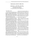
Besides, to evaluate the differentiation prediction of
NSCs, we assessed the time-dependent prediction of
collected data cells. The results are illustrated in Fig. 2,
indicating that after 1 day of cultivation, the model detected
the differentiation into Astrocyte, Neuron and
SOÁ 01 (CS.01) 2024
TAÏP CHÍ KHOA HOÏC COÂNG NGHEÄ THOÂNG TIN VAØ TRUYEÀN THOÂNG 7

Duy Hang Nguyen, Duc Van Khuat, Thang Huu Nguyen, Tuan Anh Tran
Oligodendrocyte cells with high accuracy of 93.86%,
90.89% and 80.6%, respectively.
Transplanting NSCs offers promising options for CNS
recovery, but guiding their differentiation into specific cell
types is tough. Biomarkers are commonly used to track the
changes, but the exact process of neurogenesis, especially
the early stages of neuron formation, remains unclear. This
makes it challenging to identify the direction of
differentiation early on. A reliable identification process is
necessary to develop effective treatments for
neurodegenerative diseases and neurological injuries,
regardless of the treatment pathways. Though advanced
tools aid data collection, understanding the data is difficult
due to current device limitations. Existing methods rely
heavily on human understanding, making it tough to
identify small changes in cell shape or predict drug
interactions [17]. These results of our proposed model
illustrate the efficacy of the CNN model in accurately
predicting the differentiation of NSCs into the specified
cell types. The high accuracy rates and robust performance
metrics demonstrate the model’s potential for precise
identification and classification of cell types, thereby
presenting promising prospects for the advancement of
cell-based treatments and therapies for various central
nervous system disorders.
Fig. 2: Accuracy of each testing set brightfield model
Table 8: Comparison of the accuracy performance of the
proposed CNN architecture with other method for
predicting 1 day of nscs differential cultivation.)
Ref
Astrocyte
Neuron
Oligodendrocyte
[17]
95.86%
82.73%
80.59%
Our
93.86%
90.89%
80.6%
To evaluate the performance of our model in comparison
with other research, we compare the accuracy performance
of the proposed CNN architecture to that of the reference
method for predicting the 1-day differential cultivation of
NSCs, as presented in Table 8. Regarding specific cell
types, the proposed CNN architecture achieves an accuracy
of 93.86% for Astrocyte prediction, slightly below the
reference method's 95.86%. However, it outperforms the
reference with 90.89% accuracy for Neuron prediction
compared to 82.73%. Both methods exhibit similar
accuracy for Oligodendrocyte prediction, with the
proposed CNN architecture at 80.6% and the reference
method at 80.59%. Besides, our CNN architecture is
simpler than the one presented in research [17]. These
results suggest the competitiveness of the proposed CNN
architecture in predicting NSCs differentiation,
particularly excelling in Neuron prediction, while
maintaining comparable accuracy in other cell types.
IV. CONCLUSION
In summary, this paper has introduced a method
utilizing CNN techniques to accurately predict the
differentiation of Neural Stem Cells into Astrocytes,
Neurons, and Oligodendrocytes, employing single-cell
brightfield images. With its capacity to predict NSC
differentiation within a day, the model presents a
promising avenue for investigating the effects of various
substances on NSCs. The results demonstrate the efficacy
and reliability of the proposed approach, paving the way
for improved understanding and monitoring of NSC
differentiation dynamics. This technique holds promising
implications for the advancement of cell-based therapies
for various central nervous system disorders.
ACKNOWLEDGMENT
NAVER partly supports this work. Khuat Van Duc was
funded by the Master Scholarship Programme of NAVER
Corporation and Posts and Telecommunications Institute
of Technology.
REFERENCES
[1] Vieira, Mariana S., et al. "Neural stem cell differentiation into
mature neurons: mechanisms of regulation and
biotechnological applications." Biotechnology advances 36.7
(2018): 1946-1970.
[2] Madl, Christopher M., Sarah C. Heilshorn, and Helen M.
Blau. "Bioengineering strategies to accelerate stem cell
therapeutics." Nature 557.7705 (2018): 335-342.
[3] Ahmed, Tanvir. "Neural stem cell engineering for the
treatment of multiple sclerosis." Biomedical Engineering
Advances (2022): 100053.
[4] Luarte, Alejandro, et al. "Potential therapies by stem cell-
derived exosomes in CNS diseases: focusing on the
neurogenic niche." Stem cells international 2016 (2016).
[5] Studer, Lorenz. "Strategies for bringing stem cell-derived
dopamine neurons to the clinic—the NYSTEM
trial." Progress in brain research 230 (2017): 191-212.
[6] Ladran, Ian, et al. "Neural stem and progenitor cells in health
and disease." Wiley Interdisciplinary Reviews: Systems
Biology and Medicine 5.6 (2013): 701-715.
[7] Liu, Kai, et al. "Neuronal intrinsic mechanisms of axon
regeneration." Annual review of neuroscience 34 (2011):
131-152.
[8] Lee, Ha-Rim, et al. "Functional group-dependent induction of
astrocytogenesis and neurogenesis by flavone
derivatives." Biomolecules 9.12 (2019): 812.
[9] Deng, Wenwen, et al. "EMSCs build an all‐in‐one niche via
cell–cell lipid raft assembly for promoted neuronal but
suppressed astroglial differentiation of neural stem
cells." Advanced Materials 31.10 (2019): 1806861.
[10] Li, Xiaoran, et al. "A collagen microchannel scaffold carrying
paclitaxel-liposomes induces neuronal differentiation of
neural stem cells through Wnt/β-catenin signaling for spinal
0
10
20
30
40
50
60
70
80
90
100
0.5D 1D 2D 1D 3D 5D 1D 2D 3D
Astrocyte Neuron Oligodendrocyte
Accuracy (%)
SOÁ 01 (CS.01) 2024
TAÏP CHÍ KHOA HOÏC COÂNG NGHEÄ THOÂNG TIN VAØ TRUYEÀN THOÂNG 8