AIDS Research and Therapy
Research
Comparison of brachial and carotid artery ultrasound for assessing
extent of subclinical atherosclerosis in HIV: a prospective
cohort study
Adefowope Odueyungbo*
1,2,3
,MarekSmieja
1,4,5,10
,LehanaThabane
1,2,3,10
,
Fiona Smaill
4,10
, Kevin Gough
6,10
,JohnGill
7,10
,ToddAnderson
8,10
,
Dawn Elston
4,10
,SandySmith
5,10
, Joseph Beyene
1,9,10
and Eva Lonn
5,10
Address:
1
Department of Clinical Epidemiology and Biostatistics, McMaster University, Hamilton ON, Canada,
2
Centre for Evaluation of
Medicines, St Joseph's Healthcare Hamilton, Hamilton ON, Canada,
3
Biostatistics Unit, Father Sean O'Sullivan Research Centre, St Joseph's
Healthcare Hamilton, Hamilton ON, Canada,
4
Department of Pathology and Molecular Medicine, McMaster University, Hamilton ON, Canada,
5
Department of Medicine, McMaster University, Hamilton ON, Ontario, Canada,
6
Department of Medicine, University of Toronto, Toronto ON,
Canada,
7
Department of Medicine, University of Calgary, Calgary AB, Canada,
8
Department of Cardiac Sciences and Libin Cardiovascular
Institute, University of Calgary, Calgary AB, Canada,
9
Department of Public Health Sciences, University of Toronto, Toronto ON, Canada and
10
Canadian HIV Vascular Study Group, Canada
E-mail: Adefowope Odueyungbo* - odueyuao@mcmaster.ca; Marek Smieja - smiejam@mcmaster.ca; Lehana Thabane - ThabanL@mcmaster.ca;
Fiona Smaill - Smaill@mcmaster.ca; Kevin Gough - GoughK@smh.toronto.on.ca; John Gill - John.Gill@calgaryhealthregion.ca;
Todd Anderson - todd.anderson@calgaryhealthregion.ca; Dawn Elston - elstond@mcmaster.ca; Sandy Smith - sandy@ccc.mcmaster.ca;
Joseph Beyene - joseph@utstat.toronto.edu; Eva Lonn - lonnem@mcmaster.ca
*Corresponding author
Published: 11 June 2009 Received: 14 December 2008
AIDS Research and Therapy 2009, 6:11 doi: 10.1186/1742-6405-6-11 Accepted: 11 June 2009
This article is available from: http://www.aidsrestherapy.com/content/6/1/11
©2009 Odueyungbo et al; licensee BioMed Central Ltd.
This is an Open Access article distributed under the terms of the Creative Commons Attribution License (http://creativecommons.org/licenses/by/2.0),
which permits unrestricted use, distribution, and reproduction in any medium, provided the original work is properly cited.
Abstract
Background: Non-invasive surrogate measures which are valid and responsive to change are
needed to study cardiovascular risks in HIV. We compared the construct validity of two
noninvasive arterial measures: carotid intima medial thickness (IMT), which measures anatomic
disease; and brachial flow-mediated vasodilation (FMD), a measure of endothelial dysfunction.
Methods: A sample of 257 subjects aged 35 years or older, attending clinics in five Canadian centres,
were prospectively recruited into a study of cardiovascular risk among HIV subjects. The relationship
between baseline IMT or FMD and traditional vascular risk factors was studied using regression analysis.
We analyzed the relationship between progression of IMT or FMD and risk factors using fixed-effects
models. We adjusted for use of statin medication and CD4 count in both models.
Results: Baseline IMT was significantly associated with age (p < 0.001), male gender (p = 0.034), current
smoking status (p < 0.001), systolic blood pressure (p < 0.001) and total:HDL cholesterol ratio
(p = 0.004), but not statin use (p = 0.904) and CD4 count (p = 0.929). IMT progression was significantly
associated with age (p < 0.001), male gender (p = 0.0051) and current smoking status (p = 0.011), but not
statin use (p = 0.289) and CD4 count (p = 0.927). FMD progression was significantly associated with
current statin use (p = 0.019), but not CD4 count (p = 0.84). Neither extent nor progression of FMD was
significantly associated with any of the examined vascular risk factors.
Conclusion: IMT correlates better than FMD with established cardiovascular risk factors in this
cohort of HIV patients. Standardization of protocols for FMD and IMT will facilitate the comparison
of results across studies.
Page 1 of 10
(page number not for citation purposes)
BioMed Central
Open Access

Background
HIV patients may have a higher risk of developing
cardiovascular diseases than the general population
[1-3]. This higher risk may be attributed to HIV infection
or to individual drugs (or drug classes) used in treating
the infection [1,4]. In particular, studies have shown that
protease inhibitors [4] and nucleoside reverse transcrip-
tase inhibitors such as abacavir and didanosine are
associated with increased risk of myocardial infarction in
HIV patients [5].
Cardiovascular disease is often characterized by devel-
opment of atherosclerosis, in which plaque is accumu-
lated on the inside of arterial walls [6]. The reference
standard for assessing extent of atherosclerosis is
coronary angiography, which is costly, invasive and has
occasional complications such as vascular injury [7].
Inexpensive, reproducible, validated, non-invasive mea-
surement of sub-clinical atherosclerosis involves the use
of ultrasound (US) methods for imaging the carotid and
branchial arteries [8-10]. Summary measures obtained
from arterial wall thickness have been used as surrogates
of extent, severity and progression of atherosclerosis in
numerous studies of cardiovascular health involving
diverse patient populations [10]. Examples of such
measures include carotid intimal medial thickness
(IMT), brachial artery flow-mediated vasodilation
(FMD) and plaque area [10,11].
Carotid IMT is a measure of anatomic disease, used to
identify and determine the extent of early arterial wall
changes or structural vascular abnormalities [10,12-14].
Increased carotid IMT is a strong predictor of acute
coronary events [10,14,15], and is significantly asso-
ciated with established cardiovascular risk factors among
various study populations [1,9,10,13,14,16-18].
Brachial FMD is a non-invasive and validated measure of
endothelial function [19,20]. The endothelium helps to
maintain vascular health by releasing both paracrine and
autocrine factors such as nitric oxide (also called endothelium-
derived relaxing factor). Nitric oxide (NO) promotes smooth
muscle relaxation, inhibition of platelet aggregation and
adhesion, vasodilation and increased blood flow [21,22].
Thus, endothelial generation of NO is protective against
atherogenesis [22]. A reduction in endothelial release of NO
indicates endothelial dysfunction and is regarded as an early
evidence of atherosclerosis [21-25]. Individuals with cor-
onary artery disease (CAD) may exhibit impaired brachial
FMD responses in the brachial arteries [11,20,26].
Impaired brachial FMD has been shown to be signifi-
cantly associated with cardiovascular risk factors in some
[11,24,27], but not all, studies [13,28]. Also, there are
conflicting results regarding the association between
brachial FMD and cardiovascular events in various
patient populations [20,29].
Non-invasive surrogate measures which are valid and
responsive to change are needed to study cardiovascular
risks associated with HIV or HIV treatment regimens.
There are limited data on the relationship between
extent/progression of carotid IMT or brachial FMD and
traditional vascular risk factors in HIV patients. Further,
the relationship between carotid IMT and brachial FMD
has not been well studied in HIV patients. In this study,
we compare the validity and responsiveness to change of
two ultrasound measures: 12-segment carotid artery IMT
and brachial artery FMD in Canadian HIV vascular study
participants. We also investigate the relationship
between these two measures.
Methods
Study design and study population
HIV patients aged 35 years or older, attending university-
affiliated clinics in five Canadian centers (Hamilton,
Toronto, Calgary, Quebec City and Vancouver) are being
recruited into an ongoing five-year, prospective, multi-
center cohort study to evaluate the association between
atherosclerotic progression, anti-retroviral drug regimen,
immune reconstitution and standard cardiovascular risk
factors. Subjects are recruited regardless of cardiovascular
risk factors or past cardiac history. The study was
approved by research ethics boards of each study site,
and informed consent was obtained from all participants.
All participants provide a medical history and undergo
yearly high-resolution ultrasound using a standardized
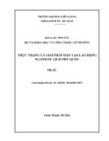
protocol and centralized reading. As of March 2008, 257
subjects had baseline measurements for carotid IMT and
brachial FMD, with 168 patients having one-year follow-
up assessments. Measurement of carotid IMT is ongoing,
but brachial FMD was discontinued after one-year follow-
up due to cost considerations. For this ancillary study, two
datasets were created namely: (1) cross-sectional data
consisting of 257 patients with baseline carotid IMT and
brachial FMD; and (2) progression data consisting of 168
patients with baseline and follow-up measurements for
carotidIMTandbrachialFMD(Figure1).
Clinical characteristics
Data on demographic and certain clinical characteristics
of subjects were collected at each centre using ques-
tionnaires administered by research staff, or by chart
review. Blood pressure was measured twice using a
mercury sphygmomanometer, and results averaged.
Lipids (total and HDL cholesterol and triglycerides)
were measured after overnight fast. LDL-cholesterol
concentration was calculated by the Friedewald formula.
AIDS Research and Therapy 2009, 6:11 http://www.aidsrestherapy.com/content/6/1/11
Page 2 of 10
(page number not for citation purposes)
CD4-T-lymphocyte counts were obtained by FACS
analysis performed by the Hamilton Regional Laboratory
Medicine Program, and plasma HIV viral load were
measured by Chiron bDNA assay at the Central Public
Health Laboratory in Toronto, Ontario.
Ultrasound methods
Ultrasound imaging and readings are conducted by trained
personnel using high resolution B-mode ultrasonography,
standardized protocol and centralized reading. The ultra-
sound laboratory in each study site uses imaging systems
equipped with 7.5 to 10 MHz linear phase-arrayed vascular
transducers. The same imaging system is used for all
ultrasound imaging within each center. Ultrasound mea-
surements are recorded on S-VHS tapes, which are later
digitized and analyzed offline at the Core Carotid Ultra-
sound Laboratory (Hamilton, Ontario) by a certified reader
blinded to patients' clinical information.
Patients were advised to fast and abstain from caffeine/
vasoactive medications 12 hours prior to measurement, and
were advised to avoid cigarette smoking (second-hand
inclusive) at least four hours prior to imaging. Imaging for
carotid IMT was done before brachial FMD on the same day.
(A) 12-segment carotid intimal medial thickness (IMT)
Carotid IMT identifies and quantitates early arterial wall
changes or structural vascular abnormalities [10,12,13]. A
rigorously-standardized, reliable, validated method of '12-
segment carotid IMT' developed by Lonn et al [8,30] was
used to assess the global extent of atherosclerosis in patients.
Images of six well-defined segments (near and far wall of the
common carotid, the bifurcation and the internal carotid)
were obtained in each of the left and right carotid arteries
using high resolution B-mode ultrasonography.
Ultrasound measurements were recorded on S-VHS
tapes, which were later digitized and analyzed using
the Image-Pro V4.5.1 software (Glen Burnie, Maryland).
For each segment a minimum of three frames were
measured. The maximum of all measurements from each
segment were summed-up and divided by 12 to obtain
the "12-segment mean-maximal carotid IMT" [8].
Twelve-segment mean-maximal carotid IMT is higher in
individuals with CAD [8,30].
(B) Brachial flow-mediated vasodilation (FMD)
Brachial FMD was measured using an extensively
validated and reliable method [13,31-33]. End-diastolic
ultrasound images of the brachial artery diameter
(longitudinally and slightly above the antebrachial fossa
or upper arm) were obtained at rest and during
vasodilator response induced by passive hyperemia
(endothelium-dependent dilation).
Each patient rested in a quiet room for 10 minutes, after
which sequential images of the brachial artery were
obtained within a 45 second interval. Subsequently, a
blood pressure cuff was inflated around the right lower arm
to at least 200 mm Hg, resulting in occlusion of blood flow
to the upper arm. The cuff was released after five minutes,
resulting in a marked increase in blood flow due to
resistance vessel dilation. The increase in blood flow
stimulates the release of NO which mediates the dilation
of conduit vessels. Peak brachial artery dilation occurs
approximately one minute after cuff release [26]. Another
set of sequential images was obtained during peak dilation.
The ultrasound image frames obtained were recorded on
S-VHS tapes, from which brachial artery diameters were
calculated using Dynamic Endothelial Assessment (DEA)
software (Montreal, Quebec). Average diameter of
brachial artery (before and after dilation) was obtained
from nine sequential images taken at rest and 12 taken
during peak artery dilation. Percent flow mediated
dilation was expressed as
FMD% average diameter at peak dilation average diameter a
=−tt rest
average diameter at rest
()
⎡
⎣
⎢⎤
⎦
⎥*100
Conduit vessel dilation is attenuated (smaller %FMD) in
individuals with CAD [26].
Twelve-segment carotid IMT and brachial FMD have
been standardized and validated in previous studies at
the Core Carotid Ultrasound Laboratory (Hamilton,
Ontario), with intraclass correlation > 90% and coeffi-
cient of variation < 5% for repeat examinations [13,30].
Statistical analysis
Continuous variables are expressed as mean (standard
deviation), while categorical variables are expressed as
count (percent) unless otherwise stated.
Figure 1
Flowchart of patients.
AIDS Research and Therapy 2009, 6:11 http://www.aidsrestherapy.com/content/6/1/11
Page 3 of 10
(page number not for citation purposes)

We hypothesized that "brachial FMD and carotid IMT
should correlate well with traditional vascular risk factors for
them to be considered good measures of extent, severity or
progression of atherosclerosis". This formed the basis for
assessment of construct validity. Multiple linear regression
models were used to examine the association between
baseline carotid IMT or brachial FMD and the well-validated
traditional "Framingham" cardiovascular risk factors of age,
male gender, current smoking status, systolic blood pressure
(SBP) and total:HDL cholesterol ratio using the cross-
sectional data. Goodness-of-fit was evaluated by plotting
the residuals from models to assess the normality assump-
tion. The distribution of residuals should approximate the
normal distribution for good model fit. We also used the co-
efficient of determination (R
2
) to quantify the proportion of
variation in the dependent variable explained by the
independent variables included in the multiple regression
models [34].
Fixed effects models were used to study the relationship
between progression of carotid IMT or brachial FMD and
known cardiovascular risk factors using the progression
data. Fixed effects models are useful for longitudinal
data in which changes in time-varying covariates such as
age, total:HDL cholesterol and SBP may affect the
repeated outcome of interest [35]. There is no reason
to assume that these quantities are constant over time.
Further, the correlation between baseline and follow-up
response is incorporated into model specification by
assuming a plausible correlation structure. We assumed a
"continuous time" version of the auto-regressive (AR(1))
correlation structure (available only for mixed/fixed
effects models in SAS
©
software), to adjust for irregula-
rities in follow-up times [36]. The reason is that many
scheduled follow-up visits were not feasible due to
circumstances beyond the control of investigators, thus
resulting in differential follow-up times for patients. A
time variable was created by designating the first visit for
each patient as (t
1
= 1) and follow-up visits as
tt
21
=+
⎛
⎝
⎜⎞
⎠
⎟
⎧Date of second visit - Date of first visit
365
⎨⎨
⎩
⎫
⎬
⎭
The time component is closer to reality by making it a
continuous, rather than a discrete, variable. Model fit
was assessed using the "Null Model Likelihood Ratio
Test" [37]. The "Null Model Likelihood Ratio Test" is a
likelihoodratiotestofwhetherthemodelwithaspecified
covariance structure fits better than a model where
repeated responses are assumed independent.Aninde-
pendent covariance structure is often implausible for
repeated measures data. A p-value < 0.05 for the
likelihood ratio test shows that the fitted model is better
than an independent covariance structure model [37].
Model adequacy was also evaluated using Akaike's
Information Criterion (AIC) to compare between "con-
tinuous time" and "fixed time" AR(1) structures. A
smaller AIC indicates better fit [37].
We evaluated the nature of the relationship between
baseline carotid IMT and brachial FMD using Pearson
correlation co-efficient.
Patients were classified as very low, low, medium/high
risk if individual Framingham risk scores were < 5%, 5–
9% and ≥10% respectively [38]. The medium and high
risk categories were combined due to limited numbers of
subjects in these categories. Framingham risk scores
quantify the 10-year risk of developing "hard" coronary
heart disease including myocardial infarction and
coronary death [38]. Framingham risk score is a strong
predictor of coronary heart disease [38]. One-way
analysis of variance (ANOVA) models were used to
cross-sectionally examine differences in brachial FMD or
carotid IMT by Framingham risk group classification.
We adjusted for current use of statin medication and
CD4 count in each regression model. All statistical tests
were conducted at 5% significance level. Graphs and
analysis results were obtained using SPSS Version 15.0
(SPSS Inc., Chicago, Illinois, USA) and SAS Version 9.1
(SAS Institute Inc., Cary, NC, USA).
The authors had full access to the data and take
responsibility for its integrity. All authors have read
and agree to the manuscript as written.
Results
Baseline and follow-up characteristics
Cross-sectional data
There were 257 patients in the baseline extent data with 232
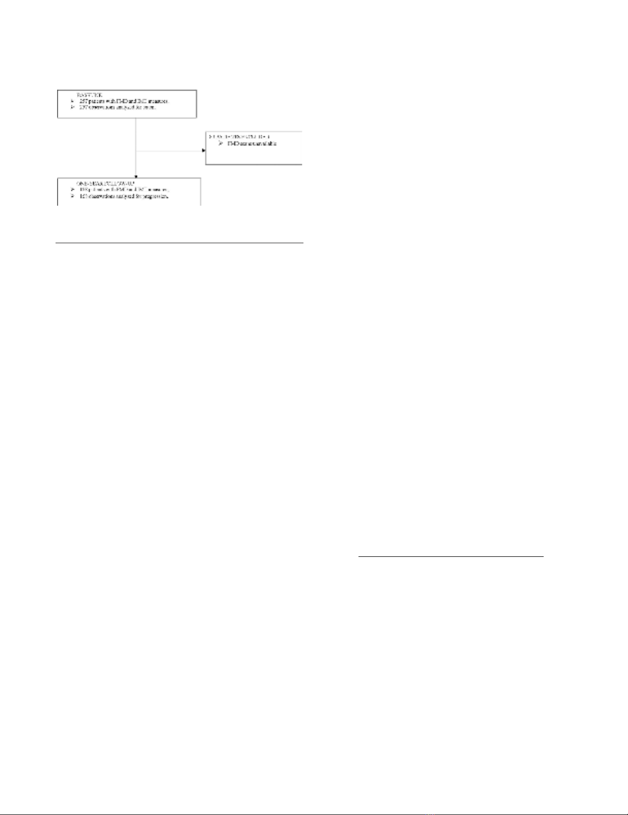
(90.3%) males and 25(9.7%) females. Carotid IMT ranged
from 0.47 mm to 2.24 mm, with mean(SD) of 0.81(0.23)
mm. Brachial FMD ranged from -7.36% to 29.96%, with
mean(SD) of 4.95(4.50)%. We found a weak inverse
relationship between carotid IMT and brachial FMD at
baseline (r = -0.126; p = 0.043; see Figure 2). Other patient
characteristics are listed in Table 1.
Stratifying by Framingham risk group, dose-response
relationships were found between risk group classifica-
tion and carotid IMT or brachial FMD (Table 2). Carotid
IMT differed significantly between risk groups from
ANOVA analysis (p < 0.001). Brachial FMD did not
differ significantly across the risk groups from ANOVA
results (p = 0.227).
Of the 257 patients assessed at baseline, information on
anti-retroviral therapy was available for 253 individuals.
AIDS Research and Therapy 2009, 6:11 http://www.aidsrestherapy.com/content/6/1/11
Page 4 of 10
(page number not for citation purposes)
There were 85 (34%) patients who were currently on
Abacavair, 106 (42%) were on Zidovudine, 61 (24%)
on Stavudine, 21 (8%) on Didanosine, 98 (39%) on
Efavirenz, 21 (8%) on Nelfinavir and 21 (8%) on
Nevirapine. However, we did not test the effects of HIV
medications on Carotid IMT/brachial FMD as that was
not part of our main goal, which was to validate these
measures against traditional risk factors.
Progression data
There were 168 patients in the progression dataset with
151(89.9%) males and 17(10.1%) females. Median
(interquartile range) follow-up time was 1.02 (0.43)
years. At baseline, carotid IMT varied from 0.47 mm to
1.57 mm with mean(SD) of 0.82(0.22) mm, while
brachial FMD varied from -6.81% to 29.96% with mean
(SD) of 5.10(4.58)%. At one-year follow-up, the
measures ranged from 0.50 mm to 1.57 mm with
mean(SD) of 0.84(0.23) mm and -13.61% to 25.52%
with mean(SD) of 4.40(4.96)% respectively. On average,
carotid IMT progressed at 0.02(standard error (SE) =
0.01) mm/year while brachial FMD decreased at 0.84
(SE = 0.79)%/year. Summary statistics for other variables
are listed in Table 3. Summary data for patients excluded
from the progression analyses are summarized in
Table 4. Patient distribution appears to be comparable
in both included and excluded data, except for viral load
and current statin use.
Examining the data cross-sectionally at baseline and
follow-up, there was a dose-response relationship between
carotid IMT and risk group classification (Table 5).
Carotid IMT differed significantly by risk group classifi-
cation at baseline and follow-up (p < 0.001 respectively
in each case). There was neither a dose-response relation-
ship nor significant difference in brachial FMD across
Figure 2
Carotid IMT versus brachial FMD at baseline.
Table 2: Baseline characteristics for extent data by Framingham
risk group
Risk group Number of
subjects
IMT
(mm)
FMD
(%)
Very low (< 5%) 88 0.68 (0.13) 5.58 (5.45)
Low (5 to 9%) 64 0.78 (0.16) 4.86 (3.59)
Medium/High (10% and above) 105 0.93 (0.27) 4.47 (4.08)
NB)EntriesforIMTandFMDarereportedasmean(standard
deviation); IMT increases significantly with increasing Framingham risk
(p < 0.001)
Table 1: Baseline characteristics for extent data (n = 257)
Variable Estimate
Male* 232 (90.3)
Age (years)
#
46.48 (7.86)
Carotid Artery Intima Media Thickness (IMT, mm)
#
0.81 (0.23)
Flow Mediated Vasodilation (FMD, %)
#
4.95 (4.50)
Total:HDL Cholesterol
#
5.28 (1.33)
Systolic Blood Pressure (mm Hg)
#
120.5 (15.6)
Current Smoking Status* 1 96 (37.5)
Current STATIN use* 1 18 (7.0)
CD4 Count
#
479.9 (270.6)
Log
10
Viral Load
#
2.2 (1.2)
NB) 1 = current smoker/user; * = count(%); # = mean(standard
deviation)
Table 4: Baseline characteristics of excluded cases (n = 89)
Variable Baseline
Male* 81(91)
AGE (years)
#
45.16 (6.80)
IMT (mm)
#
0.79 (0.26)
FMD (%)
#
4.67 (4.36)
SBP (mm Hg)
#
120.8 (15.6)
Total: HDL Cholesterol
#
5.04 (1.18)
Current smoking status* 1 36 (40.9)
Current STATIN use* 1 9 (10.1)
CD4 Count
#
451.14 (275.51)
Log
10
Viral Load
#
2.4 (1.3)
NB) 1 = current smoker/user; * = count(%); # = mean(standard
deviation)
Table 3: Baseline and follow-up characteristics for progression
data (n = 168)
Variable Baseline Follow-up
Male* 151 (89.9)
AGE (years)
#
47.19 (8.29) 48.25 (8.34)
IMT (mm)
#
0.82 (0.22) 0.84 (0.23)
FMD (%)
#
5.10 (4.58) 4.40 (4.96)
SBP (mm Hg)
#
120.4 (15.7) 121.1 (13.7)
Total: HDL Cholesterol
#
5.40 (1.39) 5.18 (1.17)
Current smoking status* 1 60 (35.7)
Current STATIN use* 1 9 (5.4)
CD4 Count
#
495.0 (267.6) 571.3 (883.2)
Log
10
Viral Load
#
2.0 (1.1) 2.1 (1.2)
NB) 1 = current smoker/user; * = count(%); # = mean(standard
deviation)
AIDS Research and Therapy 2009, 6:11 http://www.aidsrestherapy.com/content/6/1/11
Page 5 of 10
(page number not for citation purposes)


![Liệu pháp nội tiết trong mãn kinh: Báo cáo [Mới nhất]](https://cdn.tailieu.vn/images/document/thumbnail/2024/20240705/sanhobien01/135x160/4731720150416.jpg)