130
Tạp chí Y Dược học - Trường Đại học Y Dược Huế - Tập 8, số 6 - tháng 11/2018
JOURNAL OF MEDICINE AND PHARMACY
ĐÁNH GIÁ KẾT QUẢ PHẪU THUẬT NHỔ RĂNG KHÔN HÀM DƯỚI
LỆCH NGẦM CÓ SỬ DỤNG LASER CÔNG SUẤT THẤP
Nguyễn Thị Mai Hương1, Trần Tấn Tài2, Hồng Quốc Khanh3
(1) Học viên CK cấp II Răng Hàm Mặt, Trường Đại học Y Dược, Đại học Huế
(2) Khoa Răng Hàm Mặt, Trường Đại học Y Dược Huế
(3) Bệnh viện Răng Hàm Mặt thành phố Hồ Chí Minh
Tóm tắt
Đặt vấn đề: Phẫu thuật nhổ răng khôn lệch ngầm là thủ thuật phổ biến nhất trong nha khoa với độ khó
phụ thuộc vào vị trí răng mọc lệch. Liệu pháp laser sau phẫu thuật có thể giúp kích thích tái tạo tế bào và
mô, qua đó giúp giảm triệu chứng đau hậu phẫu. Mục tiêu của nghiên cứu này là nhằm khảo sát đặc điểm
lâm sàng và X quang của răng khôn hàm dưới lệch ngầm, đánh giá kết quả của phẫu thuật nhổ răng khôn
hàm dưới lệch ngầm có sử dụng laser công suất thấp. Đối tượng và phương pháp nghiên cứu: Dữ liệu lâm
sàng và X quang được thu thập từ 90 bệnh nhân (tuổi trung bình 28,13 ± 5,38), cần nhổ răng khôn hàm dưới
lệch - ngầm, phân bố ngẫu nhiên vào 3 nhóm: nhóm 1 laser chiếu trong miệng sau phẫu thuật, nhóm 2 laser
chiếu ngoài mặt sau phẫu thuật và nhóm 3 là nhóm chứng không kích hoạt tia. Đánh giá mức độ đau, sưng,
há miệng hạn chế tại thời điểm 24 giờ, 48 giờ, và 7 ngày sau phẫu thuật. Kết quả: Tương quan với cành đứng
xương hàm dưới: loại II chiếm ưu thế (88,9%), loại III (11,1%). Tương quan về độ sâu R8 với mặt nhai R7: vị
trí B (81,1%), vị trí C (18,9%). Tương quan R8 với trục R7 kế cận: nằm ngang (58,9%), nghiêng gần (40%) và
nghiêng xa (1,1%). Có sự giảm đáng kể (p<0,05) về mức độ đau, sưng và khít hàm giữa các nhóm được điều
trị với laser công suất thấp so với nhóm chứng tại thời điểm ngày thứ nhất và ngày thứ hai sau phẫu thuật.
Kết luận: Chiếu laser công suất thấp hậu phẫu trong miệng và ngoài mặt giúp cải thiện tình trạng sưng, đau,
khít hàm sau phẫu thuật răng khôn hàm dưới lệch ngầm so với nhóm chứng.
Từ khóa: Răng khôn hàm dưới lệch ngầm, liệu pháp laser công suất thấp.
Abstracts
ASSESSMENT ON RESULTS OF SURGICAL EXTRACTION OF
IMPACTED LOWER THIRD MOLARS WITH POST-OPERATIVE
LOW - LEVEL LASER THERAPY
Nguyen Thi Mai Huong1, Tran Tan Tai2, Hong Quoc Khanh3
(1) Postgraduate Students of Hue University of Medicine and Pharmacy, Hue University
(2) Faculty of Odonto-stomatology, Hue University of Medicine and Pharmacy
(3) Odonto-stomatology Hospital, Ho Chi Minh city
Background: The most frequently performed surgical procedure in dentistry is impacted third molar
extraction with difficulty varies according to the location of the tooth. Laser therapy after surgery can
accelerate cell and tissue reconstruction along with relieve post-operative pain. The objective of this study
was to investigate the clinical and radiographic characteristics of impacted lower third molar and to evaluate
the results of surgical extraction of impacted lower third molar with post-surgical low-level laser therapy
(LLLT). Subjects and Methods: Clinical and radiographic data from 90 patients (average age 28.13 ± 5.38)
subjected to a surgical extraction of lower third molar were pooled and divided randomly into 3 groups:
group 1 received LLLT immediately after surgery intraorally, group 2 treated with LLLT immediately after the
extraction extraorally. Patients received routine management with nonactivated laser were inserted in the
control group. Assessments of pain, swelling and trismus level were carried out at 24, 48 hours and on the 7th
day after surgery. Results: Correlation of wisdom teeth to ramus and adjacent teeth mainly type II (88.9%),
type III accounted for 11.1%. Relative depth of wisdom teeth in the bone mainly position B (81.1%), position
C (18.9%). Correlation of wisdom teeth axis to adjacent teeth: horizontal (58.9%), mesioangular impactions
(40%) and distoangular impaction (1.1%). There were statistically significant decreases in the level of pain,
edema and interincisal opening between the laser-treated groups and the control group on the 1st and 2nd
- Địa chỉ liên hệ: Trần Tấn Tài, email: taihangdr@gmail.com
- Ngày nhận bài: 15/10/2018; Ngày đồng ý đăng: 9/11/2018, Ngày xuất bản: 17/11/2018
DOI: 10.34071/jmp.2018.6.17

131
Tạp chí Y Dược học - Trường Đại học Y Dược Huế - Tập 8, số 6 - tháng 11/2018
JOURNAL OF MEDICINE AND PHARMACY
postoperative day. Conclusions: Intraoral and extraoral post-surgical low-level laser therapy treatment was
useful in reducing swelling, pain and trismus compared to placebo group in impacted third molar surgery.
Keywords: Impacted lower third molar, low- level laser therapy
1. ĐẶT VẤN ĐỀ
Răng khôn là răng mọc sau cùng trên cung hàm
do đó thường bị thiếu chỗ, dẫn đến tình trạng mọc
lệch lạc và gây ra nhiều biến chứng ảnh hưởng không
nhỏ đến chất lượng cuộc sống của bệnh nhân. Phẫu
thuật nhổ răng khôn hàm dưới lệch ngầm là thủ
thuật phổ biến nhưng tương đối xâm lấn. Khoảng
63% bệnh nhân cảm thấy đau đớn dữ dội trong 24
giờ đầu sau phẫu thuật, nhất là trong khoảng 3-5 giờ
sau khi thuốc tê hết tác dụng [6]. Các triệu chứng
sưng nề và hạn chế há miệng cũng gây không ít khó
chịu cho bệnh nhân.
Hiện nay, điều trị nội khoa hậu phẫu là phương
pháp được áp dụng rộng rãi, tuy hiệu quả nhưng lại
gây một số tác dụng phụ không mong muốn, tăng
nguy cơ biến chứng trên bệnh nhân có bệnh lý toàn
thân. Laser công suất thấp đã được sử dụng nhiều
trong y khoa điều trị và được chứng minh là có tác
động điều hòa quá trình viêm, giúp giảm sưng đau
và thúc đẩy quá trình lành thương mà không gây bất
kỳ tác dụng phụ nào.
Trên phương diện điều trị hậu phẫu nhổ răng,
việc chiếu laser trong và ngoài miệng, trước và sau
khâu đều cho kết quả khác nhau [6]. Dựa trên những
bằng chứng khoa học sẵn có, với mong muốn tìm
kiếm một phương pháp điều trị hỗ trợ triệu chứng
tối ưu cho bệnh nhân sau phẫu thuật nhổ răng khôn
hàm dưới lệch ngầm, chúng tôi thực hiện đề tài này
nhằm 2 mục tiêu:
1/ Khảo sát đặc điểm lâm sàng, X quang ở bệnh
nhân có răng khôn hàm dưới lệch ngầm.
2/ Đánh giá kết quả của phẫu thuật nhổ răng
khôn hàm dưới lệch ngầm có sử dụng laser công
suất thấp.
2. ĐỐI TƯỢNG VÀ PHƯƠNG PHÁP NGHIÊN CỨU
2.1. Đối tượng nghiên cứu
-Tiêu chuẩn chọn: Bệnh nhân trong độ tuổi 18-
40 tuổi, có răng khôn hàm dưới lệch ngầm cần phẫu
thuật, có sức khỏe toàn thân tốt và đồng ý tham gia
nghiên cứu.
-Tiêu chuẩn loại trừ: Bệnh nhân thuộc chống chỉ
định điều trị với laser công suất thấp, hút thuốc lá,
có thai hoặc đang cho con bú. Bệnh nhân đang dung
thuốc điều trị bệnh lý toàn thân hoặc tại chỗ.
2.2. Phương pháp nghiên cứu
- Thiết kế nghiên cứu: nghiên cứu mô tả, tiến
cứu, can thiệp lâm sàng có đối chứng.
- Cỡ mẫu: n = 90 bệnh (90 răng), phân bố ngẫu
nhiên lần lượt vào 2 nhóm nghiên cứu và 1 nhóm
chứng.
- Phương pháp tiến hành:
+ Tất cả bệnh nhân được phẫu thuật theo đúng
qui trình kỹ thuật của bệnh viện Răng Hàm Mặt
thành phố.
+ Laser diode AsGaAl bước sóng 810 nm, công suất
0,5W ± 20%, chế độ phát liên tục trong 30 giây mỗi vị
trí, mật độ năng lượng 4 J/cm2. Đầu phát tia laser được
giữ cách mô đích (ổ răng, bề mặt niêm mạc hoặc bề
mặt da) 1cm, kích thước đầu chiếu 400 µm, vòng tròn
tác động của tia laser lên mô có đường kính 2 cm. Qui
trình chiếu tia được thực hiện ngay sau khi kết thúc
phẫu thuật lấy răng khôn.
+ Nhóm I – chiếu tia trong miệng (n=30): đầu dò
laser được chiếu tại 4 vị trí: (1) ngay giữa ổ nhổ răng,
(2) phần ba cổ phía lưỡi, (3) phần ba giữa phía lưỡi,
(4) phần ba chóp phía lưỡi.
+ Nhóm II – chiếu tia ngoài mặt (n=30): giữ đầu
dò tại 4 vị trí dọc cơ cắn: (1) phần tư dưới của cơ cắn
(gần điểm bám tận ở xương hàm dưới), (2) phần tư
giữa-dưới của cơ cắn, (3) phần tư giữa-trên cơ cắn,
(4) phần tư trên cùng của cơ cắn (gần nguyên ủy ở
cung tiếp).
+ Nhóm III – nhóm chng (n=30): giữ đầu dò
theo đúng qui trình (ngẫu nhiên chọn lựa qui trình
trong miệng hoặc ngoài mặt), tuy nhiên không kích
hoạt tia laser.
+ Nghiên cứu dùng mẫu bệnh án thống nhất.
Các mốc giải phẫu, số đo trước và sau phẫu thuật
do một nhân viên y tế được huấn luyện định chuẩn
trực tiếp thu thập dữ liệu.
+ Đo độ sưng và độ há miệng tối đa được thực
hiện trên cùng một tư thế ghế nha 45 độ, vào ngày
thứ 1, thứ 2 và thứ 7 sau phẫu thuật.
+ Mức độ đau theo thang VAS và Likert 7: bệnh
nhân tự đánh giá theo mẫu thống nhất vào mỗi 2 giờ
trong 6 giờ đầu tiên sau khi hết cảm giác tê môi, và
vào thời điểm 24 giờ, 48 giờ sau phẫu thuật. Bệnh
nhân tự ghi nhận số lượng viên thuốc giảm đau đã
uống trong 2 ngày sau phẫu thuật.
+ Bảng câu hỏi OHIP-14: nhân viên y tế đọc câu
hỏi khi bệnh nhân đến tái khám vào ngày thứ 1, thứ
2 và thứ 7 sau phẫu thuật. Bệnh nhân tự chọn các
mức độ tương ứng với tình trạng của mình.
-Biến số nghiên cứu:
+ Đặc điểm lâm sàng: tuổi, nhóm tuổi, giới tính,
132
Tạp chí Y Dược học - Trường Đại học Y Dược Huế - Tập 8, số 6 - tháng 11/2018
JOURNAL OF MEDICINE AND PHARMACY
nghề nghiệp, lý do đến khám, sự hiện diện của răng
khôn hàm dưới mọc lệch trong khoang miệng.
+ Đặc điểm X quang: mức độ lệch ngầm R8 theo
Pell&Gregory, chiều hướng mọc răng theo Winter,
tình trạng ảnh hưởng R7.
+ Kết quả phẫu thuật R8 có hỗ trợ laser công suất
thấp: đánh giá đau theo thang Likert 7, VAS và số
viên thuốc giảm đau; đánh giá sưng mặt bằng thước
dây theo D1 (góc mắt ngoài-góc hàm), D2 (bình tai-
khóe mép), D3 (bình tai-điểm trước nhất của cằm);
đánh giá độ há miệng tối đa bằng thước kẹp (mm);
đánh giá chất lượng cuộc sống của BN sau phẫu
thuật bằng bộ câu hỏi OHIP-14.
2.3. Xử lý số liệu
- Số liệu được xử lý và phân tích bằng phần mềm
SPSS 18.0.
- Kiểm định χ2, Wilcoxon, Mann Whitney, T test,
Fisher’s exact.
3.KẾT QUẢ
3.1. Đặc điểm lâm sàng và X quang của đối tượng nghiên cứu
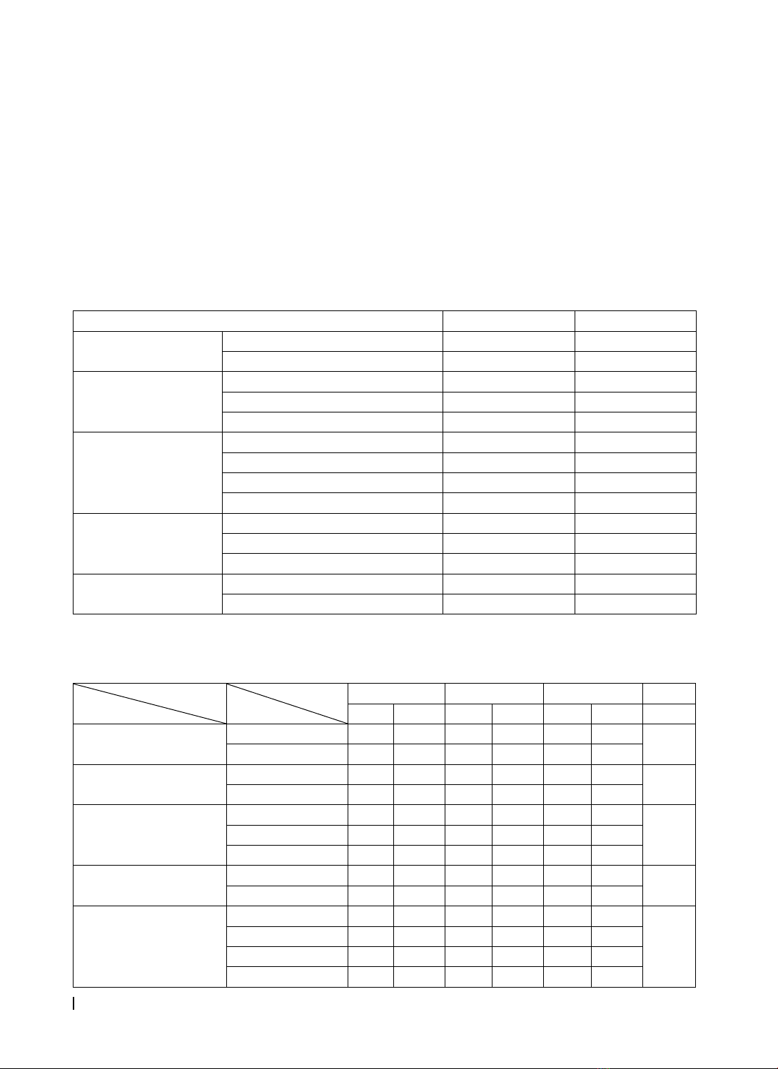
Bảng 3.1. Một số đặc điểm lâm sàng của mẫu nghiên cứu
Đặc điểm n %
Giới tính Nam 39 43,3
Nữ 51 56,7
Nhóm tuổi
18-24 25 27,8
25-35 55 61,1
> 35 10 11,1
Nghề nghiệp
CBCC 53 58,9
HSSV 10 11,1
Công nhân 10 11,1
Khác 17 18,9
Lý do đến khám
Nhét thức ăn 39 43,3
Dự phòng 29 32,2
Khác 22 24,5
Sự hiện diện R8 trong
khoang miệng
Thấy một phần R trong miệng 69 76,7
Không thấy R trong miệng 21 23,3
Bảng trên cho thấy không khác biệt về giới trong nhóm nghiên cứu, nhóm tuổi phổ biến là 25-35 tuổi, cán
bộ công chức (CBCC) là thành phần chủ yếu trong mẫu nghiên cứu. Phần lớn BN muốn nhổ răng vì khó chịu
do nhét thức ăn và đa số các răng có hiện diện một phần trong khoang miệng.
Bảng 3.2. Đặc điểm răng khôn hàm dưới lệch ngầm đánh giá trên phim toàn cảnh
Đặc điểm
Răng
Phân loại
R38 R48 Tổng số p
n%n%n%
Tương quan với cành
đứng xương hàm dưới
Loại II 33 86,8 47 90,4 80 88,9 >0,05
Loại III 5 13,2 5 9,6 10 11,1
Độ sâu so với mặt nhai
R7
Vị trí B 32 84,2 41 78,8 73 81,1 >0,05
Vị trí C 6 15,8 11 21,2 17 18,9
Độ nghiêng trục răng
khôn theo Winter
Lệch gần 17 44,7 19 36,5 36 40
>0,05Lệch xa 0 0 1 1,9 1 1,1
Nằm ngang 21 55,3 32 61,5 53 58,9
Độ lệch trục R8 dưới so
với R7
≤ 45 độ 14 36,8 15 28,8 29 32,2 >0,05
> 45 độ 24 63,2 37 71,2 61 67,8
Ảnh hưởng của R8 đến
R7 kế cận
Tiêu xương 14 36,8 26 50,0 40 44,4
>0,05
Sâu 2 5,3 0 0 2 2,2
Tiêu xương, sâu 9 23,7 6 11,5 15 16,7
Không ảnh hưởng 13 34,2 20 38,5 33 36,7
133
Tạp chí Y Dược học - Trường Đại học Y Dược Huế - Tập 8, số 6 - tháng 11/2018
JOURNAL OF MEDICINE AND PHARMACY
Đa số các răng trong mẫu có vị trí B và tương quan loại II so với cành đứng xương hàm dưới theo phân
loại Pell&Gregory và có độ nghiêng trên 45 độ so với trục R7. R8 lệch gần và nằm ngang chiếm hầu hết mẫu
nghiên cứu. 44,4% R8 gây tiêu xương R7 kế cận, tuy nhiên, tỉ lệ R8 chưa gây ảnh hưởng R7 cũng chiếm phần
khá cao (36,7%). Không có sự khác biệt giữa hai phần hàm (p>0,05).
3.2. Kết quả phẫu thuật răng khôn hàm dưới lệch ngầm có hỗ trợ của laser công suất thấp
3.2.1. Mức độ đau của bệnh nhân sau khi hết cảm giác tê môi
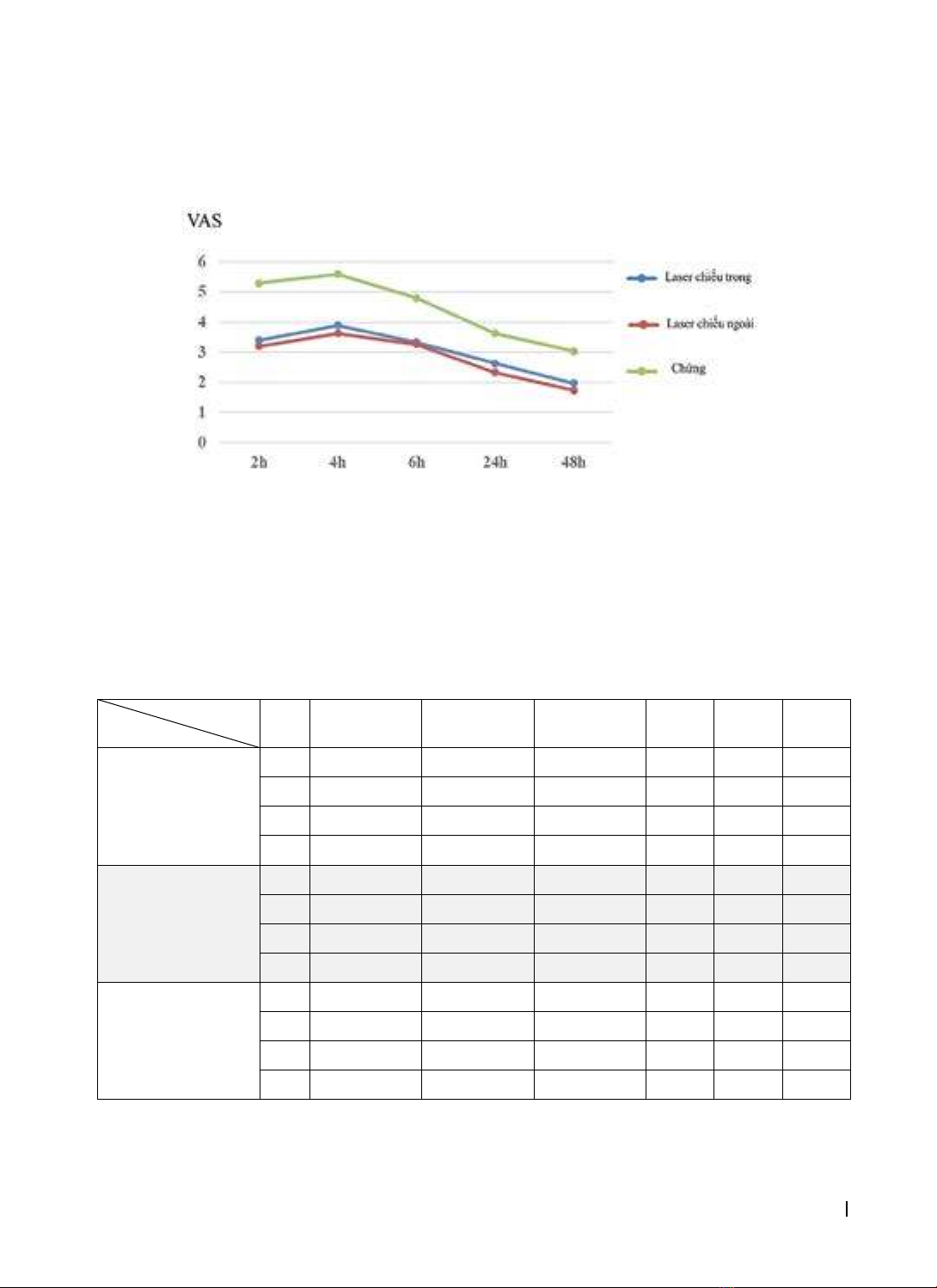
Biểu đồ 3.1. Mức độ đau giữa 3 nhóm nghiên cứu theo thang đo VAS
Vào ngày thứ hai sau phẫu thuật, đa số các bệnh nhân đều “không đau” đến “đau nhẹ”.
Điểm VAS trung bình ở nhóm có chiếu laser thấp hơn có ý nghĩa so với nhóm chứng. Điểm VAS cao nhất
ở thời điểm 4 giờ sau khi hết cảm giác tê môi, sau đó giảm dần và đến ngày thứ 2 sau phẫu thuật thì sự khác
biệt điểm VAS không còn đáng kể ở tất cả các nhóm nghiên cứu.
Số lượng trung bình viên thuốc giảm đau đã uống giữa nhóm sử dụng laser chiếu trong miệng, laser chiếu
ngoài mặt và nhóm chứng lần lượt là 0,5 – 0,43 – 1,47 viên thuốc, có sự khác biệt có ý nghĩa giữa nhóm laser
chiếu ngoài mặt và nhóm chiếu trong miệng so với nhóm chứng (p<0,001).
3.2.2. Đánh giá mức độ sưng mô mềm sau phẫu thuật nhổ răng khôn
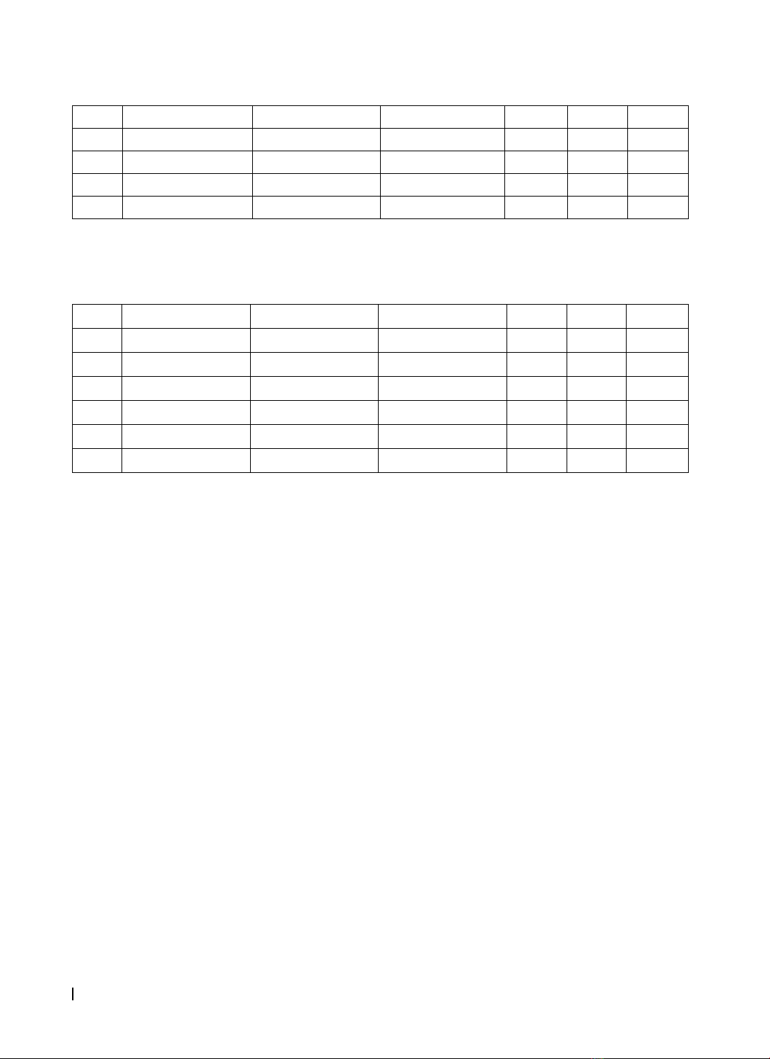
Bảng 3.3. Mức độ sưng mặt theo các khoảng cách ở 3 nhóm nghiên cứu tại các thời điểm
Mức độ sưng
Chiếu trong
(N1)
Chiếu ngoài
(N2)
Chứng
(N3) p12 p13 p23
D1
(góc mắt ngoài –
góc hàm)
T0109,23±5,18 109,3±5,31 109,5±5,02 >0,05 >0,05 >0,05
T1110,33±5,53 110,23±5,37 113,13±4,74 >0,05 <0,05 <0,05
T2110,43±5,54 110,27±5,39 113,27±4,71 >0,05 <0,05 <0,05
T7109,27±5,21 109,3±5,36 109,53±4,89 >0,05 >0,05 >0,05
D2
(bình tai – khóe
mép)
T0122,23±5,09 122,27±5,71 122,37±5,49 >0,05 >0,05 >0,05
T1123,47±5,26 123,37±5,61 126,23±5,23 >0,05 <0,05 <0,05
T2123,6±5,31 123,4±5,72 126,43±5,12 >0,05 <0,05 <0,05
T7122,23±5,14 122,27±5,77 122,4±5,37 >0,05 >0,05 >0,05
D3 (bình tai – điểm
nhô nhất của cằm)
T0153,77±2,47 153,83±3,00 153,8±2,87 >0,05 >0,05 >0,05
T1155.07±2,69 155,00±2,84 157,73±2,73 >0,05 <0,001 <0,001
T2155,2±2,77 155,07±3,06 157,9±2,75 >0,05 <0,001 <0,001
T7153,83±2,51 153,87±3,01 153,9±2,75 >0,05 >0,05 >0,05
Các khoảng cách đo được tăng vào ngày thứ nhất và thứ hai sau phẫu thuật, giảm vào ngày thứ bảy. Tại
thời điểm 7 ngày sau phẫu thuật, kích thước mô mềm đo theo 3 chiều hướng gần như đều trở về trạng thái
bình thường trên tất cả các bệnh nhân tham gia nghiên cứu. Mức độ sưng mô mềm ở nhóm có chiếu laser
thấp hơn nhóm chứng có ý nghĩa thống kê (p<0,05).
134
Tạp chí Y Dược học - Trường Đại học Y Dược Huế - Tập 8, số 6 - tháng 11/2018
JOURNAL OF MEDICINE AND PHARMACY
3.2.3. Đánh giá mức độ khít hàm sau phẫu thuật nhổ răng khôn
Bảng 3.4. Mức độ há miệng tối đa ở 3 nhóm tại các thời điểm so với trước phẫu thuật
Chiếu trong (N1) Chiếu ngoài (N2) Nhóm chứng (N3) p12 p13 p23
T049,1±2,22 49,06±2,48 49,03±2,37 >0,05 >0,05 >0,05
T147,07±2,42 47,17±2,49 44,43±2,66 >0,05 <0,001 <0,001
T246,9±2,58 47,07±2,65 44,23±2,97 >0,05 <0,001 <0,001
T749,07±2,27 49,07±2,42 48,93±2,3 >0,05 >0,05 >0,05
Có sự khác biệt có ý nghĩa thống kê về hiệu quả giảm triệu chứng khít hàm ở nhóm sử dụng laser chiếu trong
và laser chiếu ngoài so với nhóm chứng giữa các thời điểm (p < 0,001). Ngày thứ bảy sau phẫu thuật, sự khác
biệt về độ há miệng tối đa giữa các nhóm không còn khác biệt.
3.2.4. Đánh giá chất lượng cuộc sống của bệnh nhân sau phẫu thuật nhổ răng khôn
Bảng 3.5. Tổng hợp điểm bộ câu hỏi OHIP 14
Chiếu trong (N1) Chiếu ngoài (N2) Nhóm chứng (N3) p12 p13 p23
T115,43±4,87 14,73±5,19 23,77±7,51 >0,05 <0,001 <0,001
T212,37±4,54 11,73±3,28 19,57±7,07 >0,05 <0,001 <0,001
T75,27±1,28 5,23±1,10 5,33±1,32 >0,05 >0,05 >0,05
p12 <0,05** <0,05** <0,001**
p17 <0,001** <0,001** <0,001**
p27 <0,001** <0,001** <0,001**
Điểm số OHIP giảm trong cả 3 nhóm theo thời
gian. Điểm OHIP cao nhất vào ngày thứ 1 sau phẫu
thuật, giảm nhiều vào ngày thứ 2 và giảm về gần như
ban đầu vào ngày thứ 7 sau phẫu thuật.
4. BÀN LUẬN
4.1. Về đặc điểm lâm sàng và X quang của răng
khôn hàm dưới lệch ngầm
4.1.1. Tuổi và nhóm tuổi của mẫu nghiên cứu
Tuổi trung bình của mẫu nghiên cứu này là 28,13
± 5,38 tuổi. Bệnh nhân trẻ nhất 18 tuổi, lớn nhất
40 tuổi. Độ tuổi này tương đối lớn hơn so với các
nghiên cứu về răng khôn lệch ngầm có báo cáo về
đặc điểm lâm sàng này ở trong nước và trên thế giới
[2], [5]. Tuổi tiến triển có liên quan đến nhiều biến
chứng sau phẫu thuật hơn, dù vậy tuổi trung bình
của nghiên cứu này cùng với các nghiên cứu khác
đều thuộc về nhóm tuổi thanh niên.
Nhóm tuổi phổ biến của nghiên cứu là từ 25-
35 tuổi, tương đối cao hơn so với các nghiên cứu
đã tiến hành ở Việt Nam cũng như trên thế giới.
Nguyễn Đức Tịnh (2014) báo cáo nhóm tuổi 18 - 25
tuổi chiếm tỷ lệ cao nhất 59,8% [2], Braimah Ramat
(2018) báo cáo đa số bệnh nhân (43,7%) thuộc
nhóm tuổi 21-25 tuổi [5]. Nghiên cứu này loại trừ
trường hợp đến khám và nhổ răng vì lý do sưng đau
(nhằm loại trừ ảnh hưởng của thuốc đến kết quả
điều trị laser sau phẫu thuật) vốn là những nguyên
nhân cấp thường xuất hiện vào giai đoạn mọc răng
ở nhóm tuổi trẻ hơn. Ngoài ra, đối tượng khám chữa
bệnh của khoa Bảo hiểm dịch vụ đa số là cán bộ viên
chức, vì vậy độ tuổi này là phù hợp điều kiện thực tế.
4.1.2. Tỉ lệ phân bố giới tính trong mẫu nghiên
cứu
Tỉ lệ nam : nữ trong mẫu nghiên cứu là 1:1,25.
Tỷ lệ này tương đồng với nhiều nghiên cứu của Việt
Nam và thế giới[1], [2], [5]. Tỉ lệ nữ chiếm ưu thế
trong mẫu nghiên cứu có thể là do hậu quả của sự
khác biệt giữa tốc độ tăng trưởng của nam và nữ.
Ngoài ra, hình thái xương hàm dưới thanh mảnh, ít
góc cạnh và chế độ ăn tương đối “mềm” hơn ở nữ
cũng là những yếu tố góp phần làm cho tỉ lệ răng
hàm dưới lệch ngầm ở nữ cao hơn ở nam.
4.1.3. Lý do đến khám
Hầu hết bệnh nhân trong mẫu nghiên cứu này
đều đến khám và điều trị khi chưa có biến chứng
ảnh hưởng nghiêm trọng đến chất lượng cuộc sống
(32,2% nhổ răng dự phòng). Đa số các nghiên cứu
dịch tễ khảo sát các vấn đề xung quanh răng khôn
hàm dưới lệch ngầm đều cho thấy bệnh nhân chủ
yếu đến khám vì lý do đau đơn thuần hoặc đau
sưng kết hợp: Nguyễn Đức Tịnh (2014) [2] báo cáo
đau chiếm tỷ lệ cao nhất 57,3%, kế đến là sưng và
đau (29,3%); 65,2% bệnh nhân trong nghiên cứu
Braimah Ramat [5] bị sưng đau khi đến khám. Có sự
khác biệt tỉ lệ này là do trong mẫu nghiên cứu của
chúng tôi đã loại trừ lí do bệnh nhân đên khám vì lí
do sưng, đau để loại trừ yếu tố nhiễu do thuốc.