
AtCYS1, a cystatin from
Arabidopsis thaliana
, suppresses
hypersensitive cell death
Beatrice Belenghi
1,
*, Filippo Acconcia
2,
*, Maurizio Trovato
3
, Michele Perazzolli
1
, Alessio Bocedi
2
,
Fabio Polticelli
2
, Paolo Ascenzi
2
and Massimo Delledonne
1
1
Dipartimento Scientifico e Tecnologico, Universita
`degli Studi di Verona, Verona, Italy;
2
Dipartimento di Biologia,
Universita
`degli Studi ‘Roma Tre’, Rome, Italy;
3
Dipartimento di Genetica e Biologia Molecolare ‘Charles Darwin’,
Universita
`degli Studi di Roma ‘La Sapienza’, Rome, Italy
In plants, cysteine protease inhibitors are involved in the
regulation of protein turnover and play an important role
in resistance against insects and pathogens. AtCYS1 from
Arabidopsis thaliana encodes a protein of 102 amino acids
that contains the conserved motif of cysteine protease
inhibitors belonging to the cystatin superfamily (Gln-
Val-Val-Ala-Gly). Recombinant A. thaliana cystatin-1
(AtCYS1) was expressed in Escherichia coli and purified.
AtCYS1 inhibits the catalytic activity of papain
(K
d
¼4.0 ·10
)2
l
M
,atpH7.0and25C), generally
taken as a molecular model of cysteine proteases. The
molecular bases for papain inhibition by AtCYS1 have
been analysed taking into account the three-dimensional
structure of the papain–stefin B complex. AtCYS1 is
constitutively expressed in roots and in developing siliques
of A. thaliana. In leaves, AtCYS1 is strongly induced by
wounding, by challenge with avirulent pathogens and by
nitric oxide (NO). The overexpression of AtCYS1 blocks
cell death activated by either avirulent pathogens or by
oxidative and nitrosative stress in both A. thaliana sus-
pension cultured cells and in transgenic tobacco plants.
The suppression of the NO-mediated cell death in plants
overexpressing AtCYS1 provides the evidence that NO is
not cytotoxic for the plant, indicating that NO functions
as cell death trigger through the stimulation of an active
process, in which cysteine proteases and theirs proteina-
ceous inhibitors appear to play a crucial role.
Keywords:Arabidopsis thaliana; cystatin; cysteine protease;
hypersensitive response; programmed cell death.
Cysteine protease inhibitors inactivate proteases by trapping
them in a(n) (ir)reversible, tight equimolar complex [2].
Plant cystatins, homologous to animal cysteine protease
inhibitors [3], have been characterized in several monocots
and dicots, including rice, maize, soybean, Chinese cabbage
[4], chestnut, potato and tomato [5–13]. Cystatins show
different expression patterns during plant development
and defence responses to biotic and abiotic stresses [14].
Moreover, cystatins may play a role in the regulation of
protein turnover and plant defence against insect predation
and pathogens [13].
Wounding causes extensive changes in the pattern of
defence protein synthesis leading to localized resistance at
the site of the lesion. The response includes the production
of phytoalexin, enhanced lignification and suberization of
the cell wall, and systemic induction of protease inhibitors
[15,16]. Cystatin accumulation occurs after activation of
both long- and short-distance signal cascades, triggered by
accumulation of systemin or by cell wall fragments. Many
insects such as Hemiptera and Coleoptera rely on cysteine
proteases for the majority of the proteolytic activity
responsible for protein digestion in the gut [17]. Remark-
ably, cystatins have been shown to inhibit the activity of
digestive proteases from coleopteran pests in vitro,aswellas
the inhibition of larval development in vivo. Thus, cystatins
function as ‘toxins’ by targeting the major proteolytic
digestive enzymes of herbivore insects [6,11,18]. Moreover,
cysteine proteases play a fundamental role in virus replica-
tion; therefore, constitutive expression of a rice cystatin in
tobacco induces virus resistance [19].
Recently, a synthetic gene encoding the mature form of a
soybean cystatin has been reported to effectively block cell
death triggered by either oxidative stress or avirulent
Correspondence to M. Delledonne, Dipartimento Scientifico e
Tecnologico, Universita
`degli Studi di Verona, Ca¢Vignal 1,
Strada Le Grazie 15, I-37134 Verona, Italy.
Fax: + 39 045 8027929, Tel.: + 39 045 8027962,
E-mail: massimo.delledonne@univr.it, or P. Ascenzi,
Dipartimento di Biologia, Universita
`degli Studi ‘Roma Tre’,
Viale G. Marconi 446, I-00146 Roma, Italy.
Fax: + 39 06 55176321, Tel.: + 39 06 55176329,
E-mail: ascenzi@uniroma3.it
Abbreviations:AtCYS1,Arabidopsis thaliana cystatin-1; DAB,
3,3¢-diaminobenzidine; GST, glutathione S-transferase; HR, hyper-
sensitive response; IPTG, isopropyl thio-b-
D
-galactoside; NO, nitric
oxide; PCD, programmed cell death; PR, pathogenesis-related; SNP,
sodium nitroprusside; Z-Phe-Arg-AMC, N-a-benzyloxycarbonyl-
L
-phenylalanyl-
L
-arginine-(7-amido-4-methylcoumarin).
Enzymes: glucose oxidase, from Aspergillus niger (EC 1.1.3.4); lyso-
zyme, from chicken egg (EC 3.2.1.17); papain, from Carica papaya L.
(EC 3.4.22.2); thrombin, from bovine plasma (EC 3.4.21.5).
*Note: These authors contributed equally to this work.
Note: Full length chicken cystatin numbering is used throughout the
text [1].
(Received 20 February 2003, revised 22 April 2003,
accepted 23 April 2003)
Eur. J. Biochem. 270, 2593–2604 (2003) FEBS 2003 doi:10.1046/j.1432-1033.2003.03630.x

pathogens, when transiently expressed in cultured soybean
cells [20]. Thus, a role for cysteine proteases can be
envisioned in programmed cell death (PCD) by regulatory
protein degradation. Note that cysteine proteases have been
implicated in the differentiation of Zinnia elegans cells into
tracheary elements, which involves mesophyl cell death [21].
SAG12 from Arabidopsis thaliana is a senescence-associated
gene, which encodes a cysteine protease that is coordinately
expressed with hypersensitive cell death [22]. Cystatins
may therefore function as modulators of cysteine prote-
ase activity during plant growth, development and seed
maturation [23].
The activation of PCD appears to play an important role
during the hypersensitive disease-resistance response against
pathogen attack; however, it is imperative that plants
maintain the capacity to regulate this process [20]. Here, we
describe the molecular and biochemical characterization of
the A. thaliana cystatin-1 AtCYS1 that accumulates fol-
lowing wounding and during the hypersensitive response.
Moreover, the constitutive expression of AtCYS1 suppres-
ses PCD triggered by either avirulent pathogens or oxidative
and nitrosative stresses in both A. thaliana suspension
cultures and in transgenic tobacco.
Materials and methods
Materials
Papain (from Carica papaya L.), bovine thrombin,
chicken egg white lysozyme, glucose oxidase (from Asper-
gillus niger), agarose-p-amminobenzamidine, agarose-
glutathione, N-a-benzyloxycarbonyl-
L
-phenylalanyl-
L
-
arginine-(7-amido-4-methylcoumarin) (Z-Phe-Arg-
AMC), chloral hydrate, 3,3¢-diaminobenzidine (DAB),
N-lauroylsarcosine,
L
-trans-epoxysuccinyl-
L
-leucylamido(4-
guanidino)-butane, dimethylsulfoxide, dithiothreitol, iso-
propyl thio-b-
D
-galactoside (IPTG), sodium nitroprusside
(SNP), methyl jasmonate kanamycin, ampicillin, rifampicin,
Evan’s blue, trypan blue, protein molecular markers, Tris
and reduced glutathione were purchased from Sigma
Chemical Co. (St. Louis, MO, USA). Restriction enzymes
were purchased from Promega (Madison, WI, USA). All the
otherchemicalswerepurchasedfromMerckAG(Darmstadt,
Germany). All products were of analytical or reagent grade
and were used without further purification.
Southern blot analysis
Genomic DNA was isolated from A. thaliana and tobacco
leaves as reported previously [24]. In brief, 10 lgof
genomic DNA was cut with indicated restriction enzymes,
fractionated on 0.8% (w/v) agarose gels, transferred to
nylon filters and hybridized to a radioactive probe
prepared from the complete AtCYS1 cDNA. Prehybridi-
zation and hybridization were performed as described
previously [25].
Northern blot analysis
Total RNA was extracted from 4 week old A. thaliana
plants at fixed times following indicated treatment. The nitric
oxide (NO) donor SNP was prepared and infiltrated into
leaves as described previously [26]. A bacterial suspension
containing 5 ·10
6
CFUÆmL
)1
of virulent Pseudomonas
syringae pv. maculicola or the isogenic avirulent strain
carrying avrRpm1 was infiltrated into leaves as described [27].
Plants were sprayed with 45.5 l
M
methyl jasmonate pre-
pared in 0.1% (v/v) ethanol. Leaves, stems, roots, flowers
and siliques were cut and frozen directly into liquid nitrogen.
RNA from A. thaliana and tobacco frozen tissues was
extracted using the RNeasy Plant Mini Kit (QIAGEN,
Hilden, Germany) as described by the manufacturer. Then,
5lg of total RNA were separated on 1.5% (w/v) agarose gels
containing 6% (v/v) formaldehyde, blotted onto Hybond
N
+
membrane (Amersham Biosciences, Little Chalfont,
UK) according to manufacturer’s instructions, and cross-
linked by UV irradiation. For hybridization analysis, the
purified BamHI/SacI fragment (500 bp), containing the
entire AtCYS1 coding sequence, was used as probe. The level
of PR-1 transcripts in tobacco was determined using the PCR
amplification of tobacco Pr1–1a (GenBank accession num-
ber X12737) as a probe. Prehybridization and hybridization
were performed as described previously [25].
E. coli
expression and purification of recombinant
AtCYS1
The pGEX 4T-3-AtCYS1 expression vector contains
a 380 bp fragment that was obtained by PCR amplification
of the AtCYS1 cDNA using specific primers carrying a
BamHI site (forward: GGATCCGCGGATCAACAAG
CAGGAACA) and a SalI site (reverse: GTCGACTCA
CGTGGTCTGAGAGCACAC) for directional cloning
(restriction sites underlined). The amplified fragment was
subcloned into the pGEM-T vector (Promega), sequenced,
cut with BamHI and SalI, and then introduced into the
expression vector pGEX4T-3 (Amersham Biosciences) cut
with the same restriction enzymes. The resulting construct
(pGEX 4T-3-AtCYS1), which expresses AtCYS1 as a
fusion protein with the 26 kDa glutathione S-transferase
(GST), was introduced in E. coli JM101 competent cells.
Cultures of JM101 E. coli containing the pGEX
4T-3-AtCYS1 construct were grown to saturation at
30 C in Luria–Bertani broth supplemented with
50 lgÆmL
)1
ampicillin, diluted 1 : 100 in 500 mL of fresh
ampicillin-containing Luria–Bertani broth, and grown until
D
600
¼0.6. IPTG was then added to a final concentration
of 0.5 m
M
and cells were grown for additional 2.5 h at
30 C. Cells were collected by centrifugation for 15 min at
3000 g, resuspended in 10 mL of ice-cold 10 m
M
Tris/HCl
buffer, pH 8.0 (containing 1.0 m
M
EDTA and 150 m
M
NaCl), supplemented with 0.1 mL of freshly prepared
10 mgÆmL
)1
lysozyme in water, and incubated for 15 min.
Then, 0.1 mL of 1.0
M
dithiothreitol and 1.4 mL of 10%
(w/v) N-lauroylsarcosine were added and the solution
sonicated for 1 min. After sonication, the solution was
centrifuged at 30 000 gfor 20 min, and the supernatant was
recovered, supplemented with 4 mL of 10% (v/v) Triton X-
100 and brought to a final volume of 20 mL with 10 m
M
Tris/HCl buffer pH 8.0 (containing 1.0 m
M
EDTA and
150 m
M
NaCl). Then, the lysate solution was mixed with
1.0 mL bed of agarose/glutathione in NaCl/P
i
(120 m
M
NaCl, 2.7 m
M
KCl, 10.0 m
M
phosphate buffer salts,
pH 7.4) and gently shaken for 1 h, at room temperature.
2594 B. Belenghi et al.(Eur. J. Biochem. 270)FEBS 2003

After three NaCl/P
i
washes, the recombinant protein was
eluted with the elution buffer (50.0 m
M
Tris/HCl, 20.0 m
M
glutathione, pH 9.0), and then digested with 100 NIH units
of bovine thrombin per mg of fusion protein, for 4 h at
25 C. After digestion, bovine thrombin was removed by
affinity chromatography on agarose-p-aminobenzamidine
according to the supplier’s specifications, and the purified
AtCYS1 was collected. Electrophoresis analysis was per-
formed on 12% (w/v) SDS/PAGE gels according to
standard methods [28]. After staining the gels with Com-
massie Brilliant Blue, the images were acquired using a
Fluor-S Molecular Imager scanner (Bio-Rad, Hercules,
CA, USA). The correctness of the amino acid sequence was
checked by chemical sequencing.
Determination of values of
K
d
for papain inhibition
by recombinant AtCYS1
Values of K
d
for AtCYS1 binding to papain have been
determined by the inhibitory effect on papain-catalysed
hydrolysis of the fluorogenic substrate Z-Phe-Arg-AMC
[29–31]. Briefly, active papain (final concentration, 0.1 l
M
)
was incubated for 30 min with AtCYS1 (final concentra-
tion, 0.02–2.5 l
M
). Z-Phe-Arg-AMC (dissolved in dimethyl-
sulfoxide) was added (final concentration, 4.0 ·10
)5
M
),
and fluorescence (excitation wavelength 380 nm, absorption
wavelength 460 nm) was measured over 3 min, at pH 7.0
(0.1
M
sodium phosphate buffer) and 25 C. Prior to each
experiment, papain was reductively activated by incubation
with 1.0 ·10
)3
M
dithiothreitol, as already reported [29,30].
The concentration of active papain was determined by
active site titration with
L
-trans-epoxysuccinylleucyl-
amido(4-guanidino)-butane [29,32].
Molecular modelling of recombinant AtCYS1
The molecular model of AtCYS1 was built using the NMR
structure of oryzacystatin-I as a template (Protein Data
Bank accession number 1EQK) [33]. In detail, an initial
search of suitable modelling templates was performed with
BLAST
[34] on the Protein Data Bank [35]. A multiple
sequence alignment between AtCYS1 and other cystatins
with known three-dimensional structure was then obtained
using the program
CLUSTALW
[36]. The template structure
was selected on the basis of highest sequence homology and
the three-dimensional structure of AtCYS1 was built using
MODELLER
(Release 6), a program that models protein
three-dimensional structure by satisfaction of spatial
restraints [37]. Model consistency and viability were assessed
using the protein structure validation software
PROCHECK
v.3.5 [38] available online (http://www.ebi.ac.uk/Thornton/
software.html). The overall average G factor calculated by
PROCHECK
[38], a measure of how ‘normal’ the stereochem-
ical properties of the model are, is )0.18, a value well above
the threshold for ‘poor’ structures (overall average G factor
<)0.5). The complexes formed by papain with AtCYS1,
chicken egg white cystatin, oryzacystatin-I and stefin A
were modelled by superimposing the inhibitor’s structure
(AtCYS1, present study; oryzacystatin-I, PDB accession
no.: 1EQK [33]; chicken egg white cystatin, PDB accession
no.: 1A67 [39]; and stefin A, PDB accession no.: 1DVC [40])
onto the three-dimensional structure of the stefin B–papain
complex (PDB accession no.: 1STF) [41], using the fit
routines of the program
SWISS
-
PDB VIEWER
[42].
Agrobacterium strain and vector plasmid
The pBI-AtCYS1 vector plasmid (11 kb) contains a 380 bp
fragment that was obtained by PCR amplification of the
AtCYS1 cDNA, using specific primers carrying an XbaIsite
(forward: 5¢-TCTAGACTCGTGCCGCGAAAATGGCG-
3¢)andaSacI site (reverse: 5¢-GAGCTCTCACGTGGTC
TGAGAGCACAC-3¢) for directional cloning (restriction
sites underlined). The amplified fragment was subcloned
into pGEM-T (Promega), sequenced, cut with XbaIand
SacI and then introduced into the binary vector pBI121
(Clontech, Palo Alto, CA, USA) under the control of
CaMV35S promoter, replacing the uidA coding region. The
resulting binary plasmid (pBI-AtCYS1) was mobilized in
the Agrobacterium tumefaciens EHA105 disarmed strain
by electroporation at 2500 V of an A. tumefaciens culture
grown overnight and washed with 10% (v/v) glycerol [43].
Bacterial cultures were grown in the Luria–Bertani medium
[44] containing 150 mgÆL
)1
each of kanamycin and rifampi-
cin, and diluted in Murashige and Skoog liquid medium to
achieve a D
550
¼0.6 for plant transformation.
Arabidopsis thaliana
transformation
A. thaliana ecotype Col-O cell suspensions were grown and
transformed as described previously [20]. In brief, cells were
coinoculated with 5 ·10
8
A. tumefaciens EHA105 cells [45]
carrying pBI-AtCYS1 or pBI121 in 24-well culture plates
with moderate shaking at 25 C. After 48 h, the bacteria
were removed by extensive washing over Miracloth (Cal-
biochem, San Diego, CA, USA) and resuspended in the
original volume of fresh medium. An aliquot of cells
transformed with pBI121 was used for estimation of the
transformation efficiency [20]. Physiological experiments
were then performed in 12-well tissue culture plates (1 mL
per well). P. syringae pv. maculicola carrying the avrRpm1
avirulence gene was kindly provided by R. Innes (Indiana
University, IN, USA) and was grown as already described
[46]. Except where otherwise noted, reagents were added to
A. thaliana cells simultaneously, with bacteria at the indi-
cated final concentrations. The NO-donor SNP was
dissolved in water and used within 2 h.
Cell death in
Arabidopsis thaliana
suspension cultured
cells
Cell death was assayed 24 h after the indicated treatments
by incubating A. thaliana suspension cultured cells for
15 min with 0.05% (w/v) Evan’s blue. Unbound dye was
removed by extensive washing. The dye bound to dead cells
was solubilized in 50% (v/v) methanol, 1% SDS for 30 min
at 50 C and quantified by A
600
[47].
Tobacco transformation
Leaf discs of tobacco (Nicotiana tabacum L. cv. Xanthi)were
transformed according to the literature [48]. Transgenic
plants were transferred into pots and hardened in a
greenhouse. The stable integration and expression of the
FEBS 2003 AtCYS1 suppresses hypersensitive cell death (Eur. J. Biochem. 270) 2595

transgene in the regenerated plants and their progenies was
verified using PCR, Southern and Northern blot analysis.
Cell death and oxidative burst in tobacco plants
Pseudomonas syringae pv. phaseolicola (NPS3121) was
provided by K. Shirasu (John Innes Centre, Norwich,
UK). Bacteria were grown as described previously [46].
H
2
O
2
production by the oxidative burst was visualized
in situ with DAB staining as described [49]. Leaf discs were
collected 1 h and 5 h after bacterial infiltration and
immersed overnight with the DAB solution, then destained
andfixedwithasolutionof3:1ethanol/glycerol.Cell
death was visualized with trypan blue staining [50]. Leaf
discs were collected 8 h and 12 h after infiltration of leaves
with the NO-donor SNP or bacteria, respectively. Leaf discs
were immersed in a boiling solution composed of 10 mL
lactic acid, 20 mL 50% (v/v) glycerol, 0.02 g trypan blue
and 10 mL phenol for 2 min. The trypan blue solution was
decanted and the leaves were destained with 10 mL 70%
(w/v) chloral hydrate.
Papain inhibition by tobacco protein extracts
Leaves (120 mg) from untransformed and selected trans-
genic tobacco plantlets were homogenized in 500 lLof
10 m
M
Tris (pH 8.0) and centrifuged at 4 Cfor20minat
13 000 g. Then, 1 mL of 80% (v/v) ammonium sulphate
solution was added to the supernatant. Samples were
incubated for 1 h in ice and centrifuged at 4 Cfor20min
at 13 000 g. Subsequently, the supernatant was discarded
and the pellet resuspended in 100 lLof10m
M
Tris
(pH 8.0). Samples were diluted to a standard protein
concentration (400 lgÆmL
)1
) determined according to the
literature [51]. Cysteine protease inhibition was assayed as
follows. Plant protein extracts (50 lL) were mixed with
10 lL of papain solution (2.0 mgÆmL
)1
in 50 m
M
phosphate
buffer, pH 6.8, containing 4.0 m
M
cysteine) and preincu-
bated at 37 C for 15 min to allow inhibitor binding to the
protease. Next, 100 lL of azoalbumin solution (10 mgÆmL
)1
in 50 m
M
phosphate buffer, pH 6.8) were added and the
samples were incubated at 37 C for 30 min. The reaction
was stopped by the addition of 480 lLof10%(v/v)
trichloroacetic acid solution. Samples were kept on ice for
15 min and then centrifuged for 3 min at 8000 g. Aliquots
corresponding to 500 lL of supernatant were collected and
mixed with 100 lLof3.3
M
NaOH to allow staining of the
undigested substrate. Papain activity was determined by
measuring the hydrolysis of azoalbumin at 440 nm in the
absence and presence of tobacco protein extracts. For each
tobacco line, three independent protein extracts were
analysed. All assays were repeated at least twice.
Results
Isolation and molecular characterization of AtCYS1
cDNA
A search of the GenBank EST section (http://
www.ncbi.nlm.nih.gov/dbEST) for novel cystatins revealed
an A. thaliana cDNA encoding a polypeptide with a high
degree of homology to known cysteine protease inhibitors.
The clone (GenBank accession number ATTS2919) was
requested from the Arabidopsis Biological Resource Center
(The Ohio State University, Columbus, OH, USA). It was
then entirely sequenced and used to probe an A. thaliana
genomic library. A 1.4 kb fragment containing the hybrid-
izing region was subcloned and sequenced. An open reading
frame of 306 nucleotides was identified, coding for an
11 kDa polypeptide with high homology to known plant
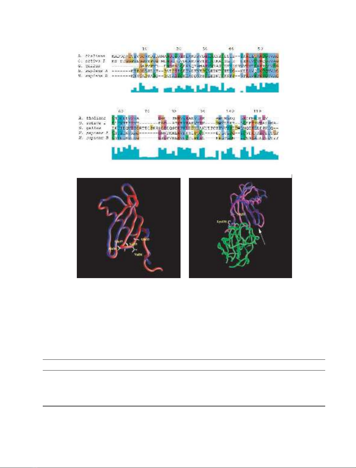
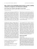
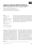
cystatins (Fig. 1A) [3].
Expression and purification of recombinant AtCYS1
E. coli JM101 cells were transformed with the construct
pGEX 4T-3-AtCYS1, which directs the synthesis of 39 kDa
GST-AtCYS1 fusion protein under the control of the
IPTG-inducible Lac promoter. Subcellular localization
experiments showed that the overwhelming majority of
the induced protein precipitated as insoluble inclusion
bodies (data not shown). By inducing pGEX 4T-3-AtCYS1
expression at 30 C in the presence of 0.5 m
M
IPTG,
however, the solubility of the fusion protein increased
dramatically, and as much as 3.6 mgÆL
)1
of soluble fusion
protein could be recovered, after centrifugation, from the
induced supernatant. The fusion protein was purified by
affinity chromatography on agarose/glutathione, and the
native 13 kDa AtCYS1 was cleaved from the fusion protein
by adding bovine thrombin. The thrombin was removed by
affinity chromatography, and the 13 kDa native AtCYS1
was recovered. The purity was higher than 95% as judged
by SDS/PAGE and chemical sequencing (data not shown).
Molecular basis for papain inhibition by recombinant
AtCYS1
AtCYS1 binding to papain follows a simple equilibrium, the
value of the Hill coefficient (n) always being equal to
1.00 ± 0.03. The K
d
value for AtCYS1 binding to papain is
(0.4 ± 0.1) ·10
)2
l
M
(pH 7.0 and 25 C; Table 1).
A
BLAST
search of the Protein Data Bank [35] reveals a
high sequence homology between AtCYS1 and oryzacysta-
tin-I [33]. In detail, 70% sequence homology is found
between the 88 residues forming the core region of oryza-
cystatin-I and the corresponding 89 residues of AtCYS1
(Fig. 1A). It must be noted that both the N-terminal 10
residues and C-terminal seven residues display a high
flexibility in the NMR structure of oryzacystatin-I [33]. For
this reason, the core region of oryzacystatin-I (residues 6–93)
was used as a template to model the corresponding region of
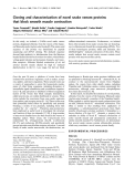
AtCYS1. Analysis of the molecular model of AtCYS1 shows
that all amino acid substitutions, including the single
insertion of Ala15A, are easily accommodated in the
structure without any gross backbone rearrangement
(Fig. 1B). In particular, all the residues forming the hydro-
phobic core of oryzacystatin-I are conserved or conserva-
tively substituted in AtCYS1. Moreover, all charge
substitutions occur on the protein surface and the position
and length of secondary structure elements (five b-strands
and one a-helix) are conserved in AtCYS1.
As shown in Table 1, values of K
d
for binding of plant
and animal cystatins to papain span 10
)1
)10
)8
l
M
(Table 1), reflecting differences in enzyme–inhibitor recog-
nition. To provide a rationale for the striking difference in
2596 B. Belenghi et al.(Eur. J. Biochem. 270)FEBS 2003
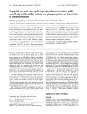
affinity observed between binding of AtCYS1, oryzacysta-
tin-I, chicken egg white cystatin, and human stefins A and B
to papain (Table 1), molecular models of the complexes of
these inhibitors with papain have been built based on the
three-dimensional structure of stefin B in complex with
papain [41].
Table 1. Dissociation equilibrium constants (K
d
) for the interaction between selected cystatins and papain.
Cystatin K
d
(l
M
) pH and temperature Reference
AtCYS1 4.0 ·10
)2
pH 7.0 and 25 C Present study
Oryzacystatin I 3.0 ·10
)2
pH 6.5 and 37 C [71]
Chicken egg white cystatin 6.0 ·10
)8
pH 7.4 and 25 C [72]
Human stefin A 1.8 ·10
)7
pH 7.4 and 25 C [73]
Human stefin B 4.9 ·10
)8
pH 7.4 and 25 C [74]
A
BC
Fig. 1. Amino acid sequence alignment of selected cystatins and schematic representation of the molecular models of AtCYS1 and AtCYS1–papain
complex. (A) Sequence alignment of Atcys1 (A. thaliana), oryzacystatin I (O. sativa I), chicken egg white cystatin (G. gallus, Gly9-truncated form
[1]), human stefin A (H. sapiens A) and human stefin B (H. sapiens B). Residues are coloured according to their chemical properties. The green
boxes under the alignment indicate the degree of sequence consensus. Note the high conservation of the Gln53-Val54-Val55-Ala56-Gly57 sequence
forming the first hairpin loop of cystatins. The alignment was obtained using the program
CLUSTALW
[36]. (B) Molecular model of AtCYS1 (blue)
superimposed onto the three-dimensional structure of oryzacystatin I (red) used as a template for modelling. (C) AtCYS1 model (blue) super-
imposed onto the three-dimensional structure of the complex formed by papain (green) and stefin B (magenta). The arrow indicates the different
conformation of the AtCYS1 second hairpin loop, as compared to stefin B. AtCYS1 is rotated by approximately 180 degrees with respect to (B).
The figure was made using
GRASP
[69]. For details, see text.
FEBS 2003 AtCYS1 suppresses hypersensitive cell death (Eur. J. Biochem. 270) 2597


%20--%3e%3cdefs%3e%3cstyle%3e%20.st0%20{%20fill:%20%23fff;%20}%20.st1%20{%20fill:%20%237800fa;%20}%20%3c/style%3e%3c/defs%3e%3cpath%20class='st1'%20d='M117.78,12.18H43.11c2.9,3.47,4.65,7.94,4.65,12.82,0,5.6-2.3,10.66-6.01,14.29h76.02l7.22-13.56-7.22-13.56Z'/%3e%3cg%3e%3cpath%20class='st0'%20d='M53.58,26.17h-.59v-1.46h.59v-4.96h2.83c1.78,0,2.67.94,2.67,2.82v5.76c0,1.87-.89,2.81-2.67,2.81h-2.83v-4.96ZM55.36,21.37v3.34h1.1v1.46h-1.1v3.34h1.01c.61,0,.91-.37.91-1.1v-5.93c0-.74-.3-1.1-.91-1.1h-1.01Z'/%3e%3cpath%20class='st0'%20d='M65.99,31.14h-1.8l-.31-2.07h-2.19l-.31,2.07h-1.64l1.82-11.39h2.62l1.82,11.39ZM65.28,18.04c-.25.46-.51.77-.75.94-.21.15-.47.22-.79.22-.26,0-.57-.07-.92-.22l-.38-.15c-.14-.05-.26-.07-.37-.07-.3,0-.53.18-.71.54l-.91-.68c.25-.46.51-.77.75-.94.21-.14.48-.21.79-.21.26,0,.57.07.92.21l.38.15c.14.05.26.07.37.07.3,0,.53-.18.71-.54l.91.68ZM61.91,27.52h1.73l-.87-5.76-.87,5.76Z'/%3e%3cpath%20class='st0'%20d='M74.53,26.89v1.52c0,1.91-.89,2.86-2.67,2.86s-2.67-.95-2.67-2.86v-5.93c0-1.91.89-2.86,2.67-2.86s2.67.95,2.67,2.86v1.11h-1.69v-1.22c0-.75-.31-1.12-.93-1.12s-.93.37-.93,1.12v6.15c0,.74.31,1.11.93,1.11s.93-.37.93-1.11v-1.63h1.69Z'/%3e%3cpath%20class='st0'%20d='M81.4,31.14h-1.8l-.31-2.07h-2.19l-.31,2.07h-1.64l1.82-11.39h2.62l1.82,11.39ZM75.9,19.2l1.52-1.91h1.71l1.51,1.91h-1.61l-.76-.95-.75.95h-1.61ZM77.32,27.52h1.73l-.87-5.76-.87,5.76ZM83.1,15.99l-1.76,1.91h-1.26l1.17-1.91h1.86Z'/%3e%3cpath%20class='st0'%20d='M84.86,19.75c1.78,0,2.67.94,2.67,2.82v1.48c0,1.87-.89,2.81-2.67,2.81h-.85v4.28h-1.79v-11.39h2.64ZM84.01,21.37v3.86h.85c.58,0,.87-.36.87-1.08v-1.71c0-.71-.29-1.07-.87-1.07h-.85Z'/%3e%3cpath%20class='st0'%20d='M93.51,19.75c1.78,0,2.67.94,2.67,2.82v1.48c0,1.87-.89,2.81-2.67,2.81h-.85v4.28h-1.79v-11.39h2.64ZM92.66,21.37v3.86h.85c.58,0,.87-.36.87-1.08v-1.71c0-.71-.29-1.07-.87-1.07h-.85Z'/%3e%3cpath%20class='st0'%20d='M98.8,31.14h-1.79v-11.39h1.79v4.88h2.03v-4.88h1.83v11.39h-1.83v-4.88h-2.03v4.88Z'/%3e%3cpath%20class='st0'%20d='M105.36,24.55h2.46v1.62h-2.46v3.34h3.09v1.63h-4.88v-11.39h4.88v1.63h-3.09v3.18ZM108.17,17.29l-1.76,1.91h-1.26l1.17-1.91h1.86Z'/%3e%3cpath%20class='st0'%20d='M112.2,19.75c1.78,0,2.67.94,2.67,2.82v1.48c0,1.87-.89,2.81-2.67,2.81h-.85v4.28h-1.79v-11.39h2.64ZM111.35,21.37v3.86h.85c.58,0,.87-.36.87-1.08v-1.71c0-.71-.29-1.07-.87-1.07h-.85Z'/%3e%3c/g%3e%3ccircle%20class='st1'%20cx='25'%20cy='25'%20r='20'/%3e%3cpath%20class='st0'%20d='M32.78,19.27c2.92,0,4.43,2.55,5.28,5.33l.71,2.17c.14.38-.33.75-.71.75h-5.61c.19-.33.24-.71.09-1.08l-.75-2.45c-.43-1.32-.99-2.64-1.79-3.77.75-.57,1.65-.94,2.78-.94h0ZM25,18.38c3.25,0,4.9,2.78,5.89,5.89l.76,2.45c.14.42-.33.8-.8.8h-11.69c-.42,0-.94-.38-.8-.8l.75-2.45c.99-3.11,2.64-5.89,5.89-5.89h0ZM25,11.35c1.74,0,3.11,1.37,3.11,3.11s-1.37,3.11-3.11,3.11-3.11-1.41-3.11-3.11,1.41-3.11,3.11-3.11h0ZM17.27,19.27c1.08,0,1.98.38,2.73.94-.8,1.13-1.37,2.45-1.74,3.77l-.8,2.45c-.14.38-.05.75.09,1.08h-5.56c-.42,0-.9-.38-.75-.75l.71-2.17c.9-2.78,2.41-5.33,5.33-5.33h0ZM17.27,12.91c1.51,0,2.78,1.27,2.78,2.83s-1.27,2.83-2.78,2.83-2.83-1.27-2.83-2.83,1.27-2.83,2.83-2.83h0ZM32.78,12.91c1.56,0,2.78,1.27,2.78,2.83s-1.23,2.83-2.78,2.83-2.83-1.27-2.83-2.83,1.27-2.83,2.83-2.83h0ZM27.07,28.56v.09c0,.57-.24,1.08-.61,1.46h0v.05c-.38.33-.9.57-1.46.57s-1.08-.24-1.46-.61h0c-.38-.38-.61-.9-.61-1.46v-.09h1.41v.09c0,.19.05.38.19.47v.05c.09.09.28.19.47.19s.38-.09.47-.19v-.05c.14-.09.24-.28.24-.47t-.05-.09h1.41ZM30.99,28.56v.09c0,1.65-.66,3.16-1.74,4.24-1.08,1.08-2.59,1.79-4.24,1.79s-3.16-.71-4.24-1.79l-.05-.05c-1.04-1.08-1.7-2.55-1.7-4.2v-.09h1.41v.09c0,1.27.47,2.4,1.27,3.25h.05c.85.85,1.98,1.37,3.25,1.37s2.4-.52,3.25-1.37c.85-.8,1.37-1.98,1.37-3.25v-.09h1.37ZM34.99,28.56v.09c0,2.78-1.13,5.28-2.92,7.07-1.79,1.79-4.29,2.92-7.07,2.92s-5.23-1.13-7.07-2.92c-1.79-1.79-2.92-4.29-2.92-7.07v-.09h1.41v.09c0,2.4.94,4.53,2.5,6.08,1.56,1.56,3.72,2.5,6.08,2.5s4.52-.94,6.08-2.5c1.56-1.56,2.5-3.68,2.5-6.08v-.09h1.41Z'/%3e%3c/svg%3e)