
Cloning and characterization of novel snake venom proteins
that block smooth muscle contraction
Yasuo Yamazaki
1
, Hisashi Koike
1
, Yusuke Sugiyama
1
, Kazuko Motoyoshi
1
, Taeko Wada
1
,
Shigeru Hishinuma
2
, Mitsuo Mita
2
and Takashi Morita
1
Departments of
1
Biochemistry; and
2
Pharmacodynamics, Meiji Pharmaceutical University, Tokyo, Japan
In this study, we isolated a 25-kDa novel snake venom
protein, designated ablomin, from the venom of the Japan-
ese Mamushi snake (Agkistrodon blomhoffi). The amino-acid
sequence of this protein was determined by peptide
sequencing and cDNA cloning. The deduced sequence
showed high similarity to helothermine from the Mexican
beaded lizard (Heloderma horridum horridum), which blocks
voltage-gated calcium and potassium channels, and ryano-
dine receptors. Ablomin blocked contraction of rat tail
arterial smooth muscle elicited by high K
+
-induced
depolarization in the 0.1–1 l
M
range, but did not block
caffeine-stimulated contraction. Furthermore, we isolated
three other proteins from snake venoms that are homolog-
ous to ablomin and cloned the corresponding cDNAs. Two
of these homologous proteins, triflin and latisemin, also
inhibited high K
+
-induced contraction of the artery. These
results indicate that several snake venoms contain novel
proteins with neurotoxin-like activity.
Keywords: snake venom; neurotoxin; helothermine; cysteine-
rich secretory proteins; ablomin.
Over the past 30 years, a plethora of toxins have been
isolated from poisonous organisms, such as snakes, scorpi-
ons, spiders, and micro-organisms. These natural toxins use
a variety of approaches to arrest the homeostatic mecha-
nisms of other living organisms, including disruption of
intracellular signal transduction and cytoskeleton organiza-
tion [1–4], and activation or inhibition of blood coagulation
factors [5–10]. Toxins that block synaptic transmission,
called neurotoxins, are widely distributed in venoms. These
toxins include the conotoxins from cone snails, agatoxins
from spiders, and scorpion toxins [11–16]. These toxins exert
their potentially lethal effects by specifically and potently
blocking a variety of ion channels, including those that
conduct Na
+
,K
+
,andCa
2+
. Therefore, neurotoxins have
been employed as useful tools to investigate the structure
and function of these ion channels [17–20]. A large number
of neurotoxin families have also been found in the venom of
Elapidae snakes. These toxins, the a-neurotoxins [21]
(represented by a-bungarotoxin [22,23], a-cobratoxin
[24–27], and erabutoxin [28,29]) potently and specifically
prevent nicotinic acetylcholine receptor activation. A second
family of snake venom neurotoxins, the dendrotoxins, are
homologous to Kunitz-type serine protease inhibitors and
act primarily by blocking neuronal K
+
channels [30,31]. In
contrast to the neurotoxin-rich venom from Elapidae
snakes, the venom from other deadly snakes, including
Viperidae and Colubridae snakes, contain surprisingly few
neurotoxins, although some neurotoxic phospholipases
have been discovered [32–36].
In this report, we describe the isolation of a novel protein,
ablomin, from the venom of the Japanese Mamushi snake
(Agkistrodon blomhoffi, a member of the Viperidae family).
When applied to arterial smooth muscle preparations from
rat-tails, ablomin blocks K
+
-stimulated contraction. This
effect is similar to that resulting from application of
calciseptine, a well-characterized neurotoxin from black
mamba (Dendroaspis polylepis polylepis).Calciseptineisa
known blocker of L-type Ca
2+
channels, a property that
underlies its ability to block K
+
-induced contractions of
aortic smooth muscle and spontaneous contractions of
uterine smooth muscle [37]. Furthermore, we demonstrate
that several snake venoms contain ablomin-like proteins,
which may constitute a novel venom protein family.
EXPERIMENTAL PROCEDURES
Materials
The lyophilized venom of A. blomhoffi was a kind gift from
S. Iwanaga (The Chemo-Sero-Therapeutic Research
Institute, Kumamoto, Japan) [38]. Other snake venoms
and venom glands were purchased from the Japan Snake
Institute (Gunma, Japan). Superdex 75 pg and 200 pg,
SP–Sepharose High Performance, and Q-Sepharose Fast
Flow columns were from Amersham–Pharmacia Biotech.
The Vydac Protein & Peptide C18 HPLC column and the
COSMOSIL 5C18 AR-300 HPLC column were the
products of JASCO (Tokyo, Japan) and Nacalai Tesque
(Kyoto, Japan), respectively. Endoprotease Lys-C was
Correspondence to T. Morita, Department of Biochemistry,
Meiji Pharmaceutical University, 2-522-1, Noshio, Kiyose,
Tokyo 204-8588, Japan,
Fax/Tel.: + 81 424 95 8479,
E-mail: tmorita@my-pharm.ac.jp
Abbreviations: CRISP, cysteine-rich secretory protein; HLTX,
helothermine; PsTx, pseudechetoxin; CAP, CRISPs Antigen 5
proteins, and Pathogenesis-related proteins.
Note: the nucleotide sequences reported here have been submitted to
GenBank database (tigrin, AY093955; ablomin, AF384218; triflin,
AF384219; latisemin, AF384220).
(Received 21 December 2001, revised 12 April 2002,
accepted 18 April 2002)
Eur. J. Biochem. 269, 2708–2715 (2002) FEBS 2002 doi:10.1046/j.1432-1033.2002.02940.x

purchased from Seikagaku Corporation (Tokyo, Japan).
Other chemicals were of analytical grade (Sigma–Aldrich,
Amersham–Pharmacia Biotech., Wako Pure Chemical Ind.
and Kanto Chemical Co.).
Purification of proteins
Tigrin was isolated from the extract of Duvernoy’s glands of
Rhabdophis tigrinus tigrinus. Ten Duvernoy’s glands were
broken into small pieces after freezing in liquid nitrogen,
and then extracted in 30 mL of 50 m
M
Tris/HCl pH 8.0 for
4hat4C. After ultracentrifugation, the supernatant was
applied onto Q-Sepharose Fast Flow column (1.6 ·10 cm)
in the same buffer, and eluted with a linear gradient from 0
to 0.5
M
NaCl. A major peak eluted at 0.05
M
NaCl, which
was subsequently purified by chromatography on Superdex
200 pg (2.6 ·60 cm column).
Ablomin was purified by three successive chromato-
graphic steps. Five hundred milligrams of lyophilized
A. blomhoffi venom was dissolved in 3 mL of 20 m
M
imidazole/HCl pH 6.8 containing 0.2
M
NaCl, and insol-
uble materials were removed by centrifugation and
filtration (0.22 lm). The filtrate was loaded onto a
Superdex 75 pg column (2.6 ·60 cm) and eluted with
the same buffer. The ablomin fractions from two gel
filtration runs (a total of 1000 mg of snake venom) were
pooled and dialyzed against 50 m
M
Tris/HCl, pH 8.0, and
applied to the Q-Sepahrose Fast Flow column
(1.6 ·15 cm). The column was eluted with a linear
gradient of NaCl from 0 to 0.4
M
at a flow rate of
2mLÆmin
)1
. Chromatographic fractions containing
ablomin were then dialyzed against 20 m
M
imidazole-
HCl, pH 6.0, and fractionated on a SP–Sepharose High
Performance column (1.6 ·11 cm). This column was
developed with a linear gradient of NaCl in the imidazole
buffer (0–0.4
M
,2mLÆmin
)1
).
For the purification of triflin, 500 mg of the venom of
Trimeresurus flavoviridis was applied to the SP–Sepharose
Fast Flow column (1.6 ·30 cm) with 10 m
M
phosphate
buffer, pH 6.8, and eluted with a linear gradient from 0 to
0.15
M
NaCl, as described previously [39]. Fractions con-
taining triflin were detected by Western blotting using anti-
tigrin serum. These fractions were pooled and fractionated
on Superdex 75 pg (2.6 ·60 cm) in a 50-m
M
Tris/HCl,
pH 8.0, containing 0.2
M
NaCl. Finally, triflin was purified
by chromatography on a Blue-Sepharose Fast Flow column
(1.6 ·15.5 cm) in 50 m
M
Tris/HCl, pH 8.0, which was
eluted with a linear gradient from 0 to 0.5
M
NaCl.
For purification of latisemin, 500 mg of the venom of
Laticauda semifasciata was loaded onto Superdex 75 pg
(2.6 ·60 cm) in a buffer containing 50 m
M
Tris/HCl,
pH 8.0, and 0.2
M
NaCl. The latisemin fractions were
loaded onto the SP–Sepharose Fast Flow column
(1.6 ·11 cm) in 10 m
M
phosphate buffer, pH 6.8, contain-
ing 0.05
M
NaCl, and eluted with a linear gradient to 0.2
M
NaCl. The latisemin fractions were re-chromatographed on
a Mono S column (0.5 ·1cm)in10m
M
phosphate buffer,
pH 6.0, with a linear gradient to 0.2
M
NaCl, and on
Heparin-Sepharose CL-6B columns (1.6 ·14 cm) with
50 m
M
Tris/HCl, pH 8.0, using a linear gradient from 0
to 0.3
M
NaCl.
All purification steps were performed at 4 Cwithan
FPLC system (Amersham–Pharmacia Biotech).
Amino-acid sequence analysis
Proteins were reduced for 3 h at room temperature with
20 m
M
dithiothreitol in the presence of 0.5
M
Tris/HCl,
pH 8.5, 6
M
guanidine hydrochloride, and 2 m
M
EDTA in
a volume of 0.5 mL. Three microliters of 4-vinylpyridine
were then added, and alkylation was allowed to proceed
for 3 h at room temperature. The S-pyridylethylated
proteins were separated from the reagents by C18
reverse-phase HPLC, and the amino-acid sequence was
determined by sequencing the peptides obtained by diges-
tion with endoprotease Lys-C. All the samples were
analyzed on Applied Biosystems protein sequencers (mod-
els 473 A and 477).
cDNA cloning of proteins
The cDNAs encoding tigrin, ablomin, and latisemin were
obtained using the RT-PCR method. Typically, venom
gland total RNA was isolated from the venom gland with
ISOGENTM (Wako Pure Chemical Industries, Japan)
according to the manufacturer’s protocol. 5¢and 3¢RACE
were carried out to determine the nucleotide sequence of the
5¢and 3¢end cDNAs with the SMARTTM RACE cDNA
amplification kit (Clontech). The amino-acid sequences of
peptides derived from purified proteins were used to design
degenerate primers. For the first amplification of tigrin and
latisemin cDNA, degenerate primers were used for both
sense and antisense primers. For ablomin cDNA, PCR was
performed with single degenerate primer (sense or antisense)
and an primer recognizing an adaptor sequence that had
been attached to the 5¢or 3¢endofcDNAs.Inthecaseof
triflin, PCR was carried out using habu cDNA library as a
template [40] with a degenerate primer and an adaptor
primer. The PCR products were subcloned into the pGEM
T-easy vector (tigrin and triflin) or pUC19 vector (ablomin
and latisemin) and sequenced with the DSQ 2000 L DNA
sequencer (Shimadzu, Japan). Primers used this study are
described as follows: tigrin, sense 5¢-AA(C,T)GT(A,C,G,T)
GA(C,T)TT(C,T)AA(C,T)TC(A,C,G,T)GA(A,G)TC-3¢
(corresponding to amino acids 1–8 in tigrin) and antisense
5¢-(A,G)TT(A,G)CA(A,G)TT(A,G)TT(A,G)TA(A,G)TC
(A,G)TC-3¢(corresponding to amino acids 187–193 in
tigrin); ablomin, sense 5¢-GGCCATTA(C,T)ACTCAG(A,G)
T(A,G)G-3¢(corresponding to amino acids 114–120 in
ablomin) and antisense 5¢-C(C,T)A(C,T)CTGAGT(A,G)
TAATGGCC-3¢(corresponding to amino acids 114–120
in ablomin); triflin, antisense 5¢-GC(A,G)TG(A,G,T)AT
(A,G,T)AT(A,G)TC(A,C,G,T)GTCCA-3¢(corresponding
to amino acids 86–91 in triflin); latisemin, sense 5¢-GA
(A,G)AA(C,T)CA(A,G)AA(A,G)GA(A,G)AT(A,C,T)G-3¢
(corresponding to amino acids 11–17 in latisemin) and
antisense 5¢-G(A,G)CA(A,G)TT(A,C,G,T)GT(A,G)AA
(C,T)TC-3¢(corresponding to amino acids 183–189 in
latisemin).
Contraction measurements on rat-tail arterial smooth
muscle
Helical strips of endothelium-free rat-tail arterial smooth
muscle were prepared as described previously [41]. All the
contraction experiments were carried out at room tempera-
ture, and all buffers were pre-oxygenated with 100% O
2
.
FEBS 2002 Novel proteins in snake venoms (Eur. J. Biochem. 269) 2709
The strips were held at 75 mg resting tension in Hepes/
Tyrode (H-T) solution (137 m
M
NaCl/2.7 m
M
KCl/1.8 m
M
CaCl
2
/1 m
M
MgCl
2
/5.6 m
M
glucose/10 m
M
Hepes, pH 7.4)
for 45 min Then, the strips were treated with H-T solutions
containing 1 l
M
prazosin, to block the effect of norepi-
nephrine via a1 adrenergic receptors, for 30 min The strips
were then exposed to 60 m
M
KCl H-T solution for 15 min
KCl H-T solution was prepared by replacing the NaCl in
H-T solution with equimolar KCl. After washing with
calcium-free H-T solution for 5 min, the smooth muscle
strips were stimulated with 20 m
M
caffeine H-T solution.
For measuring the effect of the proteins, all the H-T
solutions contained the indicated concentrations of pro-
teins.
RESULTS
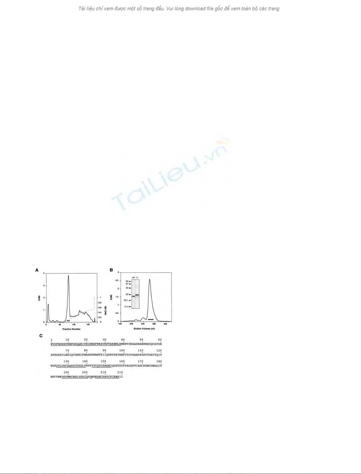
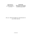
Identification, isolation and cloning of tigrin
and ablomin
During the isolation process of a prothrombin activa-
tor from the Duvernoy’s gland of Yamakagashi snake
(R. tigrinus tigrinus), we identified a large quantity of a
single chain 30-kDa protein (Fig. 1), which we named tigrin.
To permit further study, tigrin was purified two chroma-
tographic steps. The extract from Duvernoy’s glands was
first separated by anion-exchange chromatography
(Fig. 1A), and then the major peak was purified by gel
filtration (Fig. 1B). An amino-acid sequence was deter-
mined by peptide sequencing and partial cloning, revealing
that tigrin was structurally homologous to helothermine
(HLTX; 49.0% identity, Fig. 1C) from the venom of the
Mexican beaded lizard (Heloderma horridum horridum).
HLTX is known to alter a variety of ion channel acti-
vities, including voltage-gated K
+
channels, voltage-gated
Ca
2+
channels, and ryanodine receptors [42–44]. Because
we speculated that HLTX-like proteins would be wide-
spread in snake venoms, we generated an rabbit anti-tigrin
serum. We then screened several snake venoms with the
anti-tigrin serum using Western blotting or ELISAs. As a
result, we detected immunoreactive proteins in the venoms
from three snakes: A. blomhoffi,T. flavoviridis,andLaticauda
semifasciata. The immunoreactive proteins were then puri-
fied by column chromatography, using anti-tigrin serum as
a detection reagent.
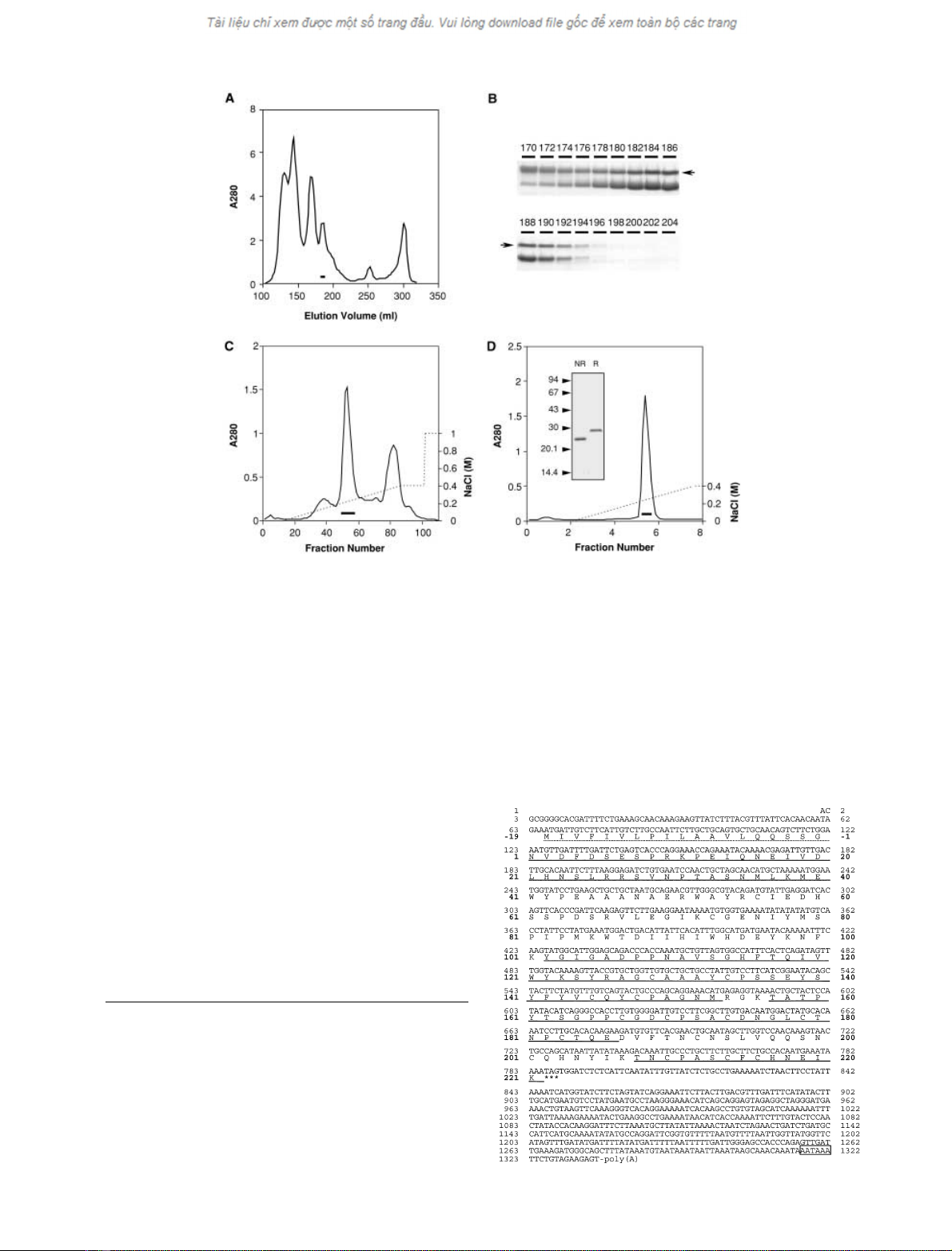
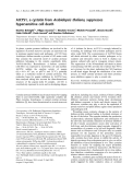
Using this procedure, we isolated a novel snake venom
protein (named ablomin) from the venom of the Mamushi
snake (A. blomhoffi) through three purification steps.
First, the crude venom of A. blomhoffi was separated by
gel filtration on a column of Superdex 75 pg (Fig. 2A).
Fractions containing ablomin were identified using with
SDS/PAGE, based upon an M
r
that was initially deter-
mined by Western blotting (Fig. 2B). These fractions were
further separated by anion-exchange chromatography on
Q-Sepharose Fast Flow column (Fig. 2C). Ablomin was
eluted at the concentration of 0.2–0.3
M
NaCl (bold line
in Fig. 2C). This fraction was then subjected to cation-
exchange chromatography on SP–Sepharose High Per-
formance (Fig. 2D). The purified ablomin migrated with a
M
r
of 26 kDa on SDS/PAGE under nonreducing condi-
tions and 29.7 kDa under reducing conditions (Fig. 2D,
inset). From this purification, we obtained 7 mg of purified
ablomin from 1 g of crude venom. The N-terminal and
partial amino-acid sequences of this protein were deter-
mined by peptide sequencing of enzymatically digested
peptides (underlined in Fig. 3). Based on the obtained
partial amino-acid sequence, we cloned ablomin cDNA
from the venom gland of A. blomhoffi by RT-PCR using
degenerate primers. The cloned ablomin cDNA was 1336
base pairs in length, encoding a 19-residue putative signal
peptide, starting at nucleotide 66, and a 221-residue
mature protein (molecular mass 24 932 Da), starting at
nucleotide 123 (Fig. 3). As expected, ablomin was quite
homologous to HLTX, with 52.8% of the deduced amino
acids identical to the corresponding residues in HLTX.
(Fig. 4).
The effects of tigrin and ablomin on rat tail arterial
contraction
We examined the effects of ablomin and tigrin on high
K
+
- or caffeine-induced contraction using helical strips of
endothelium-free rat-tail arterial smooth muscle. Ablomin
remarkably inhibited contraction evoked by treatment
with high K
+
, but not that evoked by treatment with
caffeine (Fig. 5A). In contrast, tigrin did not affect both
contraction evoked by either treatment (Fig. 6B). The
block of contraction by ablomin was concentration-
dependent to 1 l
M
(Fig. 5B) and completely reversible
after a 45-min washout of protein (data not shown).
Inhibition by ablomion was reduced at a concentration of
3l
M
(Fig. 5B). High K
+
-treatment of the artery induces
membrane depolarization and activates voltage-gated
channels, leading to smooth muscle contraction [45,46].
In contrast, caffeine exposure causes transient contraction
by activating ryanodine receptors of the sarcoplasmic
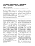
Fig. 1. Isolation of tigrin from the Duvernoy’s glands of R. tigrinus
tigrinus.(A) The extract from Duvernoy’s glands of R. tigrinus tigrinus
was fractionated on a Q-Sepharose Fast Flow column with a linear
gradient of NaCl (dotted line). (B) Major peak (bar in A) from
Q-Sepharose Fast Flow column was fractionated by gel filtration on a
Superdex 200 pg column. The pooled fraction (bar) contained purified
tigrin (inset, SDS/PAGE; R, reducing conditions; NR, nonreducing
conditions). (C) Primary structure of tigrin. The residues determined
by peptide sequencing are underlined.
2710 Y. Yamazaki et al. (Eur. J. Biochem. 269)FEBS 2002
reticulum (SR). The specific effect of ablomin on high
K
+
-induced contraction therefore suggests that this
blockage was caused by the inhibition of voltage-gated
channels, rather than interaction with contraction-related
proteins such as ryanodine receptors, myosin, or calmod-
ulin that are found in the cytoplasm [47]. In the rat-tail
artery, the intracellular Ca
2+
concentration is well corre-
lated with contraction force, and contraction evoked by
application of high extracellular K
+
is completely
dependent on the influx of extracellular Ca
2+
through
voltage-gated Ca
2+
channels [45,46,48]. In this regard, rat-
tail arterial smooth muscle cells predominantly express
L
-type Ca
2+
channels among several subtypes of Ca
2+
channels [49]. For these reasons, we hypothesize that
ablomin may target voltage-gated Ca
2+
channels on
smooth muscle. Further investigation is required to
determine the target protein(s). In previous studies, HLTX
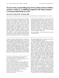
Fig. 2. Isolation of ablomin from the venom of A. blomhoffi.(A) The venom of A. blomhoffi was fractionated on a column of Superdex 75 pg. (B)
SDS/PAGE of continuous fractions eluted from Superdex 75 pg column under reduced condition. The numbers above are elution volume (mL) of
the fractions, and the arrows show ablomin. The slightly larger protein, which eluted at 170–174 mL, was determined to be a serine protease-like
venom protein by protein sequence analysis. (C) Ablomin fractions (182–192 mL in elution volume) were subjected to a Q-Sepharose Fast Flow
column and eluted with a linear gradient of NaCl (dotted line). Two-milliliter fractions were collected. The fractions indicated by bar were pooled as
ablomin. (D) The ablomin fraction from c was subjected to a SP-Sepharose High Performance column and developed with a linear gradient of NaCl
(dotted line). Two-milliliter fractions were collected. The pooled fraction (bar) contained purified ablomin. The result of SDS/PAGE of the purified
ablomin is shown in inset (NR, nonreduced; R, reduced). Seven milligrams of ablomin were obtained from 1 g of crude venom. For detailed
purification procedures, see Experimental procedures.
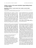
Fig. 3. Nucleotide and deduced amino-acid sequence of ablomin. The
amino-acid sequence is shown in single-letter code beneath the nuc-
leotide sequence. Nucleotide and amino acid (bold) number are shown
in the column at both sides. Translation is depicted as starting at
nucleotide 66. The putative signal peptide is dotted underlined (from
)19 to )1 in amino acid number). The underlined shows the deduced
amino-acid sequence from enzymatic-digested S-pyridylethylated
peptides. N-terminal was determined by the sequencing of intact
ablomin. The putative poly adenylation signal is boxed.
FEBS 2002 Novel proteins in snake venoms (Eur. J. Biochem. 269) 2711
has been shown to block ryanodine receptors on SR, in
addition to voltage-gated Ca
2+
channels (including L-,
N-, and P-type) [43,44]. In our current study, we used
intact arteries for measuring the activity of the protein,
which would presumably preclude access to cytoplasmic
proteins. In the previous study, HLTX was applied to
purified SR membranes and membrane-permeabilized
ventricular trabeculae [43] Therefore, these experimental
differences are likely to account for the differences in
specificity and mechanism of action.
Isolation and characterization of homologous proteins,
triflin and latisemin
In subsequent experiments, we isolated two other homol-
ogous proteins and cloned them by PCR. Triflin (221
amino-acid residues, molecular mass 24 798 Da) was puri-
fied from the venom of the Habu snake (T. flavoviridis),and
latisemin (217 amino-acid residues, molecular mass
24 272 Da), was purified from the venom of the Erabu
sea snake (Laticauda semifasciata) (Figs 4 and 6A). In
typical purification procedures (see Experimental proce-
dures), we obtained 4 mg of triflin and 2 mg of latisemin
from 500 mg of crude venom, respectively. The predicted
amino-acid sequences of triflin and latisemin are homolog-
ous to that of ablomin (83.7 and 61.5%, respectively). Like
ablomin, these proteins were capable of blocking contrac-
tion of the artery induced by high K
+
(Fig. 6B). These
findings indicate that proteins homologous to ablomin are
found in several snake venoms and represent new snake
venom proteins with neurotoxin-like activities.
DISCUSSION
Snake venom neurotoxins, represented by a-neurotoxins
and dendrotoxins, are thought to be found mostly in
Elapidae snake venoms [21,31], and only a few snake venom
neurotoxins have been isolated from Viperidae snakes
[32–36]. Recently, Brown et al. isolated a 24-kDa cyclic
nucleotide-gated ion channel blocker (designated pseudech-
etoxin; PsTx) from the venom of the Australian King
Brown snake (Pseudechis australis, Elapidae) [50]. The
N-terminal amino-acid sequence of PsTx has some identity
to those of the proteins in this study, although the complete
amino-acid sequence of PsTx has not yet been reported
(Fig. 4). These facts strongly imply that other proteins that
are homologous to ablomin may possess distinct biological
activities. In this regard, tigrin, which did not affect smooth
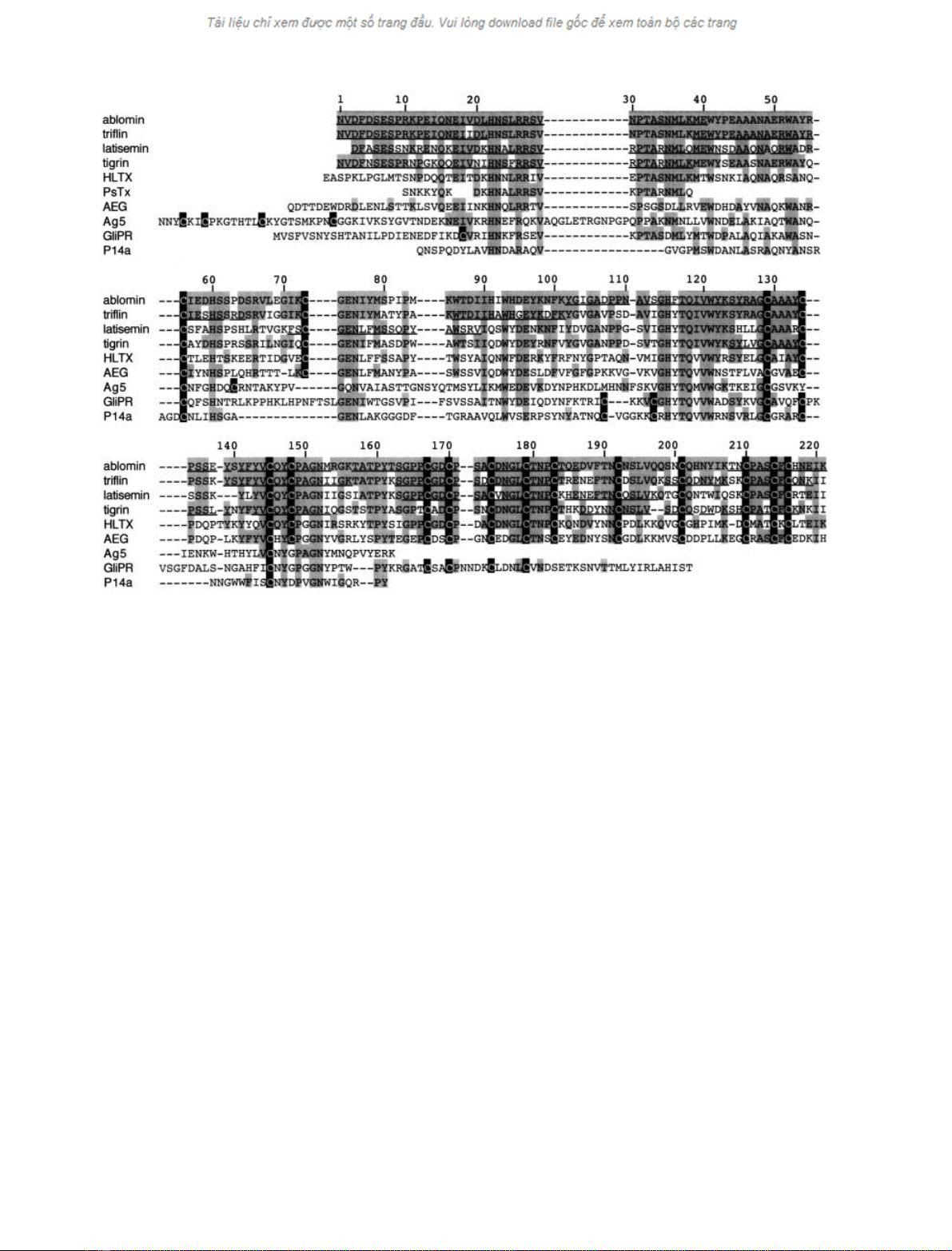
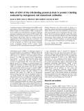
Fig. 4. Sequence alignment of ablomin and structurally related proteins. The residues conserved between the related proteins are shadowed. Gaps (–)
have been inserted to maximize similarity. All cysteine residues are shown with black shading; gray shading shows identity. The number of residues
corresponds to that of ablomin. The underline in ablomin, triflin, latisemin, and tigrin shows the amino-acid residues determined by peptide
sequencing. HLTX; helothermine, PsTx; pseudechetoxin, AEG; rat acidic epididymal glycoprotein (protein D/E), Ag5; hornet antigen 5, GliPR;
human glioma pathogenesis-related protein, P14a; tomato pathogenesis-related protein P14a. GenBank accession numbers, ablomin; AF384218,
triflin; AF384219, latisemin; AF384220, tigrin; AY093955, HLTX; U13619, AEG; M31173, Ag5; Q05108, GliPR; JC4131, P14a; P04284. Note that
the complete sequence of PsTx has not been published [50].
2712 Y. Yamazaki et al. (Eur. J. Biochem. 269)FEBS 2002


%20--%3e%3cdefs%3e%3cstyle%3e%20.st0%20{%20fill:%20%23fff;%20}%20.st1%20{%20fill:%20%237800fa;%20}%20%3c/style%3e%3c/defs%3e%3cpath%20class='st1'%20d='M117.78,12.18H43.11c2.9,3.47,4.65,7.94,4.65,12.82,0,5.6-2.3,10.66-6.01,14.29h76.02l7.22-13.56-7.22-13.56Z'/%3e%3cg%3e%3cpath%20class='st0'%20d='M53.58,26.17h-.59v-1.46h.59v-4.96h2.83c1.78,0,2.67.94,2.67,2.82v5.76c0,1.87-.89,2.81-2.67,2.81h-2.83v-4.96ZM55.36,21.37v3.34h1.1v1.46h-1.1v3.34h1.01c.61,0,.91-.37.91-1.1v-5.93c0-.74-.3-1.1-.91-1.1h-1.01Z'/%3e%3cpath%20class='st0'%20d='M65.99,31.14h-1.8l-.31-2.07h-2.19l-.31,2.07h-1.64l1.82-11.39h2.62l1.82,11.39ZM65.28,18.04c-.25.46-.51.77-.75.94-.21.15-.47.22-.79.22-.26,0-.57-.07-.92-.22l-.38-.15c-.14-.05-.26-.07-.37-.07-.3,0-.53.18-.71.54l-.91-.68c.25-.46.51-.77.75-.94.21-.14.48-.21.79-.21.26,0,.57.07.92.21l.38.15c.14.05.26.07.37.07.3,0,.53-.18.71-.54l.91.68ZM61.91,27.52h1.73l-.87-5.76-.87,5.76Z'/%3e%3cpath%20class='st0'%20d='M74.53,26.89v1.52c0,1.91-.89,2.86-2.67,2.86s-2.67-.95-2.67-2.86v-5.93c0-1.91.89-2.86,2.67-2.86s2.67.95,2.67,2.86v1.11h-1.69v-1.22c0-.75-.31-1.12-.93-1.12s-.93.37-.93,1.12v6.15c0,.74.31,1.11.93,1.11s.93-.37.93-1.11v-1.63h1.69Z'/%3e%3cpath%20class='st0'%20d='M81.4,31.14h-1.8l-.31-2.07h-2.19l-.31,2.07h-1.64l1.82-11.39h2.62l1.82,11.39ZM75.9,19.2l1.52-1.91h1.71l1.51,1.91h-1.61l-.76-.95-.75.95h-1.61ZM77.32,27.52h1.73l-.87-5.76-.87,5.76ZM83.1,15.99l-1.76,1.91h-1.26l1.17-1.91h1.86Z'/%3e%3cpath%20class='st0'%20d='M84.86,19.75c1.78,0,2.67.94,2.67,2.82v1.48c0,1.87-.89,2.81-2.67,2.81h-.85v4.28h-1.79v-11.39h2.64ZM84.01,21.37v3.86h.85c.58,0,.87-.36.87-1.08v-1.71c0-.71-.29-1.07-.87-1.07h-.85Z'/%3e%3cpath%20class='st0'%20d='M93.51,19.75c1.78,0,2.67.94,2.67,2.82v1.48c0,1.87-.89,2.81-2.67,2.81h-.85v4.28h-1.79v-11.39h2.64ZM92.66,21.37v3.86h.85c.58,0,.87-.36.87-1.08v-1.71c0-.71-.29-1.07-.87-1.07h-.85Z'/%3e%3cpath%20class='st0'%20d='M98.8,31.14h-1.79v-11.39h1.79v4.88h2.03v-4.88h1.83v11.39h-1.83v-4.88h-2.03v4.88Z'/%3e%3cpath%20class='st0'%20d='M105.36,24.55h2.46v1.62h-2.46v3.34h3.09v1.63h-4.88v-11.39h4.88v1.63h-3.09v3.18ZM108.17,17.29l-1.76,1.91h-1.26l1.17-1.91h1.86Z'/%3e%3cpath%20class='st0'%20d='M112.2,19.75c1.78,0,2.67.94,2.67,2.82v1.48c0,1.87-.89,2.81-2.67,2.81h-.85v4.28h-1.79v-11.39h2.64ZM111.35,21.37v3.86h.85c.58,0,.87-.36.87-1.08v-1.71c0-.71-.29-1.07-.87-1.07h-.85Z'/%3e%3c/g%3e%3ccircle%20class='st1'%20cx='25'%20cy='25'%20r='20'/%3e%3cpath%20class='st0'%20d='M32.78,19.27c2.92,0,4.43,2.55,5.28,5.33l.71,2.17c.14.38-.33.75-.71.75h-5.61c.19-.33.24-.71.09-1.08l-.75-2.45c-.43-1.32-.99-2.64-1.79-3.77.75-.57,1.65-.94,2.78-.94h0ZM25,18.38c3.25,0,4.9,2.78,5.89,5.89l.76,2.45c.14.42-.33.8-.8.8h-11.69c-.42,0-.94-.38-.8-.8l.75-2.45c.99-3.11,2.64-5.89,5.89-5.89h0ZM25,11.35c1.74,0,3.11,1.37,3.11,3.11s-1.37,3.11-3.11,3.11-3.11-1.41-3.11-3.11,1.41-3.11,3.11-3.11h0ZM17.27,19.27c1.08,0,1.98.38,2.73.94-.8,1.13-1.37,2.45-1.74,3.77l-.8,2.45c-.14.38-.05.75.09,1.08h-5.56c-.42,0-.9-.38-.75-.75l.71-2.17c.9-2.78,2.41-5.33,5.33-5.33h0ZM17.27,12.91c1.51,0,2.78,1.27,2.78,2.83s-1.27,2.83-2.78,2.83-2.83-1.27-2.83-2.83,1.27-2.83,2.83-2.83h0ZM32.78,12.91c1.56,0,2.78,1.27,2.78,2.83s-1.23,2.83-2.78,2.83-2.83-1.27-2.83-2.83,1.27-2.83,2.83-2.83h0ZM27.07,28.56v.09c0,.57-.24,1.08-.61,1.46h0v.05c-.38.33-.9.57-1.46.57s-1.08-.24-1.46-.61h0c-.38-.38-.61-.9-.61-1.46v-.09h1.41v.09c0,.19.05.38.19.47v.05c.09.09.28.19.47.19s.38-.09.47-.19v-.05c.14-.09.24-.28.24-.47t-.05-.09h1.41ZM30.99,28.56v.09c0,1.65-.66,3.16-1.74,4.24-1.08,1.08-2.59,1.79-4.24,1.79s-3.16-.71-4.24-1.79l-.05-.05c-1.04-1.08-1.7-2.55-1.7-4.2v-.09h1.41v.09c0,1.27.47,2.4,1.27,3.25h.05c.85.85,1.98,1.37,3.25,1.37s2.4-.52,3.25-1.37c.85-.8,1.37-1.98,1.37-3.25v-.09h1.37ZM34.99,28.56v.09c0,2.78-1.13,5.28-2.92,7.07-1.79,1.79-4.29,2.92-7.07,2.92s-5.23-1.13-7.07-2.92c-1.79-1.79-2.92-4.29-2.92-7.07v-.09h1.41v.09c0,2.4.94,4.53,2.5,6.08,1.56,1.56,3.72,2.5,6.08,2.5s4.52-.94,6.08-2.5c1.56-1.56,2.5-3.68,2.5-6.08v-.09h1.41Z'/%3e%3c/svg%3e)