RESEA R C H Open Access
Molecular characterization of two hantavirus
strains from different rattus species in Singapore
Patrik Johansson
1
, Grace Yap
2
, Hwee-Teng Low
1
, Chern-Chiang Siew
1
, Relus Kek
2
, Lee-Ching Ng
2*
, Göran Bucht
2
Abstract
Background: Hantaviruses cause human disease in endemic regions around the world. Outbreaks of hantaviral
diseases have been associated with changes in rodent population density and adaptation to human settlements
leading to their proliferation in close proximity to human dwellings. In a parallel study initiated to determine the
prevalence of pathogens in Singapore’s wild rodent population, 1206 rodents were trapped and screened. The
findings established a hantavirus seroprevalence of 34%. This paper describes the molecular characterization of
hantaviruses from Rattus norvegicus and Rattus tanezumi, the predominant rodents caught in urban Singapore.
Methodology: Pan-hanta RT-PCR performed on samples of Rattus norvegicus and Rattus tanezumi indicated that 27
(2.24%) of the animals were positive. sequence analysis of the S and M segments established that two different
hantavirus strains circulate in the rodent population of Singapore. Notably, the hantavirus strains found in Rattus
norvegicus clusters with other Asian Seoul virus sequences, while the virus strains found in Rattus tanezumi had the
highest sequence similarity to the Serang virus from Rattus tanezumi in Indonesia, followed by Cambodian
hantavirus isolates and the Thailand virus isolated from Bandicota indica.
Conclusions: Sequence analysis of the S and M segments of hantavirus strains found in Rattus norvegicus (Seoul
virus strain Singapore) and Rattus tanezumi (Serang virus strain Jurong TJK/06) revealed that two genetically
different hantavirus strains were found in rodents of Singapore. Evidently, together with Serang, Cambodian and
Thailand virus the Jurong virus forms a distinct phylogroup. Interestingly, these highly similar virus strains have
been identified in different rodent hosts. Further studies are underway to analyze the public health significance of
finding hantavirus strains in Singapore rodents.
Background
The hantavirus genus in the Bunyaviridae family con-
tains several important human pathogens that are preva-
lent worldwide. This group of viruses includes the
etiological agents of hemorrhagic fever with renal syn-
drome(HFRS),largelyseeninEuropeandAsia,and
hantaviruses causing (cardio) pulmonary syndrome
(HCPS) in the Americas. The clinical severity of hanta-
virus infections ranges from asymptomatic infections to
fulminate hemorrhagic shock and death. Hantaan virus
(HTNV) and Dobrava viruses (DOBV) are causative
agents of severe forms of HFRS and mortality rates of
up to 15% have been reported. About 20 - 30% of
HTNV infected patients develop hemorrhages [1,2].
Hantaviruses are enveloped and contain genomes
composed of three negative-stranded RNA segments;
small (S), medium (M) and large (L) segment, named
according to the size of the individual RNAs [3]. The L
segment encodes the viral RNA dependent RNA poly-
merase (RdRp), whereas M and S segments encode for
the two envelope proteins (Gn and Gc) and the nucleo-
capsid protein (N), respectively.
Transmission of hantavirus to humans occur mainly
through inhalation of aerosolized rodent excreta and
hantavirus infections are therefore limited to the geo-
graphic regions inhabited by the infected animal hosts.
Today, a wide array of hantaviruses has been detected in
numerous rodent or insectivore species [4-6].
Hantaviruses are endemic in many countries of the
world, and the trend in recent years indicates that the
natural foci have extended from rural to more urban
areas. The mainland China accounts for the majority of
* Correspondence: NG_Lee_Ching@nea.gov.sg
2
Environmental Health Institute, 11 Biopolis Way, #06-05/08, 138667,
Singapore
Johansson et al.Virology Journal 2010, 7:15
http://www.virologyj.com/content/7/1/15
© 2010 Johansson et al; licensee BioMed Central Ltd. This is an Open Access article distributed under the terms of the Creative
Commons Attribution License (http://creativecommons.org/licenses/by/2.0), which permits unrestricted use, distribution, and
reproduction in any medium, provided the original work is properly cited.

all cases reported worldwide and HTNV and Seoul virus
(SEOV) are known to be the most prevalent causative
agents of HFRS in Asia [7-9]. Specimens collected from
22 laboratories in China confirmed SEOV in 7 of 22
HFRS patients [7]. Furthermore, when comparing the
nucleotide sequences of viruses from HFRS patients and
rats captured in Beijing area, a nucleotide sequence
identity of 96.3% to 99.7% was observed, indicating that
SEOV is an important and perhaps the most common
hantavirus in China [7]. Interestingly, additional rodent
hosts and new hantaviruses are frequently discovered in
Asia including Thailand virus, isolated from the great
bandicoot rat, Bandicota (B.) indica in 1994 [10-12],
and hantavirus genetic material extracted from Rattus
(R.) tanezumi and R. norvegicus in Indonesia [13-15].
Many Rattus species are difficult to distinguish mor-
phologically and there are many changes over the years
in both genus and assignments [16]. Recently the bar-
coding technique has been proposed as a method for
the identification of species on the bases of evolutionary
divergence of a gene region such as mtDNA cytochrome
oxidase I [17] or the cytochrome b gene [18]. According
to Musser & Charlton 2005, seven groups of rodents are
recognized within the Rattus genus including the R. rat-
tus group and the R. norvegicus [19], and the R. rattus
group comprises about 21 species including R. rattus
and R. tanezumi. The R. rattus species is further divided
into two subspecies based on the chromosome number
[20]: an Ocean/European variant which Musser &
Charlton named R. rattus and an Asian type that was
named R. tanezumi.
Inthelate80’s in Singapore, Wong and co-workers
found evidence of hantavirus infections in both rodents
and humans and one hantavirus strain (R36) was iso-
lated from R. norvegicus [21].Theyalsoanalyzedthe
seroprevalence in patients and found that 8.3% of Den-
gue Hemorrhagic Fever (DHF) suspects were seroposi-
tive to hantaviruses. However, for the last 15 years, only
one case with classical manifestations of HFRS, con-
firmed by serology, have been reported in Singapore
[22]. In a parallel study initiated to determine the sero-
prevalence of rodent-borne pathogens in Singapore’s
wild rodent population, 1206 rodents were trapped and
screened (unpublished data). Findings in that study indi-
cate that one-third of R. norvegicus and one-fifth of the
R. tanezumi rodents were seropositive towards the
SEOV nucleocapsid protein. Of the seropositive rodents,
5.5 and 26% were also tested positive by PCR,
respectively.
This paper describes the subsequent screening of the
animals using PCR, and the characterization of two
genetically different hantavirus strains denoted Seoul
virus strain Singapore and Serang virus strain Jurong
TJK/06 of samples from R. norvegicus and R. tanezumi,
respectively. Phylogenetic analysis, nucleotide and amino
acid sequence identity were also determined.
Materials and methods
Ethics Statement
All animals were handled in strict accordance with good
animal practice, as defined by The Animal Research
Ethics Committee of DSO National Laboratories, 20
Science Park Drive, Singapore 118230.
Rodent identification
The rodents were identified through PCR based amplifi-
cation and sequencing of cytochrome b gene fragments
obtained by using the primers mcytb1 and mcytHb (Ken
Aplin, personal communication) before phylogenetic-
based species identification was conducted. Obtained
rodent sequences are available from the authors or
accessible from GenBank (GQ274946 - GQ274949).
Serology
Indirect Enzyme-linked immunosorbent assay (ELISA)
was performed using a recombinant truncated nucleo-
capsid protein of the Seoul virus (M34881) and serum
samples of collected rodents. The analysis was done as
described earlier [23]. The calculated cut-off value
(0.135) was set as 3 times the value of sera derived from
negative lab. rats (R. norvegicus).
Design of primers for Pan-hantavirus RT-PCR
Primers with the potential to detect all known rodent-
borne hantaviruses were designed towards conserved
regions identified from multiple alignments of hanta-
virus L segment sequences by the BioEdit program
package [24]. Using the primer pair Han-
taL_2_fwd_3312-40 5’- TYTTTGARTTTGCHCAY-
CAYTCWGATGATGC and HantaL_2_rev_3481-53 5’-
TCATGNARRTTRAACATRCTYTTCCACA for PCR,
tissue samples (lungs and kidneys) of all rodents were
analyzed, as described below. The PCR was done in Mg
++
free PCR buffer supplemented with 2 mM of MgCl
2
,
0.2mMofdNTP,1μM of the primers and 1.25 U of
AmpliTaq® (Applied Biosystems Inc., USA), in a total
volume of 25 μl. After a preincubation for 5 min at 95°
C, 35 cycles at 53°C for 30 sec, 72°C for 45 sec and 45
sec at 95°C were done. After a final incubation for 10
min at 72°C the result was examined by agarose gel
electrophoresis. A resulting PCR product of 170 bp was
considered PCR positive.
Extraction of RNA, reverse transcription and sequencing
RNA was extracted from tissue samples of kidneys and/
or lungs of individual rodents using Qiagen RNeasy
Mini kit (Qiagen, Inc., Germany) or the TRIzol reagent
(Invitrogen Inc., USA), according to manufacturer’s
instructions. Reverse transcription of RNA was per-
formed with SuperScript III (Invitrogen Inc., USA) and
random hexamers. For direct sequencing of overlapping
amplimers, numerous PCR primers were constructed
Johansson et al.Virology Journal 2010, 7:15
http://www.virologyj.com/content/7/1/15
Page 2 of 9

using the PriFi software [25]. Specific cDNA fragments
obtained by PCR were purified using QIAquick Gel
Extraction kit or QIAquick PCR Purification Kit (Qia-
gen, Inc., Germany) before sequencing was conducted.
Thereafter, the sequences of the segments were con-
firmed using a new set of primers targeting the deter-
mined cDNA sequences. However, the 15-20
nucleotides at the 5’and 3’termini of the S and M seg-
ments were set by the primers used for PCR. Finally, the
resulting sequence data was assembled as chromato-
grams, using SeqMan (LaserGene Inc., USA) and edited
with the BioEdit software package [24]. With the excep-
tion of the conserved 3’and 5’pan-handle sequence the
S and M and partial L sequences of these hantavirus
strains of Singapore have been made available (Genbank
accession no’s: GQ274936 - GQ274945).
Phylogenetic analysis
The resulting sequences were aligned by ClustalW [26],
as implemented in the BioEdit software package before
bootstrapped maximum parsimony and trees were cal-
culated using the SeqBoot, DNApars (setting: search for
best tree, more thorough search, use input order, multi-
ple data sets 1000 and 10 jumbles) and Consence soft-
wares. Branch lengths of the trees were calculated using
DNAml of the Phylip 3.67 software package as distribu-
ted by the author [27]. The tree was visualized by the
TreeView software [28]. The similarity plots were per-
formed by Stuart Ray’s SimPlot 3.5.1 software using a
window of 200 bp and steps of 20 bp, GapStrip: on,
Kimura (2-parameter), T/t: 2.0 [29].
Results
Characterization of the rodent samples
The phylogeny-based analysis of the cytochrome b gene
identifies the rodent hosts of the Seoul virus strain Sin-
gapore (RN41 and 46) as R. norvegicus. Furthermore,
the rodent hosts carrying the virus strain Jurong TJK/06
(RT49 and 50), (in this paper denoted R. tanezumi)
clearly clusters with R. rattus diardi,R. tanezumi and R.
kandianus of the diardii clade of the R. rattus complex
(Additional file 1, Figure S1) as described by Robins et.
al [18].
Serological and RT-PCR screening of rodent samples
RT-PCR screening of kidney samples from rodents with
Pan-hantavirus primers for the L segment, together with
a succeeding PCR targeting the S segment revealed that
2.1% (21/996) of R. norvegicus and 4.5% (7/156) of R.
tanezumi were PCR positive for hantavirus RNA. An
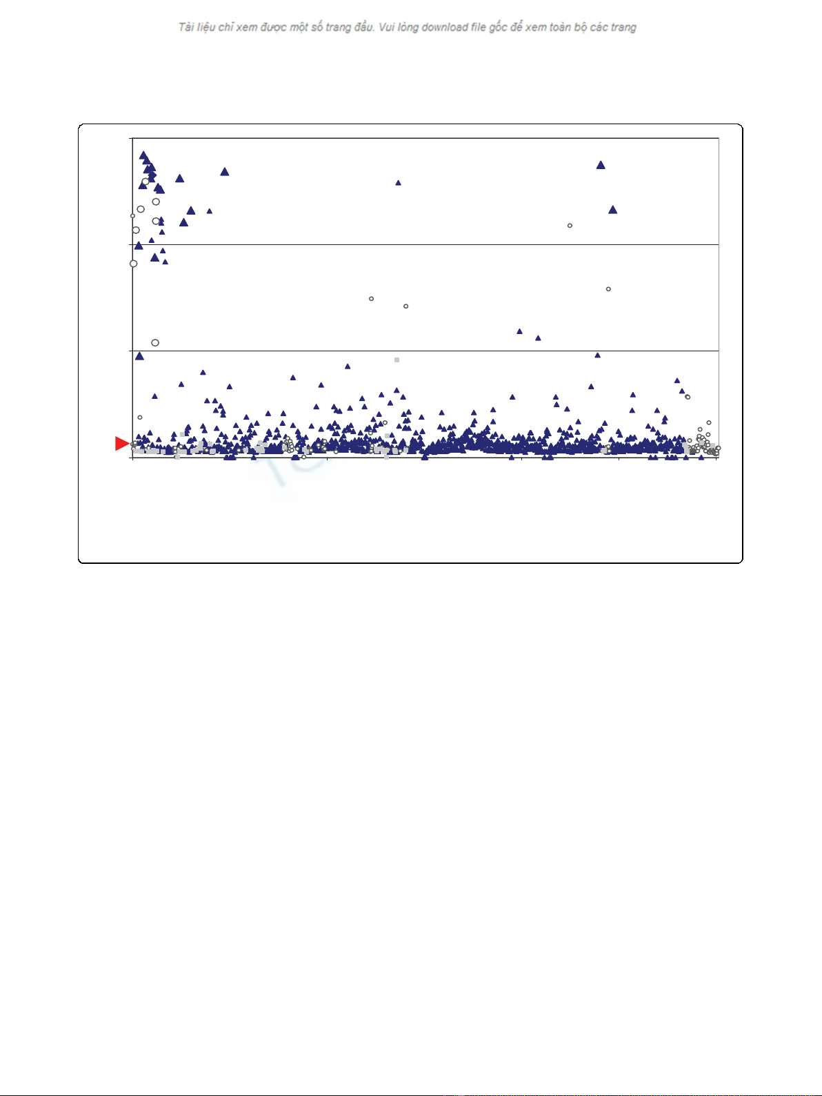
observation was made when comparing the optical den-
sity OD values of the ELISA with the PCR results. It
was noticed that rodents with the highest OD values
were more likely to be PCR positive too. Among the
rodents with OD values more than 1.0, 70% (7/10) were
also found PCR positive (Fig 1). In contrast, none of the
17 R. tanezumi and only 2 of the 335 seropositive R.
norvegicus having OD values less than 1.0 were also
PCR positive. The exceptions included one rodent with
a slightly lower ELISA OD value (0.96) and one ELISA
negative but PCR positive R. norvegicus. It is likely that
the latter rodent was infected close to the time of
trapping.
Sequence analysis
For hantavirus sequence analysis, two PCR positive lung
samples of each of the two rodent species (R. norvegicus
and R. tanezumi) were selected: rodent # 41, 46 and 49,
50, respectively. When comparing the complete open
reading frames of the S segments from the two rodent
species of Singapore with other complete or partial han-
tavirus S segment sequences (Additional file 2, Table
S1), the hantavirus of the Singaporean R. tanezumi
(strain Jurong) showed the highest nucleotide sequence
identity (95.0%) to the Serang virus, obtained of R. tane-
zumi from Indonesia [13]. In addition, the Jurong
sequence also demonstrated a high nucleotide sequence
identity to partial sequences of the S segment of rodent
samples of Cambodia (85.8%) [11] and full length S
sequences of the Thailand virus (83.6%), isolated from
B. indica [30].
Sequence comparisons of M segments also revealed
the Serang virus as most similar (94.5%) to the Jurong
strain. However, M and L segment of the Cambodian
hantavirus strains are not available in public databases
and only 343 bp and 412 bp of the Serang virus are cur-
rently accessible for sequence comparisons. As shown in
Additional file 2, Table S1, analyses of nucleotide and
amino acid sequences of the S and M segments strongly
suggest that the Serang and Cambodian isolates,
together with the Thailand virus share the closest iden-
tity to the Jurong virus strains of Singapore. When com-
paring the L segment sequences of the Jurong strains
with the Serang virus, a nucleotide sequence identity of
91.2% was found, slightly lower than between the corre-
sponding S or M segments. Though, the deduced amino
acid sequences of the S segment encoding N, the M seg-
ment encoding Gn and Gc and the L segments encoding
RdRp are nearly identical over the overlapping regions.
An analogous high nucleotide sequence identity was
also noticed when comparing hantavirus S segment
sequences of hantaviruses of R. norvegicus. The nucleo-
tide sequence identity between hantavirus strains of
Seoul Singapore and Cambodian Seoul virus strains was
foundtobebetween97and99%andthehighestiden-
tity was observed towards the Cambodian strain 41
(Camb 41) with 99.1% identity over the overlapping S
segment sequences.
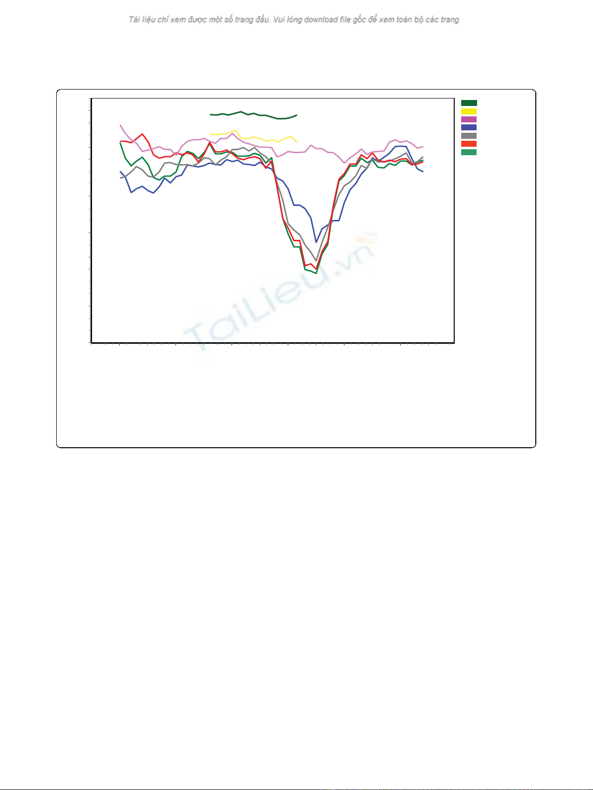
Results of similarity plot (SimPlot)
SimPlot comparisons of the complete open reading
frames of the Jurong virus with the Seoul/Hantaan/
Johansson et al.Virology Journal 2010, 7:15
http://www.virologyj.com/content/7/1/15
Page 3 of 9
Dobrava types of viruses demonstrated a significant
non-conserved region of approximately 300 nt near the
middle of the S segment [29]. However, such drop in
nucleotide sequence identity was not noticed towards
the Thailand virus sequences or against partial nucleo-
tide sequences of the corresponding Cambodian strains
or towards the Indonesian Serang virus (Fig 2). More-
over, comparisons of the amino acid sequences revealed
a similar trend, although the decrease in similarity in
the non-conserved region was less prominent (data not
shown).
Due to the lack of available sequences in public data-
bases, a comprehensive SimPlot comparison of the M
segment could only be performed towards the Thailand
virus along with a subset of less related hantaviruses. As
expected, the M segment of the Jurong strain was most
similar to the Thailand virus, especially when comparing
the amino acid sequence of the glycoprotein Gn. In
addition, two highly conserved regions consisting of
approximately 300 amino acids each were found near
the middle of the glycoproteins of the Jurong and Thai-
land viruses (data not shown).
Phylogenetic analysis
Phylogenetic analysis of S segment sequences showed
that the Jurong virus strains and the Thailand virus
isolates cluster together (Fig 3). Other hantaviruses car-
ried by rodents of the Cricetidae family, Arvicolinae,
Neotominae or Sigmodontinae subfamilies (e.g. Puumala
virus, Tula virus, Sin Nombre and Andes virus) or han-
taviruses carried by family Muridae subfamily Murinae,
such as Seoul virus, Hantaan virus and Dobrava-Belgrad
virus are genetically more distant (Fig 3). When partial
sequences were included to the phylogenetic analysis, a
novel clade consisting of the Thailand, Jurong, Serang
and Cambodian hantaviruses was found. According to
the phylogenetic trees and distance matrix, these
sequences are clearly strains of the same hantavirus spe-
cies. A common origin seems evident in such case.
As indicated by the phylogenetic trees for the viral S
and M segments (Fig 3 and 4, respectively) the Jurong
virus strains discovered in Singapore rats is genetically
similar and form a clade with the Serang virus of Indo-
nesia, Cambodian strains (e.g. Camb174, Camb117,
Camb96) and hantavirus sequences of B. indica from
Thailand (Thailand virus). Interestingly, the four closely
related viruses; Jurong, Cambodia, Serang, and Thailand
are carried by two or more different rodent species.
Hantavirus strains of R. norvegicus, such as the Seoul
Singapore and corresponding Cambodian isolates (e.g.
Camb32, 41, 58 and 180) cluster closely together with
0
1
2
3
0 200 400 600 800 1000 1200
Rodent #
Elisa absorbance units
Figure 1 Hantavirus positive rodents. Scatter plot displaying along the horizontal axis the individual rodents captured in Singapore between
2006-2008 and along the vertical axis, absorbance units determined by ELISA using serum samples of indicated rodents. Dark blue triangles
indicate R. norvegicus and open circles R. tanezumi. Other rodent species are pointed out as grey squares. Individual dots are shown as Large or
Small data points indicating PCR positive or negative rodents, respectively. The calculated cut-off value (0.135) is indicated by a red arrow.
Johansson et al.Virology Journal 2010, 7:15
http://www.virologyj.com/content/7/1/15
Page 4 of 9
other Seoul virus sequences, see Fig 3. The closest iden-
tity of the Seoul Singapore strain was observed towards
the Cambodian strain 41 (Camb 41) with 99.1% identity
for the overlapping S segment sequences.
Discussion
There are increasing numbers of studies from Asia
reporting prevalence of hantaviruses in rodent and
human populations, suggesting emerging hantaviral
infections in this part of the world [31]. Moreover, stu-
dies conducted, particularly in Southeast Asia, indicate
that hantavirus diversity is expanding [8]. Under these
circumstances, we initiated a series of studies that aimed
to provide a clearer perspective of the hantavirus situa-
tion in Singapore, as well as other rodent-borne patho-
gens (on going study).
Due to difficulties in the morphological identification
of rodent species, genetic barcoding may be used as an
additional help for rodent classification. By comparing
genes such as mtDNA cytochrome oxidase I or the cyto-
chrome b gene, the rodent hosts of the Seoul virus Sin-
gapore strains and the viruses of the Jurong strain were
identified as rodents of the R. norvegicus and rodents of
the R. diardii clade, respectively. An interesting point to
note is that rats identified as R. tanezumi are found
within three different clades in the paper published by
Robins et al, 2007 [18]. The three clades are the mono-
specific tanezumi clade, the tiomanicus clade and the
diardii clade together with R. rattus diardi and R. kan-
dianus, members of which came from Sri Lanka, Penin-
sular Malaysia, Java and northern Sulawesi (Additional
file 1, Figure S1). Evidently, R. rattus diardi,R. kandia-
nus and Rtanezumisequences are so similar, when
found in the diardii clade, indicating that they are prob-
ably representatives of the same species.
In this study, we have identified and characterized two
hantaviruses from the lungs of R. tanezumi of the diardii
clade or R. rattus linage IV (Ken Aplin, personal com-
munication) and R. norvegicus. The viral strains were
denoted Jurong TJK/06 (RT49 and 50) and Seoul Singa-
pore (RN41 and 46), respectively. Nucleotide sequences
of the S and M segments as well as partial L cDNA
sequences confirmed that these two virus strains are
genetically distinct from each other. The nucleotide
1,2001,1001,0009008007006005004003002001000
100
95
90
85
80
75
70
65
60
55
50
45
40
35
30
25
20
15
10
5
0
Serang
CambS
Thailand
Hantaan
Dobrava-Belgrade
Seoul Singapore
Seoul SR11
Position (nt)
SimilarityScore
Figure 2 Nucleotide sequence identity by SimPlot comparison. SimPlot analysis of selected hantavirus genes encoding the nucleocapsid
protein. The nucleotide sequences were analyzed in a window of 200 nucleotides and steps of 20 nucleotides. The complete open reading
frames of the Singaporean hantavirus strain Jurong TJK/06 (This study) were chosen as query and Seoul SR11 (M34881), Hantaan virus
(AB027111), Dobrava-Belgrade (L41916), Seoul Singapore (This study) and Thailand virus (AM397664) as references. SimPlot comparison of the
partial sequences of the Serang virus (AM998808) and Cambodian strains (AJ427511) are shown in dark green and yellow curves in addition to
the above sequences. Before analysis these two sequences were truncated at the 5’and 3’ends into a 486 nt overlapping region of the Serang
virus and the Camb117 and Camb132 strains and compared against the corresponding Jurong TJK/06 cDNA sequence.
Johansson et al.Virology Journal 2010, 7:15
http://www.virologyj.com/content/7/1/15
Page 5 of 9