
RESEARCH Open Access
Distinctive receptor binding properties of the
surface glycoprotein of a natural Feline Leukemia
Virus isolate with unusual disease spectrum
Lisa L Bolin
1
, Chandtip Chandhasin
1,3
, Patricia A Lobelle-Rich
1
, Lorraine M Albritton
2
and Laura S Levy
1*
Abstract
Background: Feline leukemia virus (FeLV)-945, a member of the FeLV-A subgroup, was previously isolated from a
cohort of naturally infected cats. An unusual multicentric lymphoma of non-T-cell origin was observed in natural
and experimental infection with FeLV-945. Previous studies implicated the FeLV-945 surface glycoprotein (SU) as a
determinant of disease outcome by an as yet unknown mechanism. The present studies demonstrate that FeLV-
945 SU confers distinctive properties of binding to the cell surface receptor.
Results: Virions bearing the FeLV-945 Env protein were observed to bind the cell surface receptor with significantly
increased efficiency, as was soluble FeLV-945 SU protein, as compared to the corresponding virions or soluble
protein from a prototype FeLV-A isolate. SU proteins cloned from other cohort isolates exhibited increased binding
efficiency comparable to or greater than FeLV-945 SU. Mutational analysis implicated a domain containing variable
region B (VRB) to be the major determinant of increased receptor binding, and identified a single residue, valine
186, to be responsible for the effect.
Conclusions: The FeLV-945 SU protein binds its cell surface receptor, feTHTR1, with significantly greater efficiency
than does that of prototype FeLV-A (FeLV-A/61E) when present on the surface of virus particles or in soluble form,
demonstrating a 2-fold difference in the relative dissociation constant. The results implicate a single residue, valine
186, as the major determinant of increased binding affinity. Computational modeling suggests a molecular
mechanism by which residue 186 interacts with the receptor-binding domain through residue glutamine 110 to
effect increased binding affinity. Through its increased receptor binding affinity, FeLV-945 SU might function in
pathogenesis by increasing the rate of virus entry and spread in vivo, or by facilitating entry into a novel target cell
with a low receptor density.
Background
Feline leukemia virus (FeLV) is a naturally occurring
gammaretrovirus that infects domestic cats. The out-
come of FeLV infection is variable, including malignant,
proliferative and degenerative diseases of lymphoid,
myeloid and erythroid origin. Determinants of disease
outcome are not well understood, but likely involve
both viral and host factors. FeLV, like other natural ret-
roviruses, does not occur as a single genomic species
but as a closely related, genetically complex family.
Sequence variation among natural isolates occurs most
commonly in the viral long terminal repeat (LTR) and
in the surface-exposed envelope glycoprotein (SU) [1,2].
An unusual natural isolate, designated FeLV-945, was
previously identified as the predominant isolate in a
geographic and temporal cohort of naturally infected
cats [3,4]. The predominant disease presentation in the
cohort was a multicentric lymphoma of non-T-cell ori-
gin detected in twelve cases, one of which was the origi-
nal source of FeLV-945. The cohort also included four
cases of thymic lymphoma, one case of mast cell leuke-
mia, two cases of myeloproliferative disease and two
cases of anemia [3-5]. FeLV-945 has been classified as a
member of the FeLV-A subgroup, based on host range
and analysis of superinfection interference and on
* Correspondence: llevy@tulane.edu
1
Department of Microbiology and Immunology and Tulane Cancer Center,
Tulane University School of Medicine, 1430 Tulane Avenue SL-38, New
Orleans, LA, 70112, USA
Full list of author information is available at the end of the article
Bolin et al.Retrovirology 2011, 8:35
http://www.retrovirology.com/content/8/1/35
© 2011 Bolin et al; licensee BioMed Central Ltd. This is an Open Access article distributed under the terms of the Creative Commons
Attribution License (http://creativecommons.org/licenses/by/2.0), which permits unrestricted use, distribution, and reproduction in
any medium, provided the original work is properly cited.

sequence similarity of the envelope protein [3,6]. Mem-
bers of FeLV-A are ecotropic in host range and utilize
feTHTR1, a thiamine transporter on the target cell sur-
face, as a receptor for entry [7].
FeLV-945 differs in sequence from a prototype mem-
ber of FeLV subgroup A, FeLV-A/61E, in the LTR and
in the SU gene [3,6,8,9]. Infection with 61E/945L, a
mutant in which the FeLV-945 LTR was substituted for
that of FeLV-A/61E, resulted in the relatively rapid
induction of thymic lymphoma of T-cell origin. Thus,
introduction of the FeLV-945 LTR induced the same
tumor as FeLV-A/61E, but did so more rapidly [9]. By
contrast, infection with 61E/945SL, a mutant in which
both the FeLV-945 LTR and SU gene were substituted
for those of FeLV-A/61E, resulted in the rapid induction
of multicentric lymphoma of B-cell origin, thus recapitu-
lating the predominant disease detected in the natural
cohort [9]. Taken together, these findings implicated the
FeLV-945 LTR as a determinant of the rate of disease
induction, and FeLV-945 SU as the determinant of dis-
ease spectrum. The mechanism by which FeLV-945 SU
might influence disease outcome is not known.
As the receptor-binding protein of the virus, natural
variation in SU is associated with significant functional
impact on receptor utilization, thereby influencing cell
tropism, rate of spread, and disease outcome [1,2,10-14].
The FeLV SU protein, analogous to the closely related
murine leukemia viruses, contains two amino-terminal
hypervariable regions, designated variable region A
(VRA)andvariableregionB(VRB),thatcomprisethe
receptor binding domain [1]. Previous work has demon-
strated that the VRA domain is the primary determinant
of receptor interaction and is sufficient for receptor
binding, while the VRB domain is necessary for efficient
infection [15-21]. Secondary determinants for receptor
binding have also been identified in the carboxy-term-
inal region of SU and in a central proline-rich region
(PRR) known to mediate conformational changes
required for virus entry [17,22-24]. FeLV-945 SU differs
from that of FeLV-A/61E to a larger extent than other
known FeLV-A isolates differ among themselves [3].
Point mutations in FeLV-945 SU, relative to FeLV-A/
61E, are largely contained within protein domains hav-
ing roles in receptor recognition and entry [3,6].
In the present study, unique properties of FeLV-945
SU were characterized that may play a role in its ability
to direct disease outcome. Target cell receptor binding
was compared between the FeLV-945 and FeLV-A/61E
SU proteins. FeLV-945 SU was shown to exhibit an
increased efficiency of receptor binding as compared to
FeLV-A/61E using a variety of experimental conditions,
both when presented in virus particles and in soluble
form. The SU proteins of other isolates from the cohort
were also found to exhibit an increase in receptor
binding efficiency that was comparable to or greater
than that observed with FeLV-945 SU. Mutational ana-
lyses implicated a region containing the VRB domain of
FeLV-945 SU as the major determinant of the distinctive
receptor-binding phenotype, and identified a single
amino acid residue as primarily responsible for the
effect.
Results
Relative binding activity of virus particles bearing FeLV-
945 Env and of soluble SU proteins
Flow cytometric binding assays were first performed to
assess the relative strength of receptor binding by virus
particles bearing the Env protein of FeLV-945 or of pro-
totype FeLV-A/61E. For this purpose, equivalent infec-
tious titer of particles bearing either Env protein were
allowed to bind to feline 3201 T-lymphoid cells, after
which binding was detected using monoclonal antibody
C11D8 directed against FeLV SU. The results demon-
strated that virus particles bearing FeLV-945 SU bind to
the cell surface receptor significantly more efficiently
than do particles bearing the FeLV-A/61E SU (p <
0.001; Figure 1). While these studies suggest differential
binding properties of the viruses examined, the experi-
ment as performed cannot account for the possibility
that FeLV SU may be present in higher amounts, or
may be differentially displayed, on the surface of virus
particles in a manner as to influence receptor binding
affinity. To control for these possibilities, soluble FeLV-
945 and FeLV-A/61E SU proteins were expressed and
quantified precisely by western blot analysis using anti-
SU antibody C11D8 and an infrared dye-conjugated sec-
ondary antibody followed by densitometric analysis. The
presence of equivalent mass amounts of protein was
then verified visually using chemiluminescent western
blot analysis. Having quantified the proteins, equivalent
mass amounts were then used in flow cytometric bind-
ing assays on feline 3201 T-cells using C11D8 antibody.
By this analysis, FeLV-945 SU was observed to bind cell
surface receptor with greater efficiency than did FeLV-
A/61E SU (Figure 2A-B). Replicate binding assays, using
four independently prepared and quantified protein pre-
parations, demonstrated the increased binding of FeLV-
945 SU to be statistically significantly higher than that
of FeLV-A/61E SU (p < 0.001; Figure 2C). Enhanced
binding of FeLV-945 SU relative to FeLV-A/61E was
also observed on other feline cells lines including FEA
and AH927 cells (data not shown). Further, a statisti-
cally significant increase in cell surface receptor binding
was observed on MDTF/H2 [25], a mouse cell line engi-
neered to express the FeLV-A receptor (p < 0.001; Fig-
ure 2D). C11D8, the monoclonal antibody used to
detect SU binding in the assays described above, recog-
nizes an epitope conserved between FeLV-A/61E and
Bolin et al.Retrovirology 2011, 8:35
http://www.retrovirology.com/content/8/1/35
Page 2 of 17
FeLV-945 SU proteins [26]. To further confirm the
enhanced cell surface binding phenotype of FeLV-945
SU, binding assays were performed using an antibody
that recognizes the HA epitope tag fused to the C-ter-
minus of the soluble SU proteins. This measure also
demonstrated the binding of FeLV-945 SU to be statisti-
cally significantly greater than that of FeLV-A/61E SU
(p < 0.001; Figure 2E). To determine whether the
increased receptor binding of FeLV-945 SU could be
observed over a broad range of protein concentrations,
binding assays were performed using FeLV-A/61E or
FeLV-945 SU in equivalent mass amounts varying over
a 100-fold range. A statistically significant increase in
binding activity of FeLV-945 SU was observed at each
concentrationtestedexceptatthehighestamount(Fig-
ure 3A - E). Nonlinear regression analysis of the results
using saturation binding equations revealed a 2-fold dif-
ference in dissociation constant (K
d
; Figure 3F).
As described above, FeLV-945 is a representative iso-
late from a natural cohort of infected animals in which
the predominant disease presentation was a distinctive
multicentric lymphoma of non-T-cell origin [3-5]. In
previous studies, proviral DNA was amplified by PCR
from several cases of multicentric lymphoma (945, 922,
1046, 1049) and from a case of myeloproliferative dis-
ease (1306). Sequence analysis of the SU genes demon-
strated close relatedness but not identity to FeLV-945,
although host range and superinfection interference ana-
lysis demonstrated a phenotype consistent with FeLV
subgroup A [6]. Sequence comparison demonstrated a
set of residues in common among isolates from the
cohort that are distinct from previously characterized
SU proteins from subgroup A members FeLV-A/61E,
FeLV-A/3281 and FeLV-A/Glasgow. The latter are
nearly identical to each other despite having been iso-
lated from distant geographic locations over a period of
many years [27], but are clearly distinct from the cohort
isolates within the functional domains of SU (Figure
4A). To examine whether the observed commonalities
in SU sequence confer the increased receptor binding
activity typical of FeLV-945 on other isolates from simi-
lar disease outcome, pseudotype particles bearing Env
proteins from FeLV-945, FeLV-922, FeLV-1049, FeLV-
1306, and FeLV-1046A [6] were used for flow cyto-
metric binding assays on feline 3201 T-cells. The results
demonstrated cell surface receptor binding activity com-
parable to or significantly greater than that of pseudo-
type particles bearing FeLV-945 Env. Receptor binding
by FeLV-922 or FeLV-1046A Env pseudotypes was sig-
nificantly increased as compared to pseudotypes bearing
the other Env proteins examined (p < 0.001; Figure 4B).
Mutational analysis does not implicate the consensus VRA
domain of FeLV-945 SU as a determinant of binding
phenotype
To identify the domain(s) within FeLV-945 SU responsi-
ble for the increased binding affinity, we first considered
VRA since that domain has been previously identified as
the major determinant of receptor interaction in murine
and feline gammaretroviral SU proteins [15-21]. We
began by examining the predicted crystal structure of
FeLV-945 VRA to identify potential areas of interest as
compared to prototype FeLV-A. Crystal structure of the
receptor-binding domain of FeLV subgroup B SU has
Control
*
Geometric mean
fluorescence
0
500
1000
1500
101102103104
10
20
30
40
50
C11D8-FITC
Cell count
61E Env
945 Env
Alexa 488
61E Env
945 Env
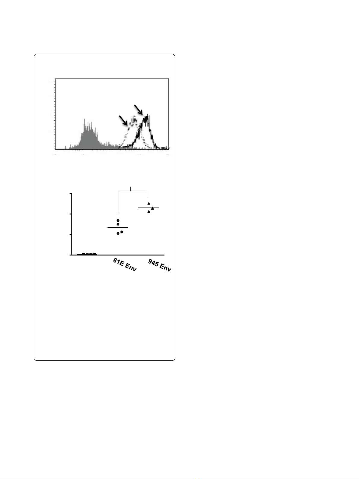
Figure 1 Comparative binding assays of virus particles bearing
the Env protein of FeLV-A/61E or of FeLV-945.A. Feline 3201
cells were incubated with equivalent numbers of virus particles
bearing the envelope protein of FeLV-A/61E (61E Env) or FeLV-945
(945 Env), followed by incubation with monoclonal antibody C11D8
to detect the surface-bound viral SU protein and then with an Alexa
Fluor 488-conjugated secondary antibody. Virus binding was
analyzed by flow cytometry. A representative histogram is shown,
demonstrating the binding activity of the particles as indicated and
a negative control in which no virus was included in the assay
(shaded). B. The geometric mean fluorescence of quadruplicate
samples from individual assays is indicated, as is the mean of
replicate experiments (horizontal bar). Asterisk indicates statistical
significance (*; p < 0.001).
Bolin et al.Retrovirology 2011, 8:35
http://www.retrovirology.com/content/8/1/35
Page 3 of 17
Geometric mean
fluorescence
*
Control 61E 945
Soluble SU Protein
0
50
100
150
200
Control 61E 945
Soluble SU Protein
*
Geometric mean
fluorescence
0
50
100
150
100101102103104
Alexa 488
Antibody: anti-SU
945
61E
100101102103104
Alexa 488
Antibody: anti-HA
61E 945
*
0
10
20
30
40
50
60
70
Control 61E 945
Soluble SU Protein
Geometric mean
fluorescence
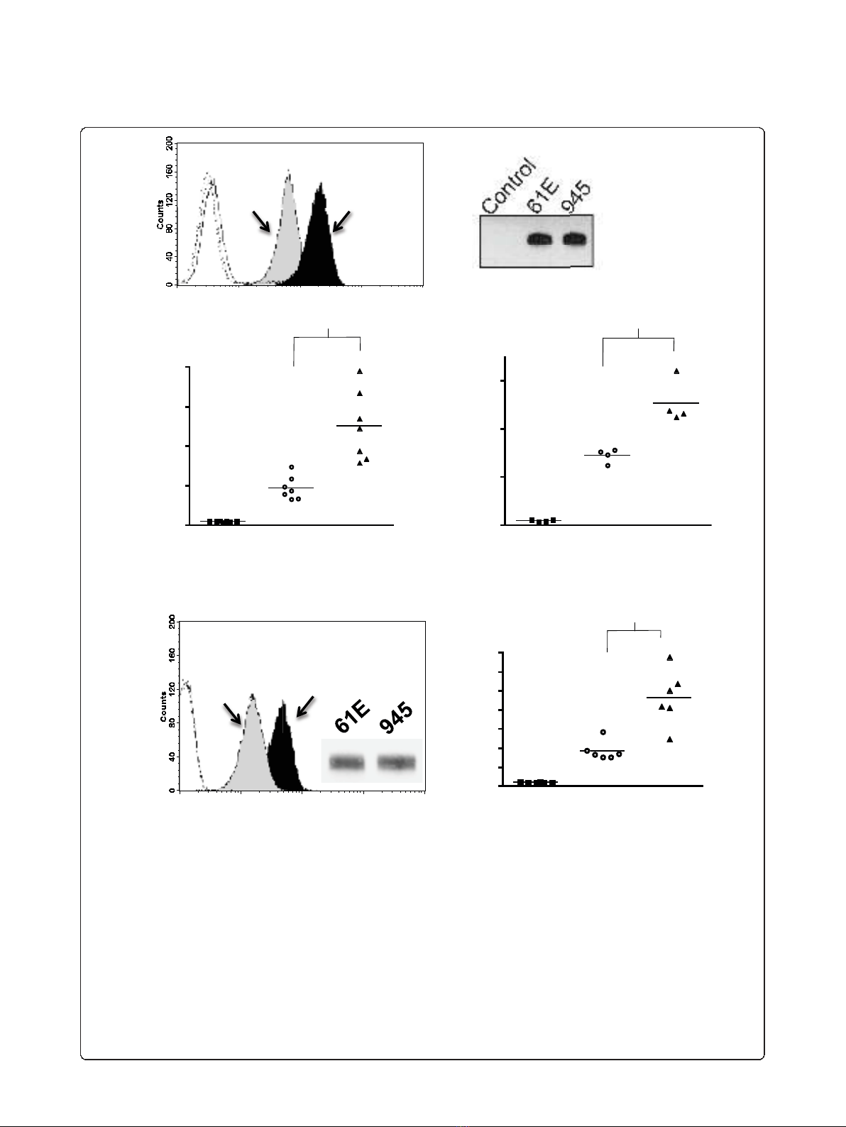
Figure 2 Comparative binding assays of soluble SU proteins of FeLV-A/61E or FeLV-945.A. A representative histogram is shown from a
comparative flow cytometric binding assay demonstrating the binding activity of FeLV-A/61E SU (61E; gray shaded) or FeLV-945 SU (945; black
shaded). Soluble SU proteins were quantified precisely using anti-SU antibody C11D8. Feline 3201 cells were incubated with equivalent mass
amounts of either SU protein for one hour, followed by incubation with C11D8 antibody to detect the surface-bound viral SU proteins and then
with an Alexa Fluor 488-conjugated secondary antibody. Negative controls (open histograms) included cell supernatants of transfections with the
empty expression vector, pCS2/Ctrl, and each SU with isotype control antibody. B. Chemiluminescent western blot analysis of equivalent mass
amounts of FeLV-A/61E and FeLV-945 SU proteins using C11D8 antibody as probe is shown to validate the precision of the infrared
quantification. Negative control was supernatants of cells transfected with pCS2/Ctrl. C-D. Geometric mean fluorescence of replicate binding
assays performed using four independently generated and quantified batches of FeLV-A/61E and FeLV-945 SU protein on either feline 3201 cells
(C) or murine MDTF/H2 cells (D) which express the FeLV-A receptor. Supernatant of mock- or pCS2/Ctrl-transfected cells were used as a negative
control. The mean of replicate experiments is represented (horizontal bar). Asterisk indicates statistical significance (*; p < 0.001). E. Flow
cytometric binding assays performed exactly as in (A) except that analysis was performed using an antibody to detect the HA tag at the C-
terminus of soluble SU proteins. Shown are a representative histogram (left), anti-HA chemiluminescent western blot analysis of equivalent mass
amounts of SU proteins to validate quantification (inset), and geometric mean fluorescence of replicate binding assays (right; p < 0.001).
Negative controls included either SU protein with isotype control antibody (open histograms).
Bolin et al.Retrovirology 2011, 8:35
http://www.retrovirology.com/content/8/1/35
Page 4 of 17
been previously described [28], although no such struc-
ture has yet been described for FeLV-A. Thus, homol-
ogy modeling of the receptor binding domain in the SU
proteins of FeLV-A/61E and FeLV-945 was performed
using the known FeLV-B SU structure [28] as a model-
ing template for the SwissModel Program [29-31] (Fig-
ure 5A). Computational models thereby generated
predict a prominent loop in the VRA domain of both
FeLV-A/61E and FeLV-945 SU that is distinct in struc-
ture from FeLV-B and is predicted to protrude on the
receptor-binding surface (Figure 5A). The predicted
structure is a cysteine-delimited loop of 31 residues that
appears similar in conformation in FeLV-945 and FeLV-
A/61E. However, the loop sequence includes five resi-
dues that diverge between FeLV-945 and FeLV-A/61E,
thereby implicating the divergent residues in the differ-
ing receptor binding phenotypes of the FeLV-945 and
FeLV-A/61E SU proteins (Figure 5B). To test the
hypothesis that the FeLV-945 sequence in the predicted
VRA domain loop confers increased binding efficiency,
site-directed mutagenesis was utilized to replace the five
divergent residues in the sequence of FeLV-A/61E SU
with those of FeLV-945, yielding a mutant SU gene
designated 61E/945-5. Soluble SU expressed by 61E/
945-5 was then prepared and quantified for use in com-
parative binding assays with SU proteins from FeLV-945
and FeLV-A/61E. The results demonstrated that the
binding phenotype of the 61E/945-5 mutant SU is statis-
tically indistinguishable from that of the FeLV-A/61E
parent protein (Figure 5C, left). Equivalent mass
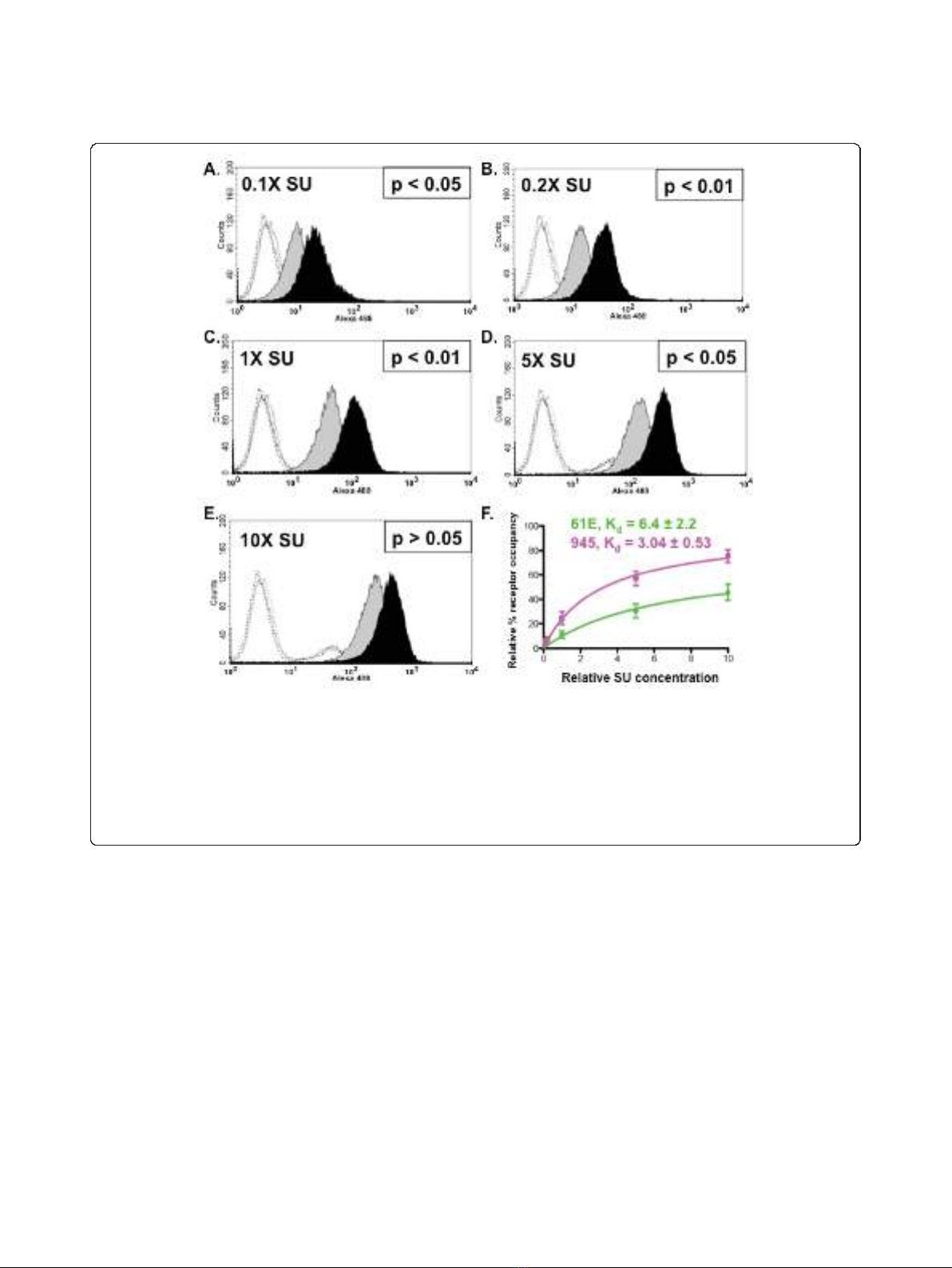
Figure 3 Increased binding activity of FeLV-945 SU is observed over a 100-fold range of SU concentration.A. - E. FeLV-945 SU or FeLV-
A/61E SU proteins in equivalent mass amounts over a 100-fold range (0.1X - 10X) were incubated with feline 3201 cells and processed for flow
cytometric binding assays as described in Figure 2. Representative histograms are shown, demonstrating the binding activity of FeLV-A/61E SU
(gray shaded) or FeLV-945 SU (black shaded). Negative controls (open histograms) included supernatants from mock-transfected cells (solid line),
FeLV-A/61E SU with isotype control antibody (dotted line), and FeLV-945 SU with isotype control antibody (dashed line). Indicated at each SU
concentration is the result of statistical analysis of replicate binding assays using four independently generated and infrared-quantified batches of
SU proteins. A statistically significant increase in geometric mean fluorescence for FeLV-945 SU binding was considered p < 0.05. F. Relative
dissociation constants (K
d
) were determined from the data shown in A - E by nonlinear regression analysis using saturation binding equations
with an assumption of one site-specific binding (GraphPad Prism5.0).
Bolin et al.Retrovirology 2011, 8:35
http://www.retrovirology.com/content/8/1/35
Page 5 of 17