
HPU2. Nat. Sci. Tech. Vol 03, issue 01 (2024), 13-19.
HPU2 Journal of Sciences:
Natural Sciences and Technology
journal homepage: https://sj.hpu2.edu.vn
Article type: Research article
Received date: 12-10-2023 ; Revised date: 04-12-2023 ; Accepted date: 06-02-2024
This is licensed under the CC BY-NC 4.0
13
A method for growing and differentiating the C2C12 muscle cell
line in the laboratory
Ngoc-Hoan Lea,*, Dinh-Toi Chub, Rina Yuc
aHanoi National University of Education, Hanoi, Vietnam
bVietnam National University, Hanoi, Vietnam
cUniversity of Ulsan, Ulsan, Republic of Korea
Abstract
Skeletal muscle-related studies have recently been applied in the combat of obesity and metabolic
disorders. Culture skeletal muscle cells in in vitro plays an important role in being a promising model
for those researches. In the present study, the C2C12 skeletal muscle cells were grown and
differentiated in in vitro. The C2C12 myoblasts were grown in Dulbecco’s modified eagle’s medium
(DMEM) containing 10% fetal bovine serum (FBS) for 4 – 5 days. The cells then reached confluent
about 70% to 100% and the medium was shifted to DMEM plus 2% horse serum which leads the
grown C2C12 cells becoming to differentiated myotubes. Myogenin mRNA levels were found to be
significantly higher in the differentiated myotubes than those in the growing cells confirming the
successful formation of the differentiated C2C12 cells. These results indicate that the C2C12 cell line
is suitable for culture in in vitro to mimic a skeletal muscle microenvironment for further
investigations.
Keywords: C2C12 skeletal muscle cells, culture, differentiation, Myogenin
1. Introduction
Obesity and obesity related metabolic disorders such as cardiovascular diseases, fatty liver
diseases, and type II diabetes are increasing rapidly and being a health issue of global concern,
including Vietnam [1], [2]. Obesity is characterized by hypertrophy of white adipose tissues which are
associated with metabolic complications [3], [4]. On another hand, skeletal muscle tissue takes up a
large percentage of body mass and, thus, dysfunction of this tissue has been shown to be closely
* Corresponding author, E-mail: hoanln@hnue.edu.vn
https://doi.org/10.56764/hpu2.jos.2024.3.1.13-19

HPU2. Nat. Sci. Tech. 2024, 3(1), 13-19
https://sj.hpu2.edu.vn 14
associated with obesity and obesity related metabolic disorders [5]. Consistent with this, skeletal
muscle dystrophy leads to a higher risk of insulin resistant and type II diabetes in human and mice [6],
[7]. Additionally, obesity-related nonalcoholic fatty liver diseases are accompanied with skeletal
muscle metabolic dysfunction [8]. These studies prove that skeletal muscle is a key tissue for the
regulation of metabolic homeostasis. Skeletal muscle is responsible for the majority of glucose uptake
in the body, and it is also a major site of fatty acid oxidation. Thus, improving skeletal muscle
metabolism can help prevent and treat obesity and related metabolic diseases such as fatty liver
diseases, type 2 diabetes, and cardiovascular disease. A recent study has indicated that exercise can
also increase muscle mass which improves skeletal muscle metabolism, as it promotes glucose uptake
and fatty acid oxidation, and thus contributes to increased insulin sensitivity [9]. Interestingly,
supplements of dietary components also improve skeletal muscle metabolic phenotype and give
positive effects on whole system metabolic homeostasis. For example, dietary supplemented with
resveratrol, a nature polyphenolic chemical, can directly alter skeletal muscle development and
metabolic phenotype which are accompanied by the lowered risks of the high-fat diet induced
metabolic disorders in the mice [10]. Taken together, those aforementioned data have suggested that
manipulation of skeletal muscle development and metabolism is a strategy to combat against obesity
and its related diseases.
However, studies of the effect of factors on skeletal muscle physiology in in vivo are usually
faced with many challenges because the body is a complex and unified system with many overlapping,
network interactions. Therefore, manipulations of in vitro studies together with in vivo are needed to
show that a certain factor is acting on the exact tissue cells. There are several skeletal muscle cell lines
that are commonly used in in vitro experiments. These include C2C12, L6, HSkMC, and Sol8 skeletal
muscle cell lines [11], [12]. Among them, the C2C12 is a mouse skeletal muscle cell line that is one of
the most commonly used cell lines for skeletal muscle studies because it has several advantages over
other cell lines, such as (1) Easy to culture: C2C12 cells are relatively easy to culture and maintain in
the laboratory, and they can be grown in a variety of culture media; (2) Well-characterized: C2C12
cells have been extensively characterized, and there is a wealth of information available on their
behaviour and properties. This makes it easier for researchers to design experiments and interpret
results; (3) Availability: C2C12 cells are widely available from commercial suppliers and can be easily
obtained for use in experiments; (4) Genetic manipulation: C2C12 cells can be genetically
manipulated using a variety of techniques, such as transfection or CRISPR/Cas9 gene editing, to study
the function of specific genes or signalling pathways in muscle development and function [13], [14].
Overall, the C2C12 cell line is a valuable tool for studying skeletal muscle development, function, and
metabolism in vitro, and its ease of use and well-characterized properties make it a popular choice
among researchers. As a result, the present study was performed to conduct a viable method to culture
C2C12 skeletal muscle cells from the growth stage to the differentiated adult stage. This provides an
in vitro skeletal muscle cell culture model for further studies, especially modelling the effects of
obesity and metabolic disorders in in vitro.
2. Experimental Methods
2.1. Cell Culture
The mouse myoblast cell line C2C12 was purchased from the American Type Culture Collection
(ATCC CRL-1772, USA). Each vial containing 3 mL freezing soluble of C2C12 myoblasts (107

HPU2. Nat. Sci. Tech. 2024, 3(1), 13-19
https://sj.hpu2.edu.vn 15
cells/mL) was stored at -20C in the refrigerator. Before culture, a vial was taken out the freezer and
thawed at room temperature from 30 minute to 1 hour. Then, the cells were diluted in the growing
medium (5×10⁵ cells/mL) and culture in the incubator at 37C in 5% CO2. Every day, the culture cells
were checked using the light microscopic and the culture medium was newly changed. When the
growing C2C12 cells reached about 70% - 100% confluent the growing medium was switched to the
differentiated medium [15]. The pictures were taken at time points of the experiment using the
microscopic camera (Olympus, Japan). The experiments were conducted in the Department of Food
science and Nutrition, University of Ulsan, South Korea.
2.2. Quantitative Real Time PCR (qRT-PCR)
At each time point, the experimental cells were collected for PCR. The cells were washed twice
to three times with PBS and lysed in Trizol reagent (Invitrogen, USA) and the final mixtures were
collected into 1.5 mL eppendorfs. Two microgram aliquots of total RNA extracted from the lysed
C2C12 cells were reverse transcribed to cDNA using M-MLV reverse transcriptase (Promega, USA).
Then, the cDNAs were carried out in duplicate with a SYBR premix Ex Taq kit (Takara Bio Inc.,
USA) by using a Thermal Cycler Dice (Takara Bio Inc., Japan). All reactions were carried out with the
same condition: 95C for 10 second (s) followed by 40 cycles of 95C for 5 s and 60C for 30 s.
Results were read with Real Time System TP800 software (Takara Bio Inc., Japan) and all values
were normalized to the levels of the control gene β-actin. Thus, for each sample the mRNA expression
level of its Myogenin was evaluated by divided by its β-actin mRNA level. The primers used in the
Real Time PCR are listed in Table 1 [16], [17].
Table 1. Mouse primers used for Real Time-PCR.
Gene
Forward primer (5' → 3')
Reverse primer (5' → 3')
Myogenin
β-actin
TGCCCAGTGAATGCAACTCC
CATCCGTAAAGACCTCTATGCCAAC
TTGGGCATGGTTTCGTCTGG
ATGGAGCCACCGATCCACA
2.3. Statistical Analysis
The results are shown as means ± standard error of the mean (SEM). Variables were compared
using Student’s t-test (Microsoft Excel Software). Comparisons were considered significant difference
at p < 0.05.
3. Results and Discussion
3.1. Thawing and growing of C2C12 myoblasts
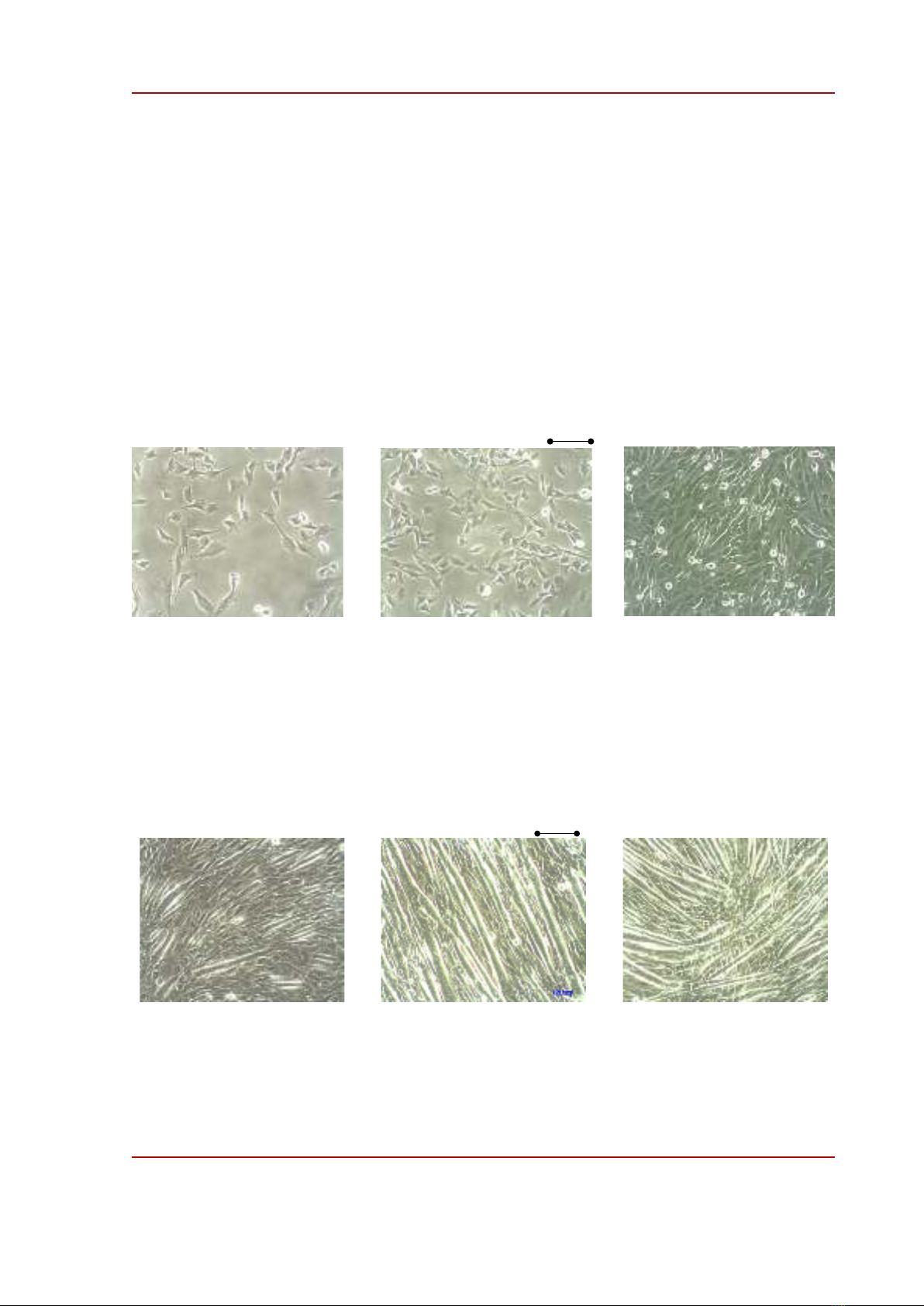
A vial containing 3 mL freezing soluble of C2C12 myoblasts (107 cells/mL) was taken out of the
-20C freezer and put outside at room temperature from 30 minute to 1 hour for thawing. After that,
the thawed C2C12 cells containing soluble was diluted (5×10⁵ cells/mL) in Dulbecco’s modified
eagle’s medium (DMEM) containing 10% fetal bovine serum (FBS) (the growing medium). This cell
containing medium was divided into 4 culture dishes (about 15-20 mL soluble in each dish). Then,
those dishes were put in the incubator at 37C in 5% CO2. After 1 day, the cells attached to the bottom
of the culture dishes and reached about 10% confluence, Figure 1a. The medium was removed, the
HPU2. Nat. Sci. Tech. 2024, 3(1), 13-19
https://sj.hpu2.edu.vn 16
cells were washed with phosphate buffered saline (PBS) two times and then the fresh growing medium
was added to the dishes. After 2-3 days of incubation, the growing cells reached about 50%
confluence, Figure 1b. From this time point, the media in incubated dishes were sucked and washed
twice with PBS, then, each 10 mL of PBS containing 0.05% Trysin and 0.02% EDTA was added in
the dishes for 2-3 minutes at room temperature. When the time finished, the solution was suctioned,
and the dishes were tapped slightly around the walls to completely detach the cells from the bottom of
the dishes. Next, 10-15 mL of the fresh growing medium was added to the dishes and pipetting and
suctioned, to completely transfer the cells from each two old dishes to a new cultured dish. These new
dishes were continuously incubated in the incubator and the cell grew fast to reach about 75% - 100%
confluence at day 4-5 (from starting day), Figure 1c. The method to detach the cells and transfer to
new cultured dish has been manipulated in culture of several cell lines [18]. This strategy to make the
cultured cells growing wells and not losing their differentiation potential [19].
Figure 1. Proliferation process of C2C12 myoblasts. Light microscopy-based images of proliferating C2C12
myoblasts at several time points. (a) The myoblasts grew at day 1. (b) C2C12 myoblasts grew ats day 2-3. (c)
C2C12 myoblasts grew at day 4-5. Magnification 100; Scale bar is 200 m.
3.2. Differentiation of C2C12 myoblasts
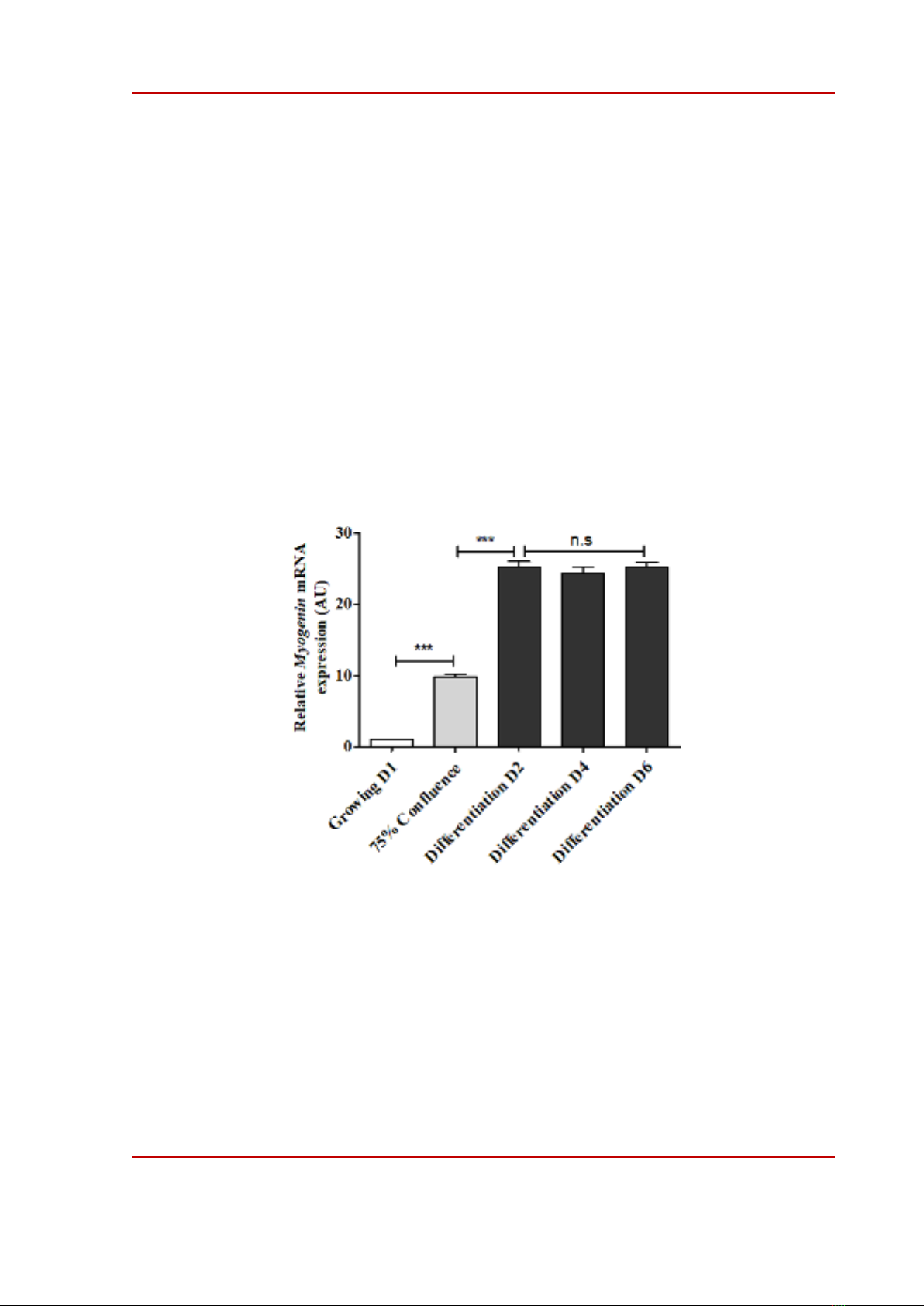
Figure 2. Differentiation process of C2C12 myotubes. Light microscopy-based images of differentiated C2C12
myotubes at several time points. (a) C2C12 myoblasts were differentiated at day 2 (D2). (b) C2C12 myoblasts
were differentiated at day 4 (D4). (c) C2C12 myoblasts were differentiated at day 6 (D6). Magnification 100;
Scale bar is 200 m.
Growing –C2C12
(Confluent about 10%)
Growing –C2C12
(Confluent about 50%)
Growing –C2C12
(Confluent about 75%)
(a) (b) (c)
200 m
C2C12 differentiation D2
200 m
(a) (b) (c)
C2C12 differentiation D4 C2C12 differentiation D6
HPU2. Nat. Sci. Tech. 2024, 3(1), 13-19
https://sj.hpu2.edu.vn 17
Myoblast differentiation is an important period in the development and maintenance of skeletal
muscle tissue. In this process, the undifferentiated myoblasts are transformed into mature and
functional muscle fibers named myotubes [20]. The confluence of culture cells refers to the percentage
of the culture dish covered by adherent culture cells, which can affect the cells’ differentiation when
switched to a differentiated medium. How much confluence of the culture precursor cells should be
used for differentiation remains variable, which depends on several factors such as the cell line, the
differentiated medium, and the expected experimental results products [21], [22]. In the present study,
when the growing myoblasts reached about 75% confluence the growing medium was replaced by the
differentiated medium that consisted of DMEM added with 2% horse serum. The culture dishes were
put in the incubator at 37C in 5% CO2 and the differentiated medium was changed every 2 days. On
day 2 of differentiation, several myoblasts fused to become bigger size myotubes as shown in Figure
2a. On day 4 of differentiation, differentiated myotubes with a typical long cylindrical morphology
were almost formed, Figure 2b. On day 6 of differentiation, the differentiated process was likely
reached to a peak when morphology of myotubes was markedly transformed and the number of
myotubes was likely at maximum, Figure 2c.
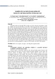
Figure 3. Expression of skeletal muscle marker. Expression of Myogenin mRNA in C2C12 muscle cells was
determined by qRT-PCR at various time points. Quantitative levels of Myogenin mRNA were normalized to the
levels of β-actin. Data are means ± SEM of three independent triplicate experiments. *** p < 0.001 compared
between the two groups. n.s is not a significant comparison. AU is an arbitrary unit. The mRNA expression level
of Myogenin of Growing D1 was considered to have a value of 1, and the mRNA expression levels of Myogenin
of the other groups were compared to the value 1 of Growing D1 group.
3.3. Determination of C2C12 growing stages
The differentiation of myoblast into mature myotubes is regulated by complex molecular signals
and factors. Among them, the myogenic regulatory factors (MRFs) such as Myogenin, MRF4, and
MyoD play a crucial role in regulation of myoblast differentiation [23]. Interestingly, a recent study
has indicated that Myogenin molecule being as a marker of myogenesis and maturation of muscle cells


![Tài liệu học tập Chuyên đề tế bào [mới nhất]](https://cdn.tailieu.vn/images/document/thumbnail/2025/20250906/huutuan0/135x160/56151757299182.jpg)

![Câu hỏi ôn tập Sinh học tế bào [chuẩn nhất]](https://cdn.tailieu.vn/images/document/thumbnail/2025/20250709/kimphuong1001/135x160/771752031316.jpg)
![Lysosome là gì? - Nguyễn Huỳnh Thịnh [Giải thích A-Z]](https://cdn.tailieu.vn/images/document/thumbnail/2015/20151217/conheokiss/135x160/463746382.jpg)
![Tài liệu giảng dạy Sinh học và di truyền [mới nhất]](https://cdn.tailieu.vn/images/document/thumbnail/2026/20260323/hoatudang2026/135x160/42181774414220.jpg)

![Giáo trình Công nghệ vi sinh (Nghề Công nghệ sinh học TC/CĐ) - Trường Cao đẳng Đà Lạt [Mới nhất]](https://cdn.tailieu.vn/images/document/thumbnail/2026/20260224/hoacattuong2026/135x160/87621772161812.jpg)


![Giáo trình Vi sinh vật học môi trường Phần 1: [Thêm thông tin chi tiết nếu có để tối ưu SEO]](https://cdn.tailieu.vn/images/document/thumbnail/2025/20251015/khanhchi0906/135x160/45461768548101.jpg)


