
RESEA R C H Open Access
Prevention of hyperglycemia-induced myocardial
apoptosis by gene silencing of Toll-like receptor-4
Yuwei Zhang
1
, Tianqing Peng
2,3
, Huaqing Zhu
2
, Xiufen Zheng
2
, Xusheng Zhang
2
, Nan Jiang
2
, Xiaoshu Cheng
4
,
Xiaoyan Lai
4
, Aminah Shunnar
2
, Manpreet Singh
2
, Neil Riordan
5
, Vladimir Bogin
6
, Nanwei Tong
1*
,
Wei-Ping Min
2,3,4*
Abstract
Background: Apoptosis is an early event involved in cardiomyopathy associated with diabetes mellitus. Toll-like
receptor (TLR) signaling triggers cell apoptosis through multiple mechanisms. Up-regulation of TLR4 expression has
been shown in diabetic mice. This study aimed to delineate the role of TLR4 in myocardial apoptosis, and to block
this process through gene silencing of TLR4 in the myocardia of diabetic mice.
Methods: Diabetes was induced in C57/BL6 mice by the injection of streptozotocin. Diabetic mice were treated
with 50 μg of TLR4 siRNA or scrambled siRNA as control. Myocardial apoptosis was determined by TUNEL assay.
Results: After 7 days of hyperglycemia, the level of TLR4 mRNA in myocardial tissue was significantly elevated.
Treatment of TLR4 siRNA knocked down gene expression as well as diminished its elevation in diabetic mice.
Apoptosis was evident in cardiac tissues of diabetic mice as detected by a TUNEL assay. In contrast, treatment with
TLR4 siRNA minimized apoptosis in myocardial tissues. Mechanistically, caspase-3 activation was significantly
inhibited in mice that were treated with TLR4 siRNA, but not in mice treated with control siRNA. Additionally, gene
silencing of TLR4 resulted in suppression of apoptotic cascades, such as Fas and caspase-3 gene expression. TLR4
deficiency resulted in inhibition of reactive oxygen species (ROS) production and NADPH oxidase activity,
suggesting suppression of hyperglycemia-induced apoptosis by TLR4 is associated with attenuation of oxidative
stress to the cardiomyocytes.
Conclusions: In summary, we present novel evidence that TLR4 plays a critical role in cardiac apoptosis. This is the
first demonstration of the prevention of cardiac apoptosis in diabetic mice through silencing of the TLR4 gene.
Introduction
Hyperglycemia is the underlying abnormality character-
izing the diabetic condition. Chronic hyperglycemia
introduces a plethora of complications such as cardio-
vascular disease, which is the most frequent cause of
death in the diabetic population [1]. Diabetic patients
have a poorer prognosis post-myocardial infarction as
well as an increased risk of subsequent heart failure
[2,3]. Studies have shown hyperglycemic patients hospi-
talized with acute coronary syndromes also have higher
mortality rates [4]. A key pathological consequence of
sustained hyperglycemia is the induction of cardiomyo-
cyte apoptosis reported in both diabetic patients and
animal models of diabetes [5]. Cardiomyocyte apoptosis
causes a loss of contractile units which reduces organ
function and provokes cardiac remodeling, which is
associated with hypertrophy of viable cardiomyocytes
[5-8]. As such, should myocardial apoptosis be inhibited,
one would expect to prevent or slow the development of
heart failure. Yet, the means by which hyperglycemia
induces apoptosis in cardiomyocytes have not been fully
understood.
Toll-like receptor 4 (TLR4) is a key proximal signaling
receptor responsible for initiating the innate immune
response. TLR4 recognizes pathogen-associated molecular
patterns and plays a vital role in myocardial dysfunction
during bacterial sepsis [9] and pressure overload-induced
* Correspondence: tongnanwei@yahoo.com.cn; mweiping@uwo.ca
1
Department of Endocrinology, West China Hospital of Sichuan University,
Chengdu, China
2
Departments of Surgery, Pathology, Medicine, Oncology, University of
Western Ontario, London, Ontario, Canada
Full list of author information is available at the end of the article
Zhang et al.Journal of Translational Medicine 2010, 8:133
http://www.translational-medicine.com/content/8/1/133
© 2010 Zhang et al; licensee BioMed Central Ltd. This is an Open Access article distributed under the terms of the Creative Commons
Attribution License (http://creativecommons.org/licenses/by/2.0), which permits unrestricted use, distribution, and reproduction in
any medium, provided the original work is properly cited.

cardiac hypertrophy. TLR4 expression is elevated in failing
and ischemic human hearts as well as in animal models of
myocardial ischemia [10,11]. In addition, recent studies
suggest TLR4 may trigger apoptosis of cardiomyocytes in
conditions of cardiac inflammation and oxidative stress
[12]. Studies have also shown that TLR4 is increased in
diabetic mice, however, the role of TLR4 in hyperglyce-
mia-induced myocardial apoptosis has not been eluci-
dated. In this study, we initially investigated the role of
TLR4 on apoptosis in cardiomyocytes under hyperglyce-
mic conditions. Subsequently, we explored the interven-
tion of apoptosis in cardiomyocytes through RNA
interference (RNAi) using small interfering RNA (siRNA)
specific to TLR4 gene. We found that TLR4 was up-regu-
lated in the myocardia of STZ-treated diabetic mice (STZ
mice), which displayed increased expression of apoptotic
genes such as Fas and caspase-3. Treatment with TLR4
siRNA attenuated apoptosis as well suppressed ROS pro-
duction and NADPH oxidase activity.
Materials and methods
Animals
C57/BL6 mice were purchased from The Jackson
Laboratory (Bar Harbor, ME, USA). All mice were male
and 6-8 weeks old. All experimental procedures were
approved by the Animal Use Sub-committee at the Uni-
versity of Western Ontario, Canada, in accordance with
the Guide for the Care and Use on Animals Committee
Guidelines.
Hyperglycemic mouse model
Adult male mice (6-8 weeks old) were intraperitoneally
injectedwithasingledoseofstreptozotocin(STZ)at
150 mg/kg body weight, dissolved in 10 mM sodium
citrate buffer (pH 4.5). On day 3 after STZ treatment,
whole blood was obtained from the mouse tail vein and
random glucose levels were measured using the One-
Touch Ultra 2 blood glucose monitoring system (Life-
Scan, Mountainview, CA). For the present study,
hyperglycemia is defined as a blood glucose measure-
ment of 20 mM or higher. Citrate buffer-treated mice
were used as a normoglycemic control (blood glucose
<12 mM).
siRNA expression vectors
Three target sequences of TLR4 gene were selected. The
oligonucleotides containing sequences specific for TLR4
(5’-GATCCCGTATTAGGAACTACCTCTATGCTTGA-
TATC CGGCATAGAGGTAGTTCCTAATATTTTTTC-
CAAA-3’and 5’-AGCTTTTGGAAAAA ATATTAGG
AACTACCTCTATGCCGGATATCAAGCATAGAGG-
TAGTTCCTAATA CGG-3’;5’-GATCCCGTTGAAAC
TGCAATCAAGAGTGTTGATATCCGCACTCTTG
ATTGCAGTTTCAATTTTTTCCAAA-3’and 5’-AGCT
TTTGGAAAAAATTGAAACT GCAATCAA-
GAGTGCGGATATCAACACTCTTGATTGCAGTTT-
CAACGG-3’;5’-GATCCCATTCGCCAAGCAATGGAAC
TTGATATCCGGTTCCATTGCTTGGCGAA TTTTT
TTCCAAA-3’and 5’-AGCTTTTGGAAAAAAATTCGC-
CAAGCAATGGAACCG GATATCAAGTTCCATTGCT
TGGCGAATGG-3’) were synthesized and annealed.
A TLR4-siRNA expression vector that expresses hairpin
shRNA under the control of the mouse U6 promoter was
constructed. A pair of annealed DNA oligonucleotides
were inserted into a pRNAT-U6.1/Neo shRNA expression
vector that had been digested with BamHI and HindIII
(Genescript, Piscataway, NJ, USA). The plasmid was
suspended in water and stored at -80°C until use.
Treatment of TLR4 siRNA
TLR4 siRNA or scrambled siRNA (50 μg) was mixed
with 40 μl of transfection reagent NANOPARTICLE
(Altogen Biosystems, Las Vegas, NV, USA) with total
volume of 500 μl of 5% glucose (W/V), as per the man-
ufacturer’s instruction. The siRNA mixture was intrave-
nously injected into the C57/BL6 mouse via the tail
vein.
Real-time PCR
TotalRNAwasisolatedfromhearttissuesusingTrizol
reagent (Invitrogen) according to the manufacturer’s
protocol. The RNA was subsequently reverse-tran-
scribed using an oligo-(dT) primer and reverse tran-
scriptase (Invitrogen). Primers used for the amplification
of murine TLR4, Fas, caspase-3 and an internal loading
control, glyceraldehyde-3-phosphate dehydrogenase
(GAPDH) were respectively, as follows: TLR4, sense 5’-
CACTGTTCTTCTCCTGCCTGAC-3’(forward), and 5’-
CCTGGGGAAAAACTCT GGATAG-3’(reverse); Fas,
5’-CAGAAATCGCCTATGGTTGTTG-3’(forward), and
5’-GCT CAGCTGTGTCTTGGATGC-3’(reverse); cas-
pase-3, 5’-TGACCATGGAGAACAACAAA ACCT-3’
(forward), and 5’-TCCGTACCAGAGCGAGATGACA-3’
(reverse); and GAPDH, 5’-TGATGACATCAAGAA
GGTGGTGAA-3’(forward) and 5’-TGGGATG-
GAAATTGT GAGGGAGAT-3’(reverse).
Real-time PCR reactions were performed using SYBR
Green PCR Master mix (Stratagene) and 80 nM of
gene-specific forward and reverse primers as described
above. The PCR reaction conditions were 95°C for
10 min, 95°C for 30 sec, 58°C for one min and 72°C for
30 sec (40 cycles). Amplification was performed accord-
ing to the manufacturer’s cycling protocol and done in
triplicate. Gene expression was calculated as 2
-ΔΔ(Ct)
[13], where Ct is cycle threshold, ΔΔ(Ct) = sample 1Δ
(Ct) -sample 2Δ(Ct); Δ(Ct) = GAPDH (Ct) - testing
gene (Ct). Data was analyzed using MX4000
Zhang et al.Journal of Translational Medicine 2010, 8:133
http://www.translational-medicine.com/content/8/1/133
Page 2 of 8

(Stratagene), Microsoft Excel 2003, and GraphPad Prism
software.
In situ detection of apoptotic cells
Apoptosis in heart tissue was detected using the Apop-
Tag in situ apoptosis detection kit (Qbiogene, Illkirch,
France), as specified by the manufacturer. Briefly, paraf-
fin embedded sections were deparaffinized and
pre-treated with proteinase K (20 μg/ml) for 15 min.
Equilibration buffer was added directly onto the speci-
men, after which terminal deoxynucleotidyl transferase
(TdT) enzyme in reaction buffer was added for 1 h at 37°
C. Sections were washed in Stop/Wash buffer for 10 min.
After incubating with anti-digoxigenin peroxidase conju-
gate for 30 min, the peroxidase substrate was added to
develop color. The samples were washed with PBS and
observed under a microscope in a blinded fashion, and
the proportion of cardiac cells undergoing apoptosis was
calculated.
Caspase-3 Activity
Caspase-3 activity in myocardial tissues was measured
by using a caspase-3 fluorescent assay kit (BIOMOL
Research Laboratory), as described previously [14].
Briefly, hearts from diabetic mice were homogenized,
and protein concentration was determined using the
Bradford method. Samples in duplicates were incubated
with caspase-3 substrate Ac-DEVD-AMC or Ac-DEVD-
AMC plus inhibitor AC-DEVD-CHO at 37°C for 2 h
before measurements were made by a fluorescent spec-
trophotometer (excitation at 380 nm, emission at 405
nm). Signals from inhibitor-treated samples served as
background.
NADPH oxidase activity assay
NADPH oxidase activity was assessed in cell lysates by
lucigenin-enhanced chemiluminescence (20 μgofpro-
tein, 100 μM NADPH, 5 μM lucigenin) with a multilabel
counter (Victor3 Wallac), as described previously [15].
Intracellular ROS measurement
The formation of ROS was measured using the ROS-
sensitive dye, 2,7-dichlorodihydro-fluorescein diacetate
(DCF-DA, Invitrogen), as an indicator. The assay was
performed on freshly dissected heart tissues. Samples
(50 μg proteins) were incubated with 10 μlofDCF-DA
(10 μM) for 3 h at 37°C. The fluorescent product
formed was quantified by spectrofluorometer at the 485/
525 nm. Changes in fluorescence were expressed as an
arbitrary unit.
Statistical analysis
Data were expressed as the mean ± SD. Differences
between two groups were compared by unpaired
Student’st-test. For multi-group comparison, data were
compared using a one-way analysis of variance
(ANOVA) followed by the Newman-Keuls test analysis.
Differences for the value of p < 0.05 were considered
significant.
Results
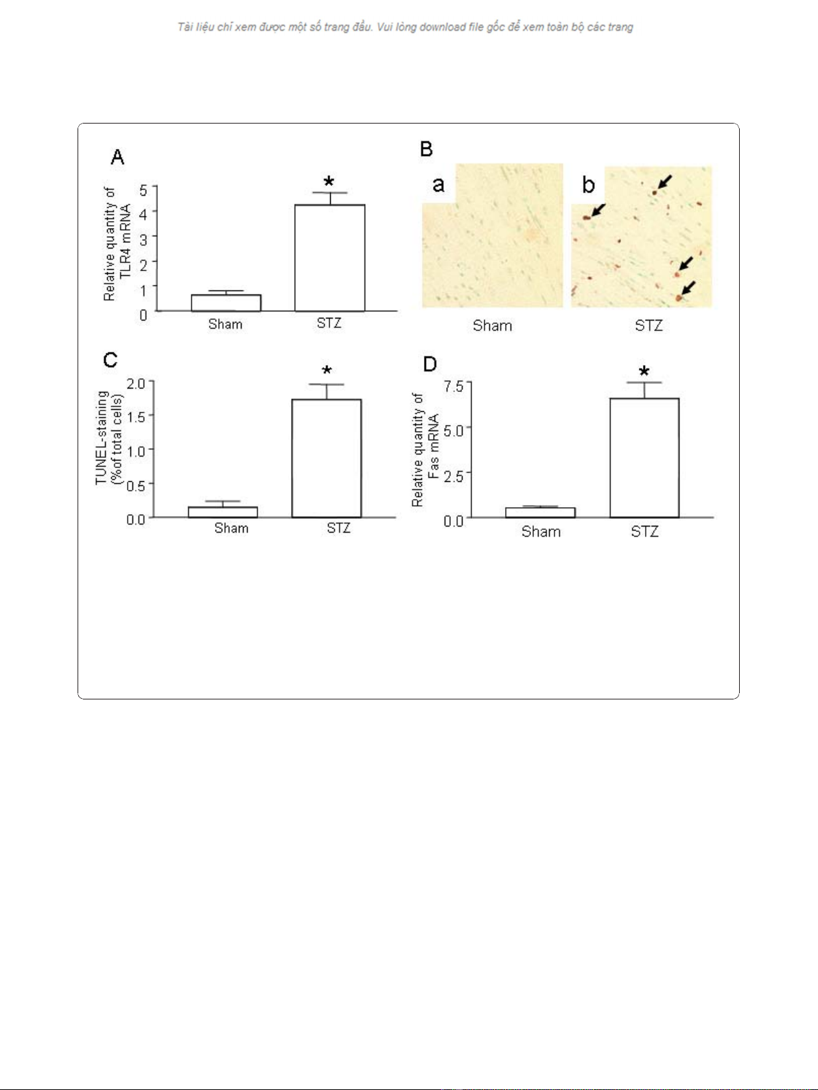
1. Up-regulation of TLR4 and apoptosis in myocardial
tissue of STZ mice
Although TLRs are reportedly up-regulated in cardio-
myocytes of diabetic patients [11], it is unclear whether
TLRs play a role in the promotion of diabetes in the
initial stages of disease or if their up-regulation is a con-
sequence of stimulation from hyperglycemia. To clarify
this, we measured TLR4 levels in mice in the early stages
of diabetes. After treatment with STZ, C57/BL6 mice
developed diabetes as evidenced by hyperglycemia (data
not shown). Significantly increased TLR4 was detected in
the myocardial tissue of STZ-mice as early as 3 days after
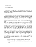
the appearance of hyperglycemia (Figure 1A).
We and others have previously demonstrated that
hyperglycemia is capable of inducing apoptosis in cardio-
myocytes [16-18]. Apoptosis is one of the earliest indica-
tors of cardiomyopathy in the diabetic heart and
accordingly, we measured apoptosis in STZ-treated mice.
Seven days after STZ treatment, substantial apoptosis was
detected in myocardial tissue (Figure 1B). Additionally,
Fas expression was significantly increased in STZ-treated
mice compared to control littermates (Figure 1D).
2. Prevention of hyperglycemia-induced apoptosis in
myocardial tissue by gene silencing of TLR4
Accumulating evidence suggests that activation of the
TLR4 pathway is associated with myocardial apoptosis
[12]. We explored whether knockdown of TLR4 may
suppress apoptosis of cardiomyocytes in STZ-mice. First,
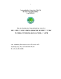
we validated in vivo gene silencing of TLR4 siRNA in
myocardial tissue. After infusion of TLR4 siRNA, the
TLR4 mRNA level was decreased by 75%, as comparing
with the mice treated with scrambled control siRNA
(Figure 2A), indicative of successful knockdown in the heart
in vivo. Treatment with TLR4 siRNA did not affect the level
of blood glucose in diabetic mice (Data not shown).
Next, we examined whether gene knockdown of TLR4
has a therapeutic effect on the prevention of myocardial
apoptosis in diabetic mice. As shown in Figure 2B,
apoptosis, as detected by the TUNEL assay, was remark-
ably attenuated in mice treated with TLR4 siRNA com-
pared with scrambled siRNA.
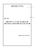
3. Inhibition of caspase-3 in myocardia after gene
silencing of TLR4
To further confirm the Fas-FasL pathway is involved in
apoptosis of cardiomyocytes, we measured the expression
Zhang et al.Journal of Translational Medicine 2010, 8:133
http://www.translational-medicine.com/content/8/1/133
Page 3 of 8
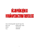
of Fas in the myocardial tissue of STZ mice. Treatment
of TLR4 siRNA resulted in the suppression of Fas expres-
sion (Figure 3A).
To understand the involvement of pro-apoptotic cas-
pases, we determined caspase-3 levels in myocardial tis-
sue. Sham-treated control mice only expressed low level
of caspase-3 while in heart tissue of STZ-treated mice,
hyperglycemia was shown to up-regulate caspase-3
expression dramatically (Figure 3B). Treatment of control
siRNA did not alter the level of caspase-3; however, treat-
ment of TLR4 siRNA effectively reversed up-regulation
of caspase-3 (Figure 3B).
To confirm caspase-3 gene suppression influences its
biological function in the apoptotic pathway, we measured
caspase-3 activity in the myocardial tissue. Caspase-3 acti-
vation was remarkably inhibited in mice treated with
TLR4 siRNA but not in mice treated with scrambled
siRNA or non-treated diabetic mice (Figure 3C).
4. Attenuation of ROS production in myocardia after gene
silencing of TLR4
It has been demonstrated that hyperglycemia may sti-
mulate the production of reactive oxygen species (ROS)
which in turn induces apoptosis in the diabetic heart
[17,19]. We measured ROS levels in the myocardia of
STZ-treated mice in order to examine the contribution
of ROS production to apoptosis and found that ROS
production was increased in mice with hyperglycemia
(Figure 4). While the treatment of scrambled siRNA did
not change the production of ROS in STZ mice, treat-
ment of TLR4 siRNA resulted in significant decrease in
ROSproductioninthediabeticheart(Figure4).
Figure 1 Up-regulation of TLR4 and increased apoptosis in the hearts of STZ mice.(A) TLR4 expression in the hearts of STZ mice. Injection
of STZ induced Type I diabetes as described in Materials and Methods. Control mice were injected with the same volume of sodium citrate
buffer (Sham). On day 7 after STZ treatment, the hearts from diabetic mice (n = 6) and sham mice (n = 6) were retrieved. Total mRNA was
extracted and used to detect the TLR4 transcripts by qPCR. (B) Determination of in situ apoptotic cells in myocardia. Apoptosis in sham-treated
mice and STZ-treated diabetic mice was detected by TUNEL assay. Representative photomicrographs of TUNEL staining in cardiomyocytes are
shown in yellow-blown signal (arrows) from (a) sham treated mice (n = 6) or (b) STZ-treated diabetic mice (n = 6). (C) Quantification of TUNEL
positive cardiomyocytes. (D) Fas expression in the hearts of STZ mice. Diabetes was induced by STZ injection as described in Materials and
Methods. On day 7 after STZ treatment, the hearts from diabetic mice (n = 6) and sham mice (n = 6) were retrieved. Total mRNA was extracted
and used to detect the Fas transcripts by qPCR. Mean ± SD are shown in A, C and D, and are representative of 3 experiments; (*) Statistical
significance when compared with sham treated mice and STZ-treated diabetic mice was denoted at p < 0.05.
Zhang et al.Journal of Translational Medicine 2010, 8:133
http://www.translational-medicine.com/content/8/1/133
Page 4 of 8
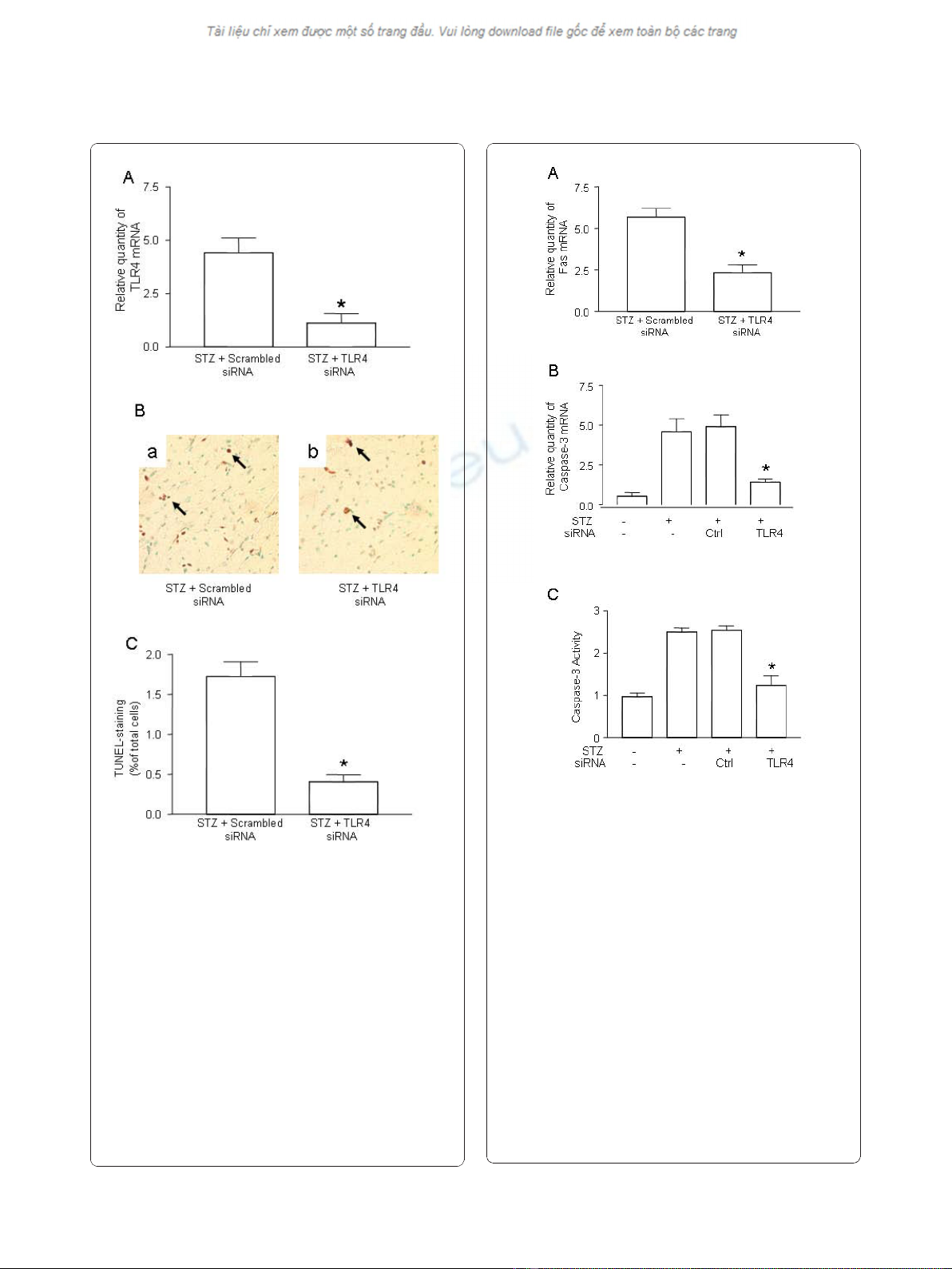
Figure 2 Suppression of TLR4 and prevention of apoptosis by
gene silencing of TLR4.(A) Suppression of TLR4 expression in the
heart of STZ mice treated with TLR4 siRNA. Diabetes was induced
by STZ injection as described in Materials and Methods. On day -1
(the day before STZ treatment), mice were intravenously injected
with 5 μg of TLR4 siRNA or scrambled control siRNA, along with
NANOPARTICLE. On day 7 after STZ treatment, the hearts from the
mice treated with TLR4 siRNA (n = 6) or scrambled siRNA (n = 6)
were retrieved. Total mRNA was extracted and used to detect the
TLR4 transcripts by qPCR. The relative quantity of TLR4 mRNA was
expressed as mean ± SD. (*) Statistical significance when compared
with scrambled siRNA treated mice was denoted as p < 0.05. (B)
Attenuation of apoptotic cells in cardiomyocyte by TLR4 siRNA.
Apoptosis in the diabetic mice treated with control siRNA (n = 6)
and TLR4 siRNA (n = 6) was detected by TUNEL assay.
Representatives of TUNEL staining in cardiomyocytes were shown in
yellow-blown signal (arrows) from the mice treated with scrambled
siRNA (a) or TLR4 siRNA (b). (C) Quantification of TUNEL positive
cardiomyocytes. Data shown are representative of 3 experiments.
Figure 3 Inhibition of caspase-3 after gene silencing of TLR4.
(A) Suppression of Fas expression in the hearts of STZ mice treated
with TLR4 siRNA. Diabetes was induced by STZ injection as
described in Materials and Methods. Diabetic mice were treated
with TLR4 siRNA (n = 6) and scrambled control siRNA (n = 6) as
described in Figure 2. On day 7 after STZ treatment, the hearts from
mice treated with TLR4 siRNA or scrambled siRNA were retrieved.
Total mRNA was extracted and used to detect Fas transcripts by
qPCR. (B) Suppression of caspase-3 expression in the heart of STZ
mice treated with TLR4 siRNA. Diabetic mice were treated with TLR4
siRNA (n = 6) and scrambled control siRNA (n = 6) as described
above. The expression of caspase-3 transcripts was detected by
qPCR. (C) Inhibition of caspase-3 activity in the heart of STZ mice
treated with TLR4 siRNA. Diabetic mice were treated with TLR4
siRNA (n = 6) and scrambled control siRNA (n = 6) as described
above. On day 7 after STZ treatment, the hearts from the mice
treated with TLR4 siRNA or scrambled siRNA were retrieved, the
protein was prepared and the caspase-3 activity was determined as
described in Methods and Materials. Relative quantity of TLR4 mRNA
and caspase-3 activity was expressed as mean ± SD. (*) Statistical
significance when compared with scrambled siRNA treated mice
was denoted as p < 0.05. Data shown are representative of 3
experiments.
Zhang et al.Journal of Translational Medicine 2010, 8:133
http://www.translational-medicine.com/content/8/1/133
Page 5 of 8


%20--%3e%3cdefs%3e%3cstyle%3e%20.st0%20{%20fill:%20%23fff;%20}%20.st1%20{%20fill:%20%237800fa;%20}%20%3c/style%3e%3c/defs%3e%3cpath%20class='st1'%20d='M117.78,12.18H43.11c2.9,3.47,4.65,7.94,4.65,12.82,0,5.6-2.3,10.66-6.01,14.29h76.02l7.22-13.56-7.22-13.56Z'/%3e%3cg%3e%3cpath%20class='st0'%20d='M53.58,26.17h-.59v-1.46h.59v-4.96h2.83c1.78,0,2.67.94,2.67,2.82v5.76c0,1.87-.89,2.81-2.67,2.81h-2.83v-4.96ZM55.36,21.37v3.34h1.1v1.46h-1.1v3.34h1.01c.61,0,.91-.37.91-1.1v-5.93c0-.74-.3-1.1-.91-1.1h-1.01Z'/%3e%3cpath%20class='st0'%20d='M65.99,31.14h-1.8l-.31-2.07h-2.19l-.31,2.07h-1.64l1.82-11.39h2.62l1.82,11.39ZM65.28,18.04c-.25.46-.51.77-.75.94-.21.15-.47.22-.79.22-.26,0-.57-.07-.92-.22l-.38-.15c-.14-.05-.26-.07-.37-.07-.3,0-.53.18-.71.54l-.91-.68c.25-.46.51-.77.75-.94.21-.14.48-.21.79-.21.26,0,.57.07.92.21l.38.15c.14.05.26.07.37.07.3,0,.53-.18.71-.54l.91.68ZM61.91,27.52h1.73l-.87-5.76-.87,5.76Z'/%3e%3cpath%20class='st0'%20d='M74.53,26.89v1.52c0,1.91-.89,2.86-2.67,2.86s-2.67-.95-2.67-2.86v-5.93c0-1.91.89-2.86,2.67-2.86s2.67.95,2.67,2.86v1.11h-1.69v-1.22c0-.75-.31-1.12-.93-1.12s-.93.37-.93,1.12v6.15c0,.74.31,1.11.93,1.11s.93-.37.93-1.11v-1.63h1.69Z'/%3e%3cpath%20class='st0'%20d='M81.4,31.14h-1.8l-.31-2.07h-2.19l-.31,2.07h-1.64l1.82-11.39h2.62l1.82,11.39ZM75.9,19.2l1.52-1.91h1.71l1.51,1.91h-1.61l-.76-.95-.75.95h-1.61ZM77.32,27.52h1.73l-.87-5.76-.87,5.76ZM83.1,15.99l-1.76,1.91h-1.26l1.17-1.91h1.86Z'/%3e%3cpath%20class='st0'%20d='M84.86,19.75c1.78,0,2.67.94,2.67,2.82v1.48c0,1.87-.89,2.81-2.67,2.81h-.85v4.28h-1.79v-11.39h2.64ZM84.01,21.37v3.86h.85c.58,0,.87-.36.87-1.08v-1.71c0-.71-.29-1.07-.87-1.07h-.85Z'/%3e%3cpath%20class='st0'%20d='M93.51,19.75c1.78,0,2.67.94,2.67,2.82v1.48c0,1.87-.89,2.81-2.67,2.81h-.85v4.28h-1.79v-11.39h2.64ZM92.66,21.37v3.86h.85c.58,0,.87-.36.87-1.08v-1.71c0-.71-.29-1.07-.87-1.07h-.85Z'/%3e%3cpath%20class='st0'%20d='M98.8,31.14h-1.79v-11.39h1.79v4.88h2.03v-4.88h1.83v11.39h-1.83v-4.88h-2.03v4.88Z'/%3e%3cpath%20class='st0'%20d='M105.36,24.55h2.46v1.62h-2.46v3.34h3.09v1.63h-4.88v-11.39h4.88v1.63h-3.09v3.18ZM108.17,17.29l-1.76,1.91h-1.26l1.17-1.91h1.86Z'/%3e%3cpath%20class='st0'%20d='M112.2,19.75c1.78,0,2.67.94,2.67,2.82v1.48c0,1.87-.89,2.81-2.67,2.81h-.85v4.28h-1.79v-11.39h2.64ZM111.35,21.37v3.86h.85c.58,0,.87-.36.87-1.08v-1.71c0-.71-.29-1.07-.87-1.07h-.85Z'/%3e%3c/g%3e%3ccircle%20class='st1'%20cx='25'%20cy='25'%20r='20'/%3e%3cpath%20class='st0'%20d='M32.78,19.27c2.92,0,4.43,2.55,5.28,5.33l.71,2.17c.14.38-.33.75-.71.75h-5.61c.19-.33.24-.71.09-1.08l-.75-2.45c-.43-1.32-.99-2.64-1.79-3.77.75-.57,1.65-.94,2.78-.94h0ZM25,18.38c3.25,0,4.9,2.78,5.89,5.89l.76,2.45c.14.42-.33.8-.8.8h-11.69c-.42,0-.94-.38-.8-.8l.75-2.45c.99-3.11,2.64-5.89,5.89-5.89h0ZM25,11.35c1.74,0,3.11,1.37,3.11,3.11s-1.37,3.11-3.11,3.11-3.11-1.41-3.11-3.11,1.41-3.11,3.11-3.11h0ZM17.27,19.27c1.08,0,1.98.38,2.73.94-.8,1.13-1.37,2.45-1.74,3.77l-.8,2.45c-.14.38-.05.75.09,1.08h-5.56c-.42,0-.9-.38-.75-.75l.71-2.17c.9-2.78,2.41-5.33,5.33-5.33h0ZM17.27,12.91c1.51,0,2.78,1.27,2.78,2.83s-1.27,2.83-2.78,2.83-2.83-1.27-2.83-2.83,1.27-2.83,2.83-2.83h0ZM32.78,12.91c1.56,0,2.78,1.27,2.78,2.83s-1.23,2.83-2.78,2.83-2.83-1.27-2.83-2.83,1.27-2.83,2.83-2.83h0ZM27.07,28.56v.09c0,.57-.24,1.08-.61,1.46h0v.05c-.38.33-.9.57-1.46.57s-1.08-.24-1.46-.61h0c-.38-.38-.61-.9-.61-1.46v-.09h1.41v.09c0,.19.05.38.19.47v.05c.09.09.28.19.47.19s.38-.09.47-.19v-.05c.14-.09.24-.28.24-.47t-.05-.09h1.41ZM30.99,28.56v.09c0,1.65-.66,3.16-1.74,4.24-1.08,1.08-2.59,1.79-4.24,1.79s-3.16-.71-4.24-1.79l-.05-.05c-1.04-1.08-1.7-2.55-1.7-4.2v-.09h1.41v.09c0,1.27.47,2.4,1.27,3.25h.05c.85.85,1.98,1.37,3.25,1.37s2.4-.52,3.25-1.37c.85-.8,1.37-1.98,1.37-3.25v-.09h1.37ZM34.99,28.56v.09c0,2.78-1.13,5.28-2.92,7.07-1.79,1.79-4.29,2.92-7.07,2.92s-5.23-1.13-7.07-2.92c-1.79-1.79-2.92-4.29-2.92-7.07v-.09h1.41v.09c0,2.4.94,4.53,2.5,6.08,1.56,1.56,3.72,2.5,6.08,2.5s4.52-.94,6.08-2.5c1.56-1.56,2.5-3.68,2.5-6.08v-.09h1.41Z'/%3e%3c/svg%3e)