
RESEARC H Open Access
Microrna profiling analysis of differences between
the melanoma of young adults and older adults
Drazen M Jukic
1,2†
, Uma NM Rao
2
, Lori Kelly
2†
, Jihad S Skaf
3
, Laura M Drogowski
1
, John M Kirkwood
4
,
Monica C Panelli
4*
Abstract
Background: This study represents the first attempt to perform a profiling analysis of the intergenerational
differences in the microRNAs (miRNAs) of primary cutaneous melanocytic neoplasms in young adult and older age
groups. The data emphasize the importance of these master regulators in the transcriptional machinery of
melanocytic neoplasms and suggest that differential levels of expressions of these miRs may contribute to
differences in phenotypic and pathologic presentation of melanocytic neoplasms at different ages.
Methods: An exploratory miRNA analysis of 666 miRs by low density microRNA arrays was conducted on formalin
fixed and paraffin embedded tissues (FFPE) from 10 older adults and 10 young adults including conventional
melanoma and melanocytic neoplasms of uncertain biological significance. Age-matched benign melanocytic nevi
were used as controls.
Results: Primary melanoma in patients greater than 60 years old was characterized by the increased expression of
miRs regulating TLR-MyD88-NF-kappaB pathway (hsa-miR-199a), RAS/RAB22A pathway (hsa-miR-204); growth
differentiation and migration (hsa-miR337), epithelial mesenchymal transition (EMT) (let-7b, hsa-miR-10b/10b*),
invasion and metastasis (hsa-miR-10b/10b*), hsa-miR-30a/e*, hsa-miR-29c*; cellular matrix components (hsa-miR-
29c*); invasion-cytokinesis (hsa-miR-99b*) compared to melanoma of younger patients. MiR-211 was dramatically
downregulated compared to nevi controls, decreased with increasing age and was among the miRs linked to
metastatic processes. Melanoma in young adult patients had increased expression of hsa-miR-449a and decreased
expression of hsa-miR-146b, hsa-miR-214*. MiR-30a* in clinical stages I-II adult and pediatric melanoma could
predict classification of melanoma tissue in the two extremes of age groups. Although the number of cases is
small, positive lymph node status in the two age groups was characterized by the statistically significant expression
of hsa-miR-30a* and hsa-miR-204 (F-test, p-value < 0.001).
Conclusions: Our findings, although preliminary, support the notion that the differential biology of melanoma at
the extremes of age is driven, in part, by deregulation of microRNA expression and by fine tuning of miRs that are
already known to regulate cell cycle, inflammation, Epithelial-Mesenchymal Transition (EMT)/stroma and more
specifically genes known to be altered in melanoma. Our analysis reveals that miR expression differences create
unique patterns of frequently affected biological processes that clearly distinguish old age from young age
melanomas. This is a novel characterization of the miRnomes of melanocytic neoplasms at two extremes of age
and identifies potential diagnostic and clinico-pathologic biomarkers that may serve as novel miR-based targeted
modalities in melanoma diagnosis and treatment.
* Correspondence: panellim@gmail.com
†Contributed equally
4
University of Pittsburgh Cancer Institute, Division of Hematology-Oncology
Hillman Cancer Center, Pittsburgh, Pennsylvania, USA
Jukic et al.Journal of Translational Medicine 2010, 8:27
http://www.translational-medicine.com/content/8/1/27
© 2010 Jukic et al; licensee BioMed Central Ltd. This is an Open Access article distributed under the terms of the Creative Commons
Attribution License (http://creativecommons.org/licenses/by/2.0), which permits unrestricted use, distribution, and reproduction in
any medium, provided the original work is properly cited.
Background
The incidence of melanoma dramatically increases with
age, and accounts for 7% of all malignancies seen in
patients between the ages of 15-29 years [1,2]. Despite
thefactthatalmost450newpatientswithmelanoma
under the age of 20 are diagnosed with melanoma each
year in the United States, published reports of this dis-
ease in young people have usually been restricted in
number and often constitute series from single institu-
tions. Two recently published large studies from the
Surveillance Epidemiology and End Results (SEER) and
National Cancer Database (NCDB) databases confirmed
and expanded previous observations that pediatric/
young adult melanoma may be clinically similar to adult
melanoma; however some differences in clinical presen-
tation and outcome such as the higher incidence of
nodal metastases in children and adolescents with
localized disease are evident, particularly in younger
patients [1-6].
The outcome of melanoma in the younger, as com-
pared to the older, populations has been shown to differ
quite substantially. In the young adult and pediatric
population the issue is complicated because of inability
even amongst experts to identify conventional melano-
mas from certain melanocytic neoplasms of uncertain
biologic behavior because of subtle overlapping histo-
morphological features. Notably in Spitzoid nevi, this
subject has been debated since the entity was first
described by Sophie Spitz in 1948 [7] because some of
these neoplasm have metastasized to regional lymph
nodes [8,9]. It has also been recently suggested that the
Spitzoid melanocytic neoplasms with nodal metastases
mayhaveabetterprognosisinyoung/pediatricage
group [10]. In many of the cases, these lesions have
been treated as malignant melanomas [11].
The aim of this study was to identify the differences
between melanoma in young and older adult popula-
tions with the ultimate goal of finding useful biomarkers
of etiology and outcome at different ages. Therefore we
have included some of the Spitzoid melanocytic neo-
plasms (as a part of the group of patients age less than
30 years old/Mel 30) that have documented sentinel
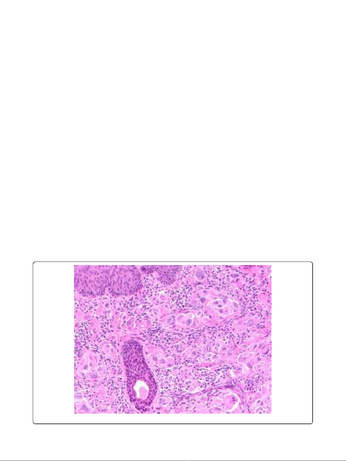
lymph node metastases. (Figure 1).
As Chen summarized [12], the use of DNA microar-
rays to monitor tumor RNA profiles has defined a mole-
cular taxonomy of cancer, which can be used to identify
new drugs and better define prognosis, with the ultimate
potential to predict patterns of drug resistance. Cellular
behavior is also governed by translational and posttran-
slational control mechanisms that are not reflected in
mRNA profiles of tumor specimens. Since microRNAs
regulate gene expression at the post-transcriptional
level, the availability of a comprehensive microRNA
(miRNAs/miR) expression profile can provide informa-
tion that is complementary to that derived from mRNA
transcriptional profiling. Thus, comprehensive micro-
RNA expression profiling can help to unravel these mas-
ter regulators of gene expression, which represent a
Figure 1 Atypical Spitz. Example of atypical Spitz neoplasm of uncertain biological significance.
Jukic et al.Journal of Translational Medicine 2010, 8:27
http://www.translational-medicine.com/content/8/1/27
Page 2 of 23

pivotal regulatory network in the transcriptional cell
machinery and have been associated with deregulation
of immune and cell cycle processes in cancer [13].
MiRNAs are a family of endogenous, small (18-25
nucleotides in length), noncoding, functional RNAs. It is
estimated that there may be 1000 miRNA genes in the
human genome (Internet address: http://www.sanger.ac.
uk/Software/Rfam/mirna/). The latest update of miR-
Base (Internet address: release 13 March 2009, http://
microrna.sanger.ac.uk/sequences/index.shtml) includes
more than 1900 annotated miR sequences.
MiRNAs are transcribed by RNA polymerase II or III
as longer primary-miRNA molecules, which are subse-
quently processed in the nucleus by the RNase III endo-
nuclease Drosha and DGCR8 (the “microprocessor
complex”) to form approximately 70 nucleotide-long
intermediate stem-loop structures called “precursor
miRNAs”(pre-miRNAs). These pre-miRNAs are trans-
ported from the nucleus to the cytoplasm, where they
are further processed by the endonuclease Dicer. Dicer
produces an imperfect duplex composed of the mature
miRNA sequence and a fragment of similar size
(miRNA*), which is derived from the opposing arm of
the pre-miRNA [14].
Only the mature-miRNA remains stable on the RNA-
induced silencing complex (RISC) and induces post-
transcriptional silencing of one or more target genes by
binding with imperfect complementarity to a target
sequence in the 3’-UTR of the target RNA with respect
to a set of general rules that are only incompletely
determined experimentally and bioinformatically to date
[15]. Identification of miRNA targets has been difficult
because only the seed sequence, about 6-8 bases of the
approximately 22 nucleotides, aligns perfectly with the
target mRNA’s3’untranslated region. The remainder of
the miRNA may bind perfectly to the target mRNA, but
more often it does not [14]. RNA interference and
related small RNA mediated pathways are central in the
silencing of gene expression, and at least 30% of human
genes are thought to be regulated by microRNAs [16].
MiRNAs are expressed in a tissue-specific manner, and
can contribute to cancer development and progression.
They are differentially expressed in normal tissues and
both hematological and solid tumors. In human solid
tumors such as hepatocellular carcinoma [17] and ovar-
ian cancer [18], the miRNA expression signature defines
neoplasm-specific dys-regulation of specific gene targets.
Despite the hundreds of miRs discovered to date, their
biological functions are incompletely understood.
Increasing evidence suggests that the expression of miR-
NAs (miRs) is deregulated in many cancers, and miRs
can control cell proliferation, differentiation and apopto-
sis [19]. The alteration of miR expression may contri-
bute to the initiation and manintanance of tumors as
their abnormal levels have important pathogenic conse-
quences: miR overexpression in tumors usually contri-
butes to oncogenesis by downregulating tumor
suppressors. For example, the mir-17-miR 92 cluster
reduces the transcription factor E2F1 in lymphomas and
miR -21 represses the tumor suppressor PTEN in hepa-
tocellular carcinoma. MiRs lost by tumors lead to onco-
gene overexpression (let -7 loss leads to expression of
KRAS, NRAS in lung carcinoma, while miR15a and 16-1
loss leads to expression of BCL-2 in CLL and cyclinD1
in prostate carcinoma [20].
The significance of microRNA differential modulation
in the diagnostic and prognostic workup of melanocytic
neoplasms, especially in relationship to the age-stratified
groups, has not, to our knowledge, been investigated.
In this article, we present profiling results in regard to
666 microRNAs evaluated in melanocytic neoplasms of
pediatric and young adults compared with older adults;
the results of which emphasize the importance of these
master regulators in the transcriptional machinery of
melanocytic neoplasms and support the notion that dif-
ferential levels of expressions of these miRs may contri-
bute to differences in phenotypic and pathologic
presentation of melanocytic neoplasms at different ages.
We performed an exploratory analysis of 666 miR on
formalin-fixed paraffin-embedded (FFPE)-primary mela-
noma tissue using the Taqman ®TLDA miRNA arrays
platform A and B (Applied Biosystems, Foster City, CA,
http://www.appliedbiosystems.com) to investigate
whether there were differentially expressed miRs
between young adult and adult melanoma specimens
(including melanocytic neoplasms of uncertain biological
potential). The comparativeprofilingwaspurposively
conducted at extremes of age, <30 and >60 years, to
clearly define age groups. Our study represents the first
attempt to perform a true intergenerational and com-
parative microRNA profiling of the primary melanocytic
neoplasms of adults and young adults.
We observed distinct miRNA profiles in the primary
melanocytic neoplasms of adults and young adults that
could also potentially be associated with the clinical
parameters of stage and nodal involvement. Our obser-
vations represent an important basis for expanded analy-
sis of the etiology and clinico-pathologic spectrum of
this disease.
Materials and methods
Patient Selection
This study included the utilization of archival melanoma
specimens obtained and was approved by the University
of Pittsburgh Cancer Institute (UPCI) Internal Review
Board (IRB): UPCI reference IRB#: PRO07120294.
Archival paraffin blocks of melanocytic neoplasms stu-
died at the UPCI were retrieved from the files of the
Jukic et al.Journal of Translational Medicine 2010, 8:27
http://www.translational-medicine.com/content/8/1/27
Page 3 of 23

Health Sciences Tissue Bank (HSTB) database and dis-
bursed by UPCI HSTB according to UPCI-IRB regula-
tions. Ten primary FFPE-tissues (including melanocytic
neoplasms of uncertain biological potential) were
obtained from two cohorts of patients respectively seg-
regated according to age: Cohort A - > 60 years and
Cohort B - <30 years and utilized for microRNA profil-
ing. These two case cohorts were separated by at least
30 years, thereby representing an adequate basis for an
intergenerational study.
Additionally, 6 benign nevi were used as homologous
controls (3 from adults and 3 from young adult patients,
respectively). A total of 26 lesions (20 test specimens +
6 controls) were analyzed. Primary diagnostic workup
and verification of the diagnosis of primary neoplasms
was performed by two independent reference
pathologists.
Total RNA was isolated from all lesions from (at aver-
age) 30 5 μm sections obtained specifically from areas
that contained at least 70% viable tumor (identified by a
pathologist). RNA quality was assessed using Nanodrop
(OD 260/280 and 260/230 (Table 1)). The overall micro-
RNA profiling of these two groups (adult and young
adult) included a total of 56 Taqman ® microRNA Low
density arrays (TLDAs). Each group included 10 mela-
nocytic neoplasm samples (older adult melanoma, AM,
pediatric and young adult melanoma PM) and 3 control
nevi specimens (adult nevi, AN, pediatric nevi, PN). The
assays were run in 3 batches for processing and a cali-
brator RNA was included in each batch for normaliza-
tion. For each specimen, 2 TLDA were run, TLDA
panel A and TLDA panel B.
Patient characteristics of specimen groups utilized for
class comparison analyses are summarized in Table 2.
The pediatric and young adult melanoma (PM) speci-
mens were obtained from 5 males and 5 females, and
the 3 control nevi (PN) from 1 male and 2 females.
Patient PM8 had a Spitzoid neoplasm of uncertain
Table 1 Summary Of RNAs Extracted From FFPE Melanoma And Nevus (Control) Specimens Obtained From Pediatric
Or Young Adults < 30 Years Of Age And Older Adults > 60 Years Of Age
Sample ID Sample
Name
FFPE Tissue
Type
Percentage Tumor or
Nevus
Total RNA yield
(ug)
ng/ul
RNA
OD 260/
280
OD 260/
230
TB08-190A PM7 Mel 80% 2.26 251 1.98 2.02
TB08-192 1H PM2 Mel 90% 0.45 50.1 1.79 1.47
TB08-239 B PM3 Mel 80% 0.72 79.61 1.87 1.23
TB09-044B PM6 Mel 75% 2.03 226 1.94 1.59
TB08-243A PM8 Mel 85% 1.85 205 1.94 1.95
TB08-231 A PM4 Mel 75% 0.31 34.97 1.81 1.35
TB08-199D PM11112 Mel 75% 1.24 103 1.9 1.65
TB08-195 2A PM5 Mel 80% 0.17 18.69 1.76 1.23
TB08-245D PM9 Mel 100% 2.37 263 1.94 1.83
TB08-477-
478C
PM10 Mel 90% 4.59 255 1.88 1.72
TB08-242A PN1 Nevus 100% 0.77 85.89 1.86 1.41
TB08-232 2A PN2 Nevus 100% 2.71 226 1.86 1.56
TB08-188A PN3 Nevus 100% 0.30 25 1.84 1.45
TB08-236 1L AM1 Mel 100% 0.93 103.09 1.88 1.6
TB08-180P 1H AM2 Mel 100% 3.23 269 2 1.86
TB08-217 1D AM3 Mel 75% 1.42 158.07 1.97 1.64
TB08-223 C AM10 Mel 70% 0.57 63 1.88 1.72
TB08-181 B AM4 Mel 95% 11.29 941 1.84 1.35
TB08-211 1J AM5 Mel 90% 0.66 55 1.89 1.66
TB08-216 F AM6 Mel 80% 0.46 51.37 1.93 1.59
TB08-219 1G AM9 Mel 75% 0.47 52 1.89 1.86
TB08-237 1G AM7 Mel 70% 1.23 136.28 1.85 1.63
TB09-043B AM8 Mel 90% 2.72 302 1.87 1.17
TB09-003 A AN1 Nevus 100% 0.90 100 1.99 1.71
TB08-233D AN2 Nevus 100% 0.36 30 1.93 1.68
TB08-234A AN3 Nevus 100% 0.12 10.4 1.8 1.22
Top group (PM/PN): young adults <30 yrs old; lower group (AM/AN): adults >60; PM = pediatric and young adult melanoma (<30 yrs); AM = adult melanoma
(>60 yrs);PN = pediatric and young adult nevus (<30 yrs); AN = adult nevus (>60 yrs); % tumor refers to the percentage of tumor in the area that was ID &
scraped for RNA isolation. Quality of RNA was established by Nanodrop OD reading.
Jukic et al.Journal of Translational Medicine 2010, 8:27
http://www.translational-medicine.com/content/8/1/27
Page 4 of 23

Table 2 Patients Characteristics
Sample
name
Mel 60/
30 or
Nevus
60/30
Age Age
range
Gender Diagnosis Site T
Stage
N
Stage
M
Stage
Stage Group
at Diagnosis-
AJCC 6th Ed.
PM7 Mel 30 21 20-29 M Melanoma, invasive and insitu, arising in
association with a nevus
Trunk cT1* pN0 cM0 Unknown
PM2 Mel 30 26 20-29 M Superficial spreading melanoma, invasive and in
situ
Back pT1b pN1a cM0 3B
PM3 Mel 30 26 20-29 F Melanoma, superficial spreading in radial growth
phase & vertical, epithelioid, nevoid and balloon
cell
Scapula pT2b pN0 cM0 2A
PM6 Mel 30 28 20-29 F Superficial spreading melanoma, invasive Thigh pT1b pN0 cM0 1B
PM8 Mel 30 28 20-29 M Highly atypical spitzoid neoplasm Arm n/a n/a n/a n/a
PM4 Mel 30 28 20-29 F Superficial spreading melanoma, invasive Shin pT1a pN0 cM0 1A
PM11112 Mel 30 29 20-29 F Superficial spreading (Spitzoid) melanoma, insitu &
invasive
Thigh pT1a pN0 cM0 1A
PM5 Mel 30 29 20-29 M Melanoma in situ (arising in compound
melanocytic nevus)
Abdomen pTis cN0 cM0 0
PM9 Mel 30 29 20-29 F Invasive and in situ melanoma, nodular. Note:
Description of superficial spreading also in
synopsis but registry only codes final diagnoses.
Buttock pT4b pN3 cM1c 4
PM10 Mel 30 29 20-29 M Superficial spreading melanoma, insitu and
invasive
Scalp pT1a cN0 cM0 1A
PN1 Nevus 30 12 10-19 F Compound, predominantly intradermal
melanocytic nevus
Forehead n/a n/a n/a n/a
PN2 Nevus 30 14 10-19 M Compound predominantly intradermal
melanocytic nevus with architectural features of
congenital onset
Scalp n/a n/a n/a n/a
PN3 Nevus 30 26 20-29 F Compound melanocytic nevus with features of a
congenital nevus, architectural disorder and mild
cytologic atypia (aka Clark’s nevus with features of
congenital onset).
Back n/a n/a n/a Unknown
AM1 Mel 60 64 60-69 F Melanoma, invasive, nevoid type. Leg pT2a pN0 cM0 1B
AM2 Mel 60 69 60-69 M Superficial spreading (outside path) and Nevoid
Melanoma, invasive
Ear pT4b pN3 cM0 3C
AM3 Mel 60 69 60-69 M Desmoplastic melanoma, invasive Forehead pT3a pN0 cM0 2A
AM10 Mel 60 72 70-79 M Malignant melanoma in situ arising in a
compound dysplastic nevus
Back pTis cN0 cM0 0
AM4 Mel 60 73 70-79 M Nodular melanoma, invasive and insitu Calf pT4b pN3 cM0 3C
AM5 Mel 60 78 70-79 F Melanoma, insitu and invasive Foot pT2b pN2c cM0 3B
AM6 Mel 60 79 70-79 M Lentingo malignant melanoma in situ with focus
invasive melanoma
Back pT1a cN0 cM0 1A
AM9 Mel 60 79 70-79 M Invasive melanoma (&Melanoma in Situ arising in
a background of dysplastic nevus
Back pT1a cN0 cM0 1A
AM7 Mel 60 82 80-89 F Desmoplastic melanoma with associated
lentiginous component
Arm pT4a pN0 cM0 2B
AM8 Mel 60 86 80-89 M Nodular melanoma (3% in situ) Flank pT2a cN0 cM0 1B
AN1 Nevus 60 62 60-69 F Compound, predominantly intradermal
melanocytic nevus with architectural features of
congenital onset
Back n/a n/a n/a n/a
AN2 Nevus 60 63 60-69 M Compound predominantly intradermal
melanocytic nevus with architectural features of
congenital onset
Flank n/a n/a n/a n/a
AN3 Nevus 60 68 60-69 M Compound melanocytic nevus with moderate
cytological atypia and congenital features.
Deltoid n/a n/a n/a n/a
PM = pediatric and young adult melanoma (<30 yrs);AM = adult melanoma (>60 yrs);PN = pediatric and young adult nevus(<30 yrs); AN = adult nevus(>60 yrs);
Mel 60: adult melanoma (>60 yrs); Mel 30: pediatric and young adult melanoma (<30 yrs); Nevus 60: adult nevus(>60 yrs); Nevus 30: pediatric and young adult
nevus(<30 yrs). TNM Staging:regardless of year of diagnosis, all cases staged according to AJCC 6th Edition. P:pathologic staging; c: clinical staging. * Not able to
stage T further as Clarks level missing in original path report.
Jukic et al.Journal of Translational Medicine 2010, 8:27
http://www.translational-medicine.com/content/8/1/27
Page 5 of 23


%20--%3e%3cdefs%3e%3cstyle%3e%20.st0%20{%20fill:%20%23fff;%20}%20.st1%20{%20fill:%20%237800fa;%20}%20%3c/style%3e%3c/defs%3e%3cpath%20class='st1'%20d='M117.78,12.18H43.11c2.9,3.47,4.65,7.94,4.65,12.82,0,5.6-2.3,10.66-6.01,14.29h76.02l7.22-13.56-7.22-13.56Z'/%3e%3cg%3e%3cpath%20class='st0'%20d='M53.58,26.17h-.59v-1.46h.59v-4.96h2.83c1.78,0,2.67.94,2.67,2.82v5.76c0,1.87-.89,2.81-2.67,2.81h-2.83v-4.96ZM55.36,21.37v3.34h1.1v1.46h-1.1v3.34h1.01c.61,0,.91-.37.91-1.1v-5.93c0-.74-.3-1.1-.91-1.1h-1.01Z'/%3e%3cpath%20class='st0'%20d='M65.99,31.14h-1.8l-.31-2.07h-2.19l-.31,2.07h-1.64l1.82-11.39h2.62l1.82,11.39ZM65.28,18.04c-.25.46-.51.77-.75.94-.21.15-.47.22-.79.22-.26,0-.57-.07-.92-.22l-.38-.15c-.14-.05-.26-.07-.37-.07-.3,0-.53.18-.71.54l-.91-.68c.25-.46.51-.77.75-.94.21-.14.48-.21.79-.21.26,0,.57.07.92.21l.38.15c.14.05.26.07.37.07.3,0,.53-.18.71-.54l.91.68ZM61.91,27.52h1.73l-.87-5.76-.87,5.76Z'/%3e%3cpath%20class='st0'%20d='M74.53,26.89v1.52c0,1.91-.89,2.86-2.67,2.86s-2.67-.95-2.67-2.86v-5.93c0-1.91.89-2.86,2.67-2.86s2.67.95,2.67,2.86v1.11h-1.69v-1.22c0-.75-.31-1.12-.93-1.12s-.93.37-.93,1.12v6.15c0,.74.31,1.11.93,1.11s.93-.37.93-1.11v-1.63h1.69Z'/%3e%3cpath%20class='st0'%20d='M81.4,31.14h-1.8l-.31-2.07h-2.19l-.31,2.07h-1.64l1.82-11.39h2.62l1.82,11.39ZM75.9,19.2l1.52-1.91h1.71l1.51,1.91h-1.61l-.76-.95-.75.95h-1.61ZM77.32,27.52h1.73l-.87-5.76-.87,5.76ZM83.1,15.99l-1.76,1.91h-1.26l1.17-1.91h1.86Z'/%3e%3cpath%20class='st0'%20d='M84.86,19.75c1.78,0,2.67.94,2.67,2.82v1.48c0,1.87-.89,2.81-2.67,2.81h-.85v4.28h-1.79v-11.39h2.64ZM84.01,21.37v3.86h.85c.58,0,.87-.36.87-1.08v-1.71c0-.71-.29-1.07-.87-1.07h-.85Z'/%3e%3cpath%20class='st0'%20d='M93.51,19.75c1.78,0,2.67.94,2.67,2.82v1.48c0,1.87-.89,2.81-2.67,2.81h-.85v4.28h-1.79v-11.39h2.64ZM92.66,21.37v3.86h.85c.58,0,.87-.36.87-1.08v-1.71c0-.71-.29-1.07-.87-1.07h-.85Z'/%3e%3cpath%20class='st0'%20d='M98.8,31.14h-1.79v-11.39h1.79v4.88h2.03v-4.88h1.83v11.39h-1.83v-4.88h-2.03v4.88Z'/%3e%3cpath%20class='st0'%20d='M105.36,24.55h2.46v1.62h-2.46v3.34h3.09v1.63h-4.88v-11.39h4.88v1.63h-3.09v3.18ZM108.17,17.29l-1.76,1.91h-1.26l1.17-1.91h1.86Z'/%3e%3cpath%20class='st0'%20d='M112.2,19.75c1.78,0,2.67.94,2.67,2.82v1.48c0,1.87-.89,2.81-2.67,2.81h-.85v4.28h-1.79v-11.39h2.64ZM111.35,21.37v3.86h.85c.58,0,.87-.36.87-1.08v-1.71c0-.71-.29-1.07-.87-1.07h-.85Z'/%3e%3c/g%3e%3ccircle%20class='st1'%20cx='25'%20cy='25'%20r='20'/%3e%3cpath%20class='st0'%20d='M32.78,19.27c2.92,0,4.43,2.55,5.28,5.33l.71,2.17c.14.38-.33.75-.71.75h-5.61c.19-.33.24-.71.09-1.08l-.75-2.45c-.43-1.32-.99-2.64-1.79-3.77.75-.57,1.65-.94,2.78-.94h0ZM25,18.38c3.25,0,4.9,2.78,5.89,5.89l.76,2.45c.14.42-.33.8-.8.8h-11.69c-.42,0-.94-.38-.8-.8l.75-2.45c.99-3.11,2.64-5.89,5.89-5.89h0ZM25,11.35c1.74,0,3.11,1.37,3.11,3.11s-1.37,3.11-3.11,3.11-3.11-1.41-3.11-3.11,1.41-3.11,3.11-3.11h0ZM17.27,19.27c1.08,0,1.98.38,2.73.94-.8,1.13-1.37,2.45-1.74,3.77l-.8,2.45c-.14.38-.05.75.09,1.08h-5.56c-.42,0-.9-.38-.75-.75l.71-2.17c.9-2.78,2.41-5.33,5.33-5.33h0ZM17.27,12.91c1.51,0,2.78,1.27,2.78,2.83s-1.27,2.83-2.78,2.83-2.83-1.27-2.83-2.83,1.27-2.83,2.83-2.83h0ZM32.78,12.91c1.56,0,2.78,1.27,2.78,2.83s-1.23,2.83-2.78,2.83-2.83-1.27-2.83-2.83,1.27-2.83,2.83-2.83h0ZM27.07,28.56v.09c0,.57-.24,1.08-.61,1.46h0v.05c-.38.33-.9.57-1.46.57s-1.08-.24-1.46-.61h0c-.38-.38-.61-.9-.61-1.46v-.09h1.41v.09c0,.19.05.38.19.47v.05c.09.09.28.19.47.19s.38-.09.47-.19v-.05c.14-.09.24-.28.24-.47t-.05-.09h1.41ZM30.99,28.56v.09c0,1.65-.66,3.16-1.74,4.24-1.08,1.08-2.59,1.79-4.24,1.79s-3.16-.71-4.24-1.79l-.05-.05c-1.04-1.08-1.7-2.55-1.7-4.2v-.09h1.41v.09c0,1.27.47,2.4,1.27,3.25h.05c.85.85,1.98,1.37,3.25,1.37s2.4-.52,3.25-1.37c.85-.8,1.37-1.98,1.37-3.25v-.09h1.37ZM34.99,28.56v.09c0,2.78-1.13,5.28-2.92,7.07-1.79,1.79-4.29,2.92-7.07,2.92s-5.23-1.13-7.07-2.92c-1.79-1.79-2.92-4.29-2.92-7.07v-.09h1.41v.09c0,2.4.94,4.53,2.5,6.08,1.56,1.56,3.72,2.5,6.08,2.5s4.52-.94,6.08-2.5c1.56-1.56,2.5-3.68,2.5-6.08v-.09h1.41Z'/%3e%3c/svg%3e)