T
P CHÍ KHOA HC
TRƯ
NG ĐI HC SƯ PHM TP H CHÍ MINH
Tp 21, S 9 (2024): 1702-1712
HO CHI MINH CITY UNIVERSITY OF EDUCATION
JOURNAL OF SCIENCE
Vol. 21, No. 9 (2024): 1702-1712
ISSN:
2734-9918
Websit
e: https://journal.hcmue.edu.vn https://doi.org/10.54607/hcmue.js.21.9.4224(2024)
1702
Research Article1
SCREENING OF Monascus purpureus STRAIN WITH MONACOLIN K
ACTIVITY AND CITRININ-FREE CHARACTERISTICS
FOR RED YEAST RICE PRODUCTION BY LC-MS/MS ANALYSIS
BY LC-MS/MS ANALYSIS
Dao Nu Dieu Hong1*, Nguyen Thi Thuy Trang1, Nguyen Thi Dung1,
Bui Le Kha Tu1, Pham Thi Thanh Tinh2, Phan Thi Phuong Trang2, Ha Thi Loan1
1Biotechnology Center of Ho Chi Minh City, Ho Chi Minh City, Vietnam
2University of Science, Vietnam National University Ho Chi Minh City, Vietnam
*Corresponding author: Dao Nu Dieu Hong – Email: dndhong.snn@tphcm.gov.vn
Received: April 11, 2024; Revised: April 28, 2024; Accepted: May 21, 2024
ABSTRACT
Red yeast rice is produced from rice using Monascus purpureus fungus, which results in the
red colour of the fermented rice. Red yeast rice has been used for a thousand years as a food
preservative and in traditional medicine to support vascular and digestive health. The compound
known as monacolin K is the main active component that helps decrease blood cholesterol. However,
the byproduct of fermentation is citrinin, which causes hepato-nephrotoxic mycotoxin. This study
used Liquid Chromatography-Mass Spectrometry (LC-MS/MS) to concurrently analyse citrinin and
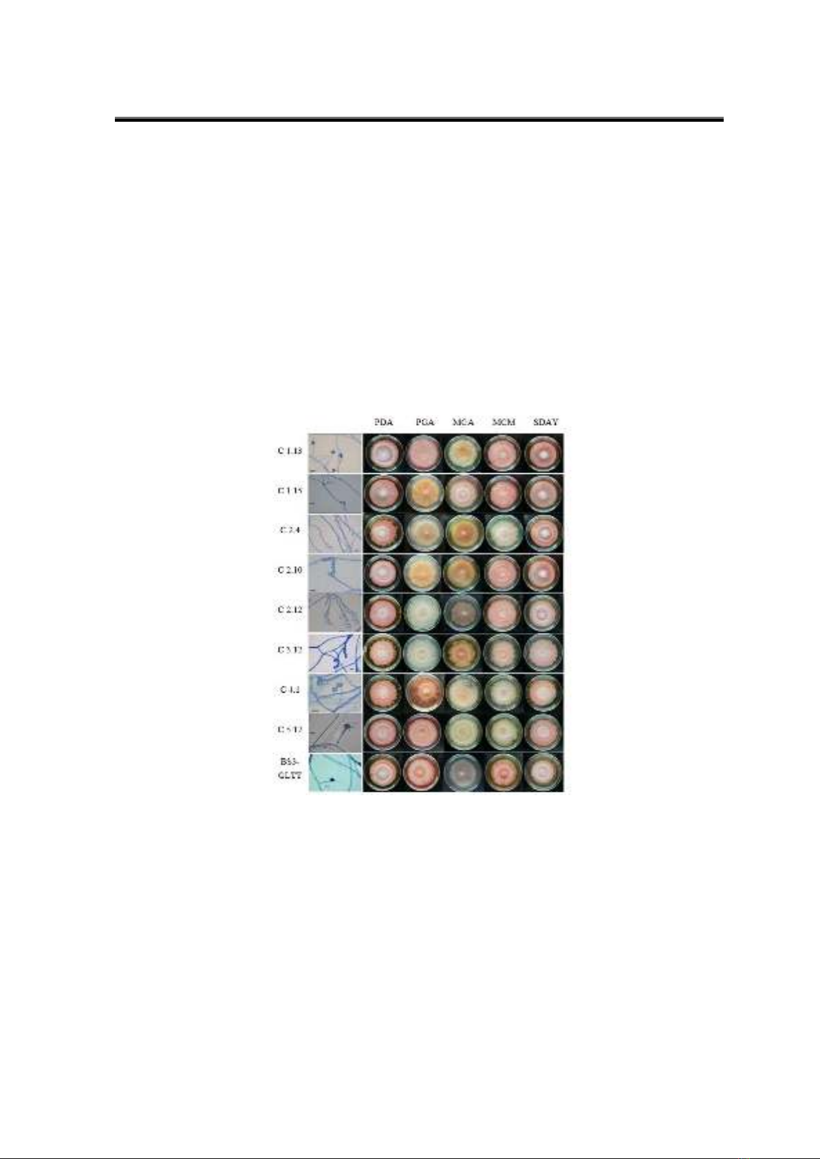
monacolin K produced by M. purpureus strain. The pigmentations were varied when cultured on
various media such as PDA, PGA, MGA, MCM, and SDAY media. MGA (61.12 1,996.20 AU/g)
and PGA (8.78 507.26 AU/g) displayed the highest levels of red pigment, while SDAY (10.55–
31.79 AU/g) showed the lowest levels. Morphological examination revealed typical features of
Monascus sp., including spherical or oval shapes and spore chains of two to four spores. Qualitative
analysis of chromatographic plates TLC revealed consistent bands of Monacolin K standard across
most strains, indicating their potential for producing this compound, with exceptions like strain BS3-
GLTT. Notably, strains C3.12, C5.17, C4.1, C1.15, and BS3-GLTT exhibited either faint or absent
spots corresponding to citrinin, suggesting their potential as citrinin-free strains. Furthermore, the
C5.17 strain identified as Monascus purpureus, under specific conditions, yielded a monacolin K
concentration of 292,32 ppm, with no detectable citrinin by LC-MS/MS analysis, highlighting its
suitability for monacolin K production.
Keywords: citrinin; monacolin K; Monascus purpureus; LC-MS/MS; Red yeast rice
1. Introduction
Monascus sp. belongs to the Monascaceae family, genus Monascus, which is one of
the species capable of producing natural bioactive pigments (Mussalbakri et al., 2017).
Cite this article as: Dao Nu Dieu Hong, Nguyen Thi Thuy Trang, Nguyen Thi Dung, Bui Le Kha Tu, Pham Thi
Thanh Tinh, Phan Thi Phuong Trang, & Ha Thi Loan (2024). Screening of Monascus purpureus strain with
monacolin K activity and citrinin-free characteristics for Red yeast rice production by LC-MS/MS analysis by
LC-MS/MS analysis. Ho Chi Minh City University of Education Journal of Science, 21(9), 1702-1712.
HCMUE Journal of Science
Vol. 21, No. 9 (2024): 1702-1712
1703
Pigments are formed through the polyketide biosynthetic pathway, in which polyketide
synthesis and fatty acid synthesis play essential roles (Rasheva et al., 1998). One of the
processed rice that brings economic and health value is red yeast rice (RYR) (Seenivasan et
al., 2008; Song et al., 2019). RYR is the product after fermenting rice with Monascus
purpureus fungus, which has many beneficial biological effects such as improving
cardiovascular circulation, reducing cholesterol to prevent metabolic disorders, and in
cosmetics for skin rejuvenation and digestive support (Fukami et al., 2021). Currently, RYR
is recognized as the leading natural therapy for high cholesterol individuals. The total
cholesterol level of the body in general, including bad LDL cholesterol and triglycerides, is
significantly reduced compared to the control group with patients using RYR (Cicero et al.,
2019). The reason cholesterol has been extensively researched is that the leading cause of
death worldwide is cardiovascular disease, accounting for 32.3%. There is an alarming
situation of lipid disorders worldwide, with 33.6 million people in the US having high lipid
levels, accounting for 15% of the adult population; in the UK, two-thirds of the population
have higher cholesterol levels than recommended; in Vietnam, 29% of adults have high lipid
levels, with the urban population accounting for 44.3%. Many complications occur, such as
48% of cases of stroke, 56% of cases of myocardial ischemia, and myocardial infarction due
to complications of atherosclerosis from lipid disorders. Monacolin K activity in Monascus
sp. is similar to statin drugs used in lipid treatment (Heber et al., 1999). Natural monacolins,
especially monacolin K, have anti-osteoporosis effects and inhibit cholesterol synthesis by
HMG-CoA enzyme. In 2007, Đức Mạnh investigated the pigment-producing and
lovastatin-producing capabilities of Monascus purpureus MD and Monascus purpureus
3403 strains, revealing the latter's ability to synthesise lovastatin hydroxy acid at 60 µg/g in
dried samples. Despite low citrinin production, its presence in both strains raised concerns
for their application in colouring agents or health food production.
2. Material and method
2.1. Microorganism and inoculum
The M. purpureus strain used in this study was obtained from rice from the HCM
Biotech collection. The cultured strain will be inoculated onto a PDA medium and then
incubated at room temperature (24-30°C) for 14 days. Spore suspension will be prepared by
adding distilled water to the PDA culture medium to obtain a spore density of 107 spores/mL.
2.2. Morphological identification
We perform a dilution series ranging from 10-2 to 10-5 using 1 gram of untreated and
fermented rice samples in physiological saline solution, followed by vortexing to
homogenise the samples. Then, pipette 100 ul onto Potato Dextrose Agar (PDA) and
Sabouraud Dextrose Agar with Yeast Extract (SDAY) plates for screening strains with red
pigment. To observe the morphological characteristics of fungal colonies, inoculate spots
onto Petri dishes containing PDA, Potato Glucose Agar (PGA), Malt Glucose Agar (MGA),
HCMUE Journal of Science
Dao Nu Dieu Hong et al.
1704
Malt Extract Agar (MEA), and SDAY media, and incubate at room temperature.
Successfully cultured strains will produce fungal hyphae. Use a moist chamber method for
microscopic observation. The growth of fungal colonies is observed under an automated
microscope (Darwesh et al., 2020; Carvalho et al., 2005).
2.3. Method for determining the content of red pigment
Fungal biomass is obtained and dried for 24 hours at 50°C, then finely ground.
Consequently, 70% ethanol is added to the sample at a ratio of 1:20, and the mixture is
agitated at 150 rpm for 2 hours at room temperature. Afterwards, the sample is centrifuged
at 7000 rpm for 10 minutes to collect the supernatant, then diluted with 70% ethanol. The
absorption at 505 nm (red pigment) is measured with the sample, using 70% ethanol as the
blank control. The total absorbed pigment (AU/g) is calculated using the formula: AU/g =
Abs x (10/0.5) x df, where Abs represents the sample absorbance and df denotes the dilution
factor (Passos et al., 2019).
2.4. Selection of Monascus strains capable of producing monacolin K using the Thin
Layer Chromatography (TLC) method
The TLC method was employed for the rapid screening of citrinin and monacolin K
using thin-layer chromatography (TLC), with silica gel thin-layer chromatography plates as
the stationary phase and chloroform: methanol (90:10, v/v) as the mobile phase. Standard
citrinin and monacolin K were simultaneously analysed. Monascus strains were identified
according to the method of Babitha et al. (2007). Fungal biomass obtained after freeze-drying
for 24 hours at 50°C was finely ground. 70% ethanol was added to the sample at a ratio of 1:20,
and the mixture was shaken at 150 rpm at room temperature for 2 hours. Subsequently, the
mixture was centrifuged at 7000 rpm for 10 minutes to collect the supernatant, which was then
diluted with 70% ethanol. Absorbance at 505 nm (red pigment) was measured with 70% ethanol
as the negative control (Seenivasana et al., 2015).
2.5. Mass spectrometry condition LC-MS/MS
Weigh 0.5 grams of dry sample into a 50 mL falcon tube. Monascus fungal spores are
extracted with 50 mL of Acetonitrile (ACN) in an ultrasonic bath at 30°C for 30 minutes.
The resulting mixture is then heated in a water bath at 60°C for 1 hour and centrifuged at
3500 rpm for 10 minutes. Add 0.01g of adsorbent material (MgSO4: Na-Acetate, adjusted)
to 1 mL of the upper phase, followed by centrifugation at 10000 rpm for 5 minutes. The
supernatant is then filtered through a 0.2 µm filter membrane. A volume of 5µL of the
filtered solution is injected into the LC-MS/MS system. Transfer the sample into a vial and
analyse it using LC-MS/MS Waters. Standards are determined with known concentrations
and peak areas. The chromatographic conditions include a C18 column, a 0.3 mL/minute
flow rate, and a sample injection volume of 5 µL. Mobile phase A consists of 10 mM
ammonium formate in H2O (0.1% HCOOH); B1: MeOH (0.1% HCOOH) for citrinin and
monacolin K (Svoboda et al., 2017; Fogarty et al., 2018). Monacolin K, ESI +, primary ion
HCMUE Journal of Science
Vol. 21, No. 9 (2024): 1702-1712
1705
405 (m/z), daughter ion 303 and 199 (m/z). Citrinin, ESI -, primary 281 (m/z), daughter ion
205 and 249 (m/z).
2.6. Method for DNA sequencing and identification
Total DNA is extracted by adding fungal fibres into a 1.5 mL Eppendorf tube
containing 500 µL of a phenol-chloroform-isoamyl alcohol and STES buffer mixture. The
mixture is centrifuged at 10000 rpm for 5 minutes to collect the supernatant. The supernatant
is transferred to a new 1.5 mL Eppendorf tube containing 300 µL of isopropanol and
CH3COONa. After centrifugation at 10000 rpm for 5 minutes, the supernatant is discarded,
and the precipitate is collected. The precipitate is washed with ethanol and dried. 50 µL of
TE buffer is added to dissolve the DNA. For the PCR reaction, the reaction mixture contains
2.5 µL of 10X.
Add 2.5 µL of dNTPs, 0.5 µL of 10µM ITS5 forward primer, 0.5 µL of 10µM reverse
primer LR7, 0.25 µL of Taq DNA polymerase, 2 µL of DNA template, and ddH2O to a total
volume of 25 µL. The PCR thermal cycling conditions are as follows: 95°C for 5 minutes,
followed by 35 cycles of 95°C for 1 minute, 56°C for 45 seconds, 72°C for 1 minute, and a
final extension step at 72°C for 7 minutes. The PCR products are then electrophoresed on a
1.5% agarose gel for 20 minutes and purified using the GeneJET PCR Purification Kit. For
PCR sequencing, the reaction mixture includes 1 µL of BigDye Terminator 3.1 Ready
Reaction Mix, 1 µL of primer, 10 - 100 ng of purified PCR product, and ddH2O to a total
volume of 10 µL. The sequencing products are cleaned using Sephadex G-50, sequenced
with an ABI 3500 Genetic Analyzer, and analysed using the BLAST program from NCBI.
Phylogenetic trees are constructed using MEGA-10 software, and species identification is
performed using the Neighbor-Joining method with a bootstrap index of 1000 (Joaquin et
al., 2016; Suharna et al., 2005).
3. Results and discussion
3.1. Morphological identification
To observe the morphological characteristics of fungal colonies, inoculate spots onto
Petri dishes containing PDA, Potato Glucose Agar (PGA), Malt Glucose Agar (MGA), Malt
Extract Agar (MEA), and SDAY media, and incubate at room temperature. A description of
fungal strains is presented in Figure 1. After 21 days of cultivation on different media,
including PDA, PGA, MGA, MCM, and SDAY, the isolated fungal strains generally
exhibited robust growth, with most mycelia densely covering the entire Petri dish surface.
Pigmentation was observed on the agar plates' upper and lower surfaces, accompanied by
distinct morphologies. The strains exhibited characteristic pigments of Monascus sp.,
predominantly red, pink, orange, yellow, and white. Specifically, fungal colonies grown on
PDA, MCM, and SDAY media mainly displayed pink pigmentation, while those on PGA and
MGA media showed deeper shades of pink, red, or orange. The morphology of fungal colonies
HCMUE Journal of Science
Dao Nu Dieu Hong et al.
1706
appeared round, with mycelia arranged in a flat or slightly raised manner at the centre, often
forming concentric rings with smooth or undulating edges.
The identified fungal strains were characterised morphologically and described in
detail in Figure 1. Morphological characteristics, including spore capsule size, morphology
of asexual spores, and the number of spores in chains, were examined. Overall, the strains
exhibited spherical or oval shapes, occurring singly or in chains of 2 to 4 spores, indicating
characteristics typical of Monascus sp. In the five different cultivation media, dextrose was
the carbon source in PDA, MCM, and SDAY, while glucose was used in PGA and MGA.
Therefore, it can be concluded that the carbon source in the cultivation media
influences pigment variation and growth in the examined strains. Studies by Kujumdzieva
et al. (1998) and Lee et al. (2001) also concluded that glucose is preferred as a substrate for
pigment and biomass production in Monascus sp. over other carbon sources.
Figure 1. Morphological characteristics of Monascus sp. on different media, including
PDA, PGA, MGA, MCM, and SDAY
3.2. Method for determining the content of red pigment
From Table 1, it can be observed that there are differences in the red pigment content
among the cultured strains and between different media within the same strain. When cultured
on various media such as PDA, MCM, MGA, SDAY and PGA media, the pigmentations were
varied PDA (11.37-41.49 AU/g), MCM (14.51-129.99 AU/g), MGA (61.12-1,996.20 AU/g),
SDAY (10.55-31.79 AU/g) and PGA (8.78-507.26 AU/g).