
RESEARC H Open Access
Papillomavirus pseudovirions packaged with the
L2 gene induce cross-neutralizing antibodies
Nicolas Combelas
1
, Emilie Saussereau
1
, Maxime JJ Fleury
1
, Tatiana Ribeiro
1,3
, Julien Gaitan
1
,
Diego F Duarte-Forero
1,2
, Pierre Coursaget
1*
, Antoine Touzé
1
Abstract
Background: Current vaccines against HPVs are constituted of L1 protein self-assembled into virus-like particles
(VLPs) and they have been shown to protect against natural HPV16 and HPV18 infections and associated lesions. In
addition, limited cross-protection has been observed against closely related types. Immunization with L2 protein in
animal models has been shown to provide cross-protection against distant papillomavirus types, suggesting that
the L2 protein contains cross-neutralizing epitopes. However, vaccination with L2 protein or L2 peptides does not
induce high titers of anti-L2 antibodies. In order to develop a vaccine with the potential to protect against other
high-risk HPV types, we have produced HPV58 pseudovirions encoding the HPV31 L2 protein and compared their
capacity to induce cross-neutralizing antibodies with that of HPV L1 and HPV L1/L2 VLPs.
Methods: The titers of neutralizing antibodies against HPV16, HPV18, HPV31 and HPV58 induced in Balb/c mice
were compared after immunization with L2-containing vaccines.
Results: Low titers of cross-neutralizing antibodies were detected in mice when immunized with L1/L2 VLPs, and
the highest levels of cross-neutralizing antibodies were observed in mice immunized with HPV 58 L1/L2
pseudovirions encoding the HPV 31 L2 protein.
Conclusions: The results obtained indicate that high levels of cross-neutralizing antibodies are only observed after
immunization with pseudovirions encoding the L2 protein. HPV pseudovirions thus represent a possible new
strategy for the generation of a broad-spectrum vaccine to protect against high-risk HPVs and associated neoplasia.
Background
The fact that cervical cancer is the second most com-
mon cause of cancer deaths in women worldwide [1],
and that virtually all cervical cancers are etiologically
linked with infection by “high risk”human papilloma-
virus (HPV) [2], has encouraged the development of
prophylactic vaccines to prevent genital infection. Fif-
teen of the HPV types infecting the mucosal epithelium
cause cervical cancer, HPV16 and 18 being the most
prevalent types detected in cervical carcinoma [1]. Papil-
lomaviruses are small non-enveloped DNA viruses and
their icosahedral capsid is constituted of L1 and L2 pro-
teins, which encapsidate a closed circular, double-
stranded DNA of about 8 kbp. The viral capsid of 50-60
nm in diameter contains 72 pentamers of L1 major
protein and 12 to 72 copies of L2 minor capsid protein
[3,4].
Immunization with L1 self-assembled into virus-like
particles (VLPs) induces high titers of neutralizing anti-
bodies and confers protection in animals against homo-
logous experimental infection [5,6]. It has also been
shown that protection is mediated by neutralizing anti-
bodies directed against conformational epitopes. These
results have led to the industrial development of vac-
cines against genital HPV types. Pre-clinical studies have
shown that the neutralizing antibodies induced by L1
VLPs are predominantly type-specific [7,8]. However,
low levels of cross-neutralization have been reported
between HPV6 and 11 and HPV 16 and 31 [9-12] and
higher levels between HPV18 and 45 [13]. Clinical trials
have shown that the immune response is associated
with protection against HPV16 and HPV18 infections
and associated lesions [14,15].
* Correspondence: coursaget@univ-tours.fr
1
Inserm U618 “Protéases et vectorisation pulmonaires”, Tours; University
François Rabelais, Tours, France and IFR 136 “Agents Transmissibles et
Infectiologie”, Tours, France
Combelas et al.Journal of Translational Medicine 2010, 8:28
http://www.translational-medicine.com/content/8/1/28
© 2010 Combelas et al; licensee BioMed Central Ltd. This is an Open Access article distributed under the terms of the Creative
Commons Attribution License (http://creativecommons.org/licenses/by/2.0), which permits unrestricted use, distribution, and
reproduction in any medium, provided the original work is properly cited.

Current HPV vaccines containing L1 VLPs promote
the generation of a strong, mainly type-specific, neutra-
lizing antibody response. Clinical trials with HPV16 and
18 vaccines have also revealed that cross-protection
against HPV types is limited to closely related types.
Protection against HPV31 lesions was clearly established
for both vaccines and protection against HPV45 lesions
for only one vaccine [15,16]. As the licensed HPV vac-
cines target only two of the 15 high-risk HPV, one strat-
egy is to combine many types of L1 VLPs to prevent
infection against multiple high-risktypes.Toaddress
this issue, a multivalent VLP vaccine is currently under
clinical trial [17]. However, the inclusion of numerous
VLP types complicates vaccine development and would
increase the risk of antigenic competition that could
result in lower protective efficacy and/or affect long last-
ing protection against certain HPV types.
The minor capsid L2 protein has emerged as another
candidate prophylactic vaccine, since immunization with
L2 in animal models of papillomavirus infection induces
cross-neutralizing antibodies that are able to mediate
broader protection than L1 VLPs [7,18-24]. Preclinical
and clinical findings [25-27] have confirmed that L2 vac-
cines induce broad-spectrum cross-neutralizing antibo-
dies. However, L2 protein and L2 peptides are less
immunogenic than L1 VLPs, and it has been reported
that the incorporation of the L2 protein into L1 VLPs
does not increase the anti-L2 response due to the
immunodominance of L1 [23]. This suggests that new
vaccine strategies have to be investigated if such an L2-
based vaccine is to be effective.
Although most investigations concerning VLPs have
dealt with vaccine development, it has also been demon-
strated that HPV VLPs can be used to generate pseudo-
virions (PsV) by packaging unrelated plasmids within
the VLPs, and they thus represent a valuable gene deliv-
ery system that could be used to induce an immune
response against the packaged de novo synthesized
transgene product [28,29].
The aims of this study were to investigate the possibi-
lity of generating an HPV vaccine by packaging a plas-
mid encoding the HPV 31 L2 protein within HPV58 L1/
L2 PsV (PsV58-31L2). The L2-pseudovirion vaccination
strategy aims to induce high-titers of conformation-
dependent antibodies to L1 similar to those observed
with monovalent HPV VLP L1 vaccines and to induce
de novo L2 expression for augmented immunogenicity
to L2 protein in order to cross-neutralize multiple HPV
types [30].
Materials and methods
Antibodies and Cell lines
CamVir-1 monoclonal antibody (MAb) (BD Biosciences,
Le Pont de Claix, France) binds to a linear epitope
which has been mapped between amino acids 203 to
209 of the HPV-16 L1 protein [31]. Rabbit anti-HPV16
L2 immune serum was kindly provided by Richard
Roden. COS-7 cells (African green monkey kidney cells,
ATCC CRL-1651) were grown in Dulbecco’s modified
Eagle’s Medium (Invitrogen, Illkirch, France) supple-
mented with 10% heat-inactivated fetal calf serum
(FCS), 100 IU/ml penicillin, and 100 μg/ml streptomycin
and 1 mM sodium pyruvate. The 293FT cell line (Invi-
trogen) is a fast growing variant of the 293 cell line that
stably expresses SV40 TAg and the neomycin resistance
gene from pCMVPORT6AT.neo plasmid. 293FT cells
weregrowninDulbecco’s modified Eagle’sMedium,
supplemented as above, plus 1% non-essential amino
acids and 500 μg/ml G418 (Invitrogen). Cell lines were
grown at 37°C in a humidified atmosphere with 5% CO
2
.
Production of HPV VLP vaccines
HPV31L1andHPV31L1/L2VLPswereproducedand
purified from Sf21 insect cells infected with recombinant
baculoviruses encoding both L1 and L2 proteins as pre-
viously described [32,33]. HPV58 L1/L2 PsV were
obtained using a cellular system with codon-modified
HPV capsid genes [34]. Briefly, HPV 58 L1 and L2
genes were designed to contain the most frequently
used codons found in highly expressed genes in Homo
sapiens (FN178626 and FN178627, respectively). L1 and
L2 genes were cloned into the mammalian bicistronic
expression vector, pIRES (BDBiosciences, Clontech).
The HPV58 L1 gene was cloned between the NheIand
EcoRI restriction sites of MCS A downstream from the
CMV IE promoter. The HPV58 L2 gene was subse-
quently cloned between the XbaIandNotI restriction
sitesofMCSBofpIRES-HPV58L1togeneratepIRES-
HPV58 L1/L2 plasmids of 9.1 kbp. Plasmids of this size
were previously shown not to be packaged when form-
ing PsV in a cellular system [35]. DNA plasmid pIRES
L2 ΔNLS (7.4 kbp) used for the production of PsV was
prepared by classical phenol/chloroform DNA prepara-
tion. This plasmid contains the DNA sequence encoding
amino acids 12 to 442 of the HPV31 L2 between the
XbaIandNotI restriction sites. This sequence was PCR-
amplified from a plasmid containing a Homo sapiens
codon-adapted full length HPV31 L2 gene [36]. This
deleted mutant of the L2 gene was selected to reduce
the amount of HPV31 L2 protein exported to the
nucleus and to prevent its incorporation into the
HPV58 PsV structure. For the generation of HPV58 PsV
in 293FT cells, cells were transfected with 0.5 μgDNA,
0.25 μg pIRES HPV31 L2 ΔNLS or 0.25 μg pCMV-GFP,
0.25 μg of pIRES-HPV58 L1/L2 and 1 μlFugene6
(Roche) per cm
2
of the culture area. Cells were har-
vested two days post-transfection, and PsV were purified
as previously described [36] and stored at -80°C until
Combelas et al.Journal of Translational Medicine 2010, 8:28
http://www.translational-medicine.com/content/8/1/28
Page 2 of 9

use. Pseudovirions were quantified by Western blotting
usingCamVir-1antibodybycomparisonwithknown
concentrations of HPV58 L1/L2 VLPs. Pseudovirions
containing HEV ORF2
108-660
(PsV31-HEV) were pro-
duced using the same procedure as described for HPV
58 PsV using previously described pIRES-HPV31 L1/L2
[36] and pcDNA3 HEV ORF2
108-660
, plasmids [29].
Immunization protocol
Six-week-old female BALB/c mice (CERJ Janvier, Le
Genest St Isle, France) were intramuscularly immunized
with the different vaccine preparations. Mice from
group 1 received saline, mice from groups 2 and 3
received 1 and 10 μg of pIRES-HPV31 L2ΔNLS plasmid
(DNA L2), respectively (Table 1). Mice from groups 4
and 5 received HPV31 L1 and HPV31 L1/L2 VLPs (31
L1L2 VLPs), respectively. Mice from group 6 received
10 μg of HPV31 L1/L2 PsV containing HEV ORF2
108-
660
expression plasmid (PsV31-HEV) [29]. Mice from
groups 7 and 8 received HPV58 L1/L2 PsV containing
GFP expression plasmid (PsV58-GFP) and HPV58 PsV
packaged with HPV31L2ΔNLS plasmid (PsV58-31L2),
respectively. In order to eliminate variations in the pseu-
dovirion DNA content, the preparations used were from
the same batch. Mice were immunized at days 0, 7 and
21. Two weeks after the last injection, serum samples
were collected and stored at -20°C. All animal proce-
dures were performed according to approved protocols
and in accordance with the recommendations for the
proper use and care of laboratory animals, and experi-
ments were approved by the regional animal ethics
commmittee (CREEA Centre-Limousin).
Expression of L2SA and detection of anti- L2 antibodies
L2 protein was expressed in insect cells as a fusion pro-
tein. In order to purify the L2 protein from insect cells,
the Streptactin (SA) coding sequence [37] including
upstream (BamHI and SalI) and downstream (HindIII)
restriction sites was synthesized by Geneart (Regensburg,
Germany) using an adapted codon usage for expression
in Spodoptera frugiperda.TheSAsequencewascloned
between SalIandHindIII sites of the pFastBacDual
expression vector (Invitrogen) in order to obtain the
pFastBacDual SA plasmid. The HPV16 L2 ORF was then
fused at the 5’end of the SA ORF. For this purpose, the
HPV16 L2ΔNLS ORF (amino acids 12 to 442) was ampli-
fied by PCR from a plasmid containing a Homo sapiens
codon adapted version of the wild type L2 gene
(FN297862) using HPV16 L2 F (CCGGATCCGCCAC-
CATGGCCAGCGCCACCCAGCTG) and HPV16 L2ΔR
(GTCGACCATGTAGTAGCTGGGGTGCAGGATG). A
forward primer was designed to introduce a BamHI site,
and a Kozak sequence upstream from the start codon
and the reverse primer contained a SalI restriction site.
The PCR product was then cloned by TA cloning into
the pCR2.1 vector (Invitrogen). Both pCR2.1-16 L2ΔNLS
and pFastBacDual SA plasmids were submitted to restric-
tion with BamHI and SalI, and the L2 gene was fused to
the Streptactin gene in order to generate the pFastBac-
Dual-16 L2ΔNLS (pFBD-L2SA).
A recombinant baculovirus encoding L2SA was gener-
ated using the Bac-to-Bac system (Invitrogen) according
to the manufacturer’s recommendations. Sf21 insect cells
weregrownat27°CinSF900IImediumsupplemented
with penicillin, streptomycin and amphotericin B (Invitro-
gen). Cells were infected at a m.o.i. of ten and grown for
four days. Cells were scraped off, centrifuged at 300 × g
and then resuspended in PBS 1× containing 0.5% Nonidet
P40 and an anti-protease cocktail (Roche, Meylan, France)
and incubated on ice for 30 min. The lysate was centri-
fuged at 4°C for 10 min at 12,000 × g. The pellet, repre-
senting the nuclear fraction, was subjected to sonification
(3 × 15 s bursts, Vibracell, Fischer Scientific, France).
L2SA protein was purified by affinity on immobilized imi-
nobiotin according to the manufacturer’s instructions
(Pierce, Ozyme, Montigny le Bretonneux, France).
Table 1 Composition of the vaccines preparations used and anti-HPV16, HPV18, HPV31 and HPV58 neutralizing
antibody titers in mice immunized with the different vaccines.
Group
N°
Proteins Gene Neutralizing titers
Name L1 L2 HPV16 HPV18 HPV31 HPV58
1 Saline - - - - - - -
2 DNA L2 (1 μg) - - HPV31 L2Δ----
3 DNA L2 (10 μg) - - HPV31 L2Δ----
4 31 L1 VLPs 31 - - - - 2,800 -
5 31 L1L2 VLPs 31 31 - - - 3,400 65
6 PsV31-HEV 31 31 HEV ORF2 - - 5,198 54
7 PsV58-GFP 58 58 GFP - - 50 4,650
8 PsV58-31L2 58 58 HPV31 L2Δ60 400 733 5,382
Cross-neutralizing titers are in bold-faced characters.
Combelas et al.Journal of Translational Medicine 2010, 8:28
http://www.translational-medicine.com/content/8/1/28
Page 3 of 9

Two hundred nanograms of L2SA were distributed in
half of the wells of a 96-well plate (Maxisorp, Nunc,
ATGC, Marne-la-Vallée, France) and incubated at 4°C
overnight. After two washes with PBS-Tween (0.1%), the
wells were saturated with PBS supplemented with 1%
FCS for 1 h at 37°C. Duplicate wells (one test and one
control) were incubated with two-fold dilutions (starting
at 1:25) of mice sera in dilution buffer (PBS 5×, 1%
Tween, 10% FCS) for 1 h at 45°C. After four washes,
peroxidase-conjugated goat anti-mouse IgG (Fc-specific)
(Sigma Aldrich) diluted 1:1,000 in PBS - Tween (1%) -
FCS (10%) was added to the wells and incubated for 1 h
at 45°C. After four washes, 0.4 mg/ml o-phenylene-dia-
mine and 0.03% hydrogen peroxide in 25 mM sodium
citrate and 50 mM Na
2
HPO
4
were added. After 30 min,
the reaction was stopped with H
2
SO
4
4N and optical
density (OD) was read at 492 nm. For data analysis, OD
values obtained in the absence of L2SA were subtracted
from OD values of test antigens. A result was consid-
ered positive when the difference in OD between test
and control wells was greater than 0.2. Individual titers
represented the reciprocal of the last dilution giving an
OD difference greater than 0.2. Values for individual
mice were the means of duplicates. Geometric mean
titers (GMTs) were calculated for each group. Animals
without detectable antibody titers (< 25) were assigned a
titer of 1 for calculation of GMTs.
Detection of anti-HPV neutralizing antibodies
Neutralization assays were performed by inhibition of
pseudoinfection of COS-7 cells by pseudovirions con-
taining the pGL3-luc plasmid (Promega, Charbonnières-
les-Bains, France). HPV16and18PsVwereproduced
by the previously published disassembly-reassembly
method [38] with some modifications [39]. L1/L2 VLPs
(100 μg) were incubated in 50 mM Tris-HCl buffer (pH
7.5) containing 20 mM DTT and 1 mM EGTA for
30 min at room temperature. At this stage, pGL3-luc
(10 μg) was added to the disrupted VLPs. The prepara-
tion was then diluted with increasing concentrations of
CaCl
2
(up to a final concentration of 5 mM) in the pre-
sence of 10 nM ZnCl
2
. Pseudovirions were then dialyzed
overnight against PBS 1× and stored at 4°C before use.
HPV31 and 58 PsV were obtained using a cellular sys-
tem with codon-modified HPV capsid genes and pGL3-
luc plasmid as described above for HPV58 pseudovirons
encoding L2.
COS-7 cells (10
4
/well) were seeded in 96-well plates
(TPP, ATGC). After 24 h incubation at 37°C, cells were
washed twice before addition of pseudovirion/sera mix-
ture. The amount of pseudovirions was adjusted to
obtain a relative luciferase activity of 0.2 RLU (Relative
Light Unit) (final dilutions in test wells: 1:500 for
HPV16, 1:50 for HPV 18, 1:800 for HPV31, and
1:10,000 for HPV 58). Mock transduced COS-7 cells
exhibit 0.00001 RLU (Luminoskan Ascent, Thermo
scientific, Courtaboeuf, France). Fifty μl of diluted pseu-
dovirions were mixed with 50 μl of mice sera diluted by
two-fold dilution in incomplete DMEM from 1:12.5 to
1:25,600 in order to obtain final serum dilutions of 1:25
to 1:51,200. After 1 h incubation at 37°C, the mixture
was added to the wells and plates were incubated 3 h at
37°C. Then 100 μlofcompleteDMEMwereadded,and
the luciferase gene expression was measured after incu-
bation for 48 h at 37°C (Firefly luciferase 1-step assay
kit, Fluoprobes, Interchim,Montluçon,France).The
results were expressed as the percentage of inhibition of
luciferase activity [36]. The data presented are the
means of 2 to 3 determinations performed in duplicate.
Neutralization titers were defined as the reciprocal of
the highest dilution of mice sera that induced at least
50% reduction in luciferase activity. Geometric mean
titers were calculated for each group. Animals without
detectable neutralizing antibodies were assigned a titer
of 1 for the calculation of GMTs.
Statistical analysis
Geometric mean titers were compared to evaluate
ELISA and neutralizing responses. Group results (10
animals per group) were compared by Student ttest
using XLStat software (Addinsoft, Paris, France).
Results
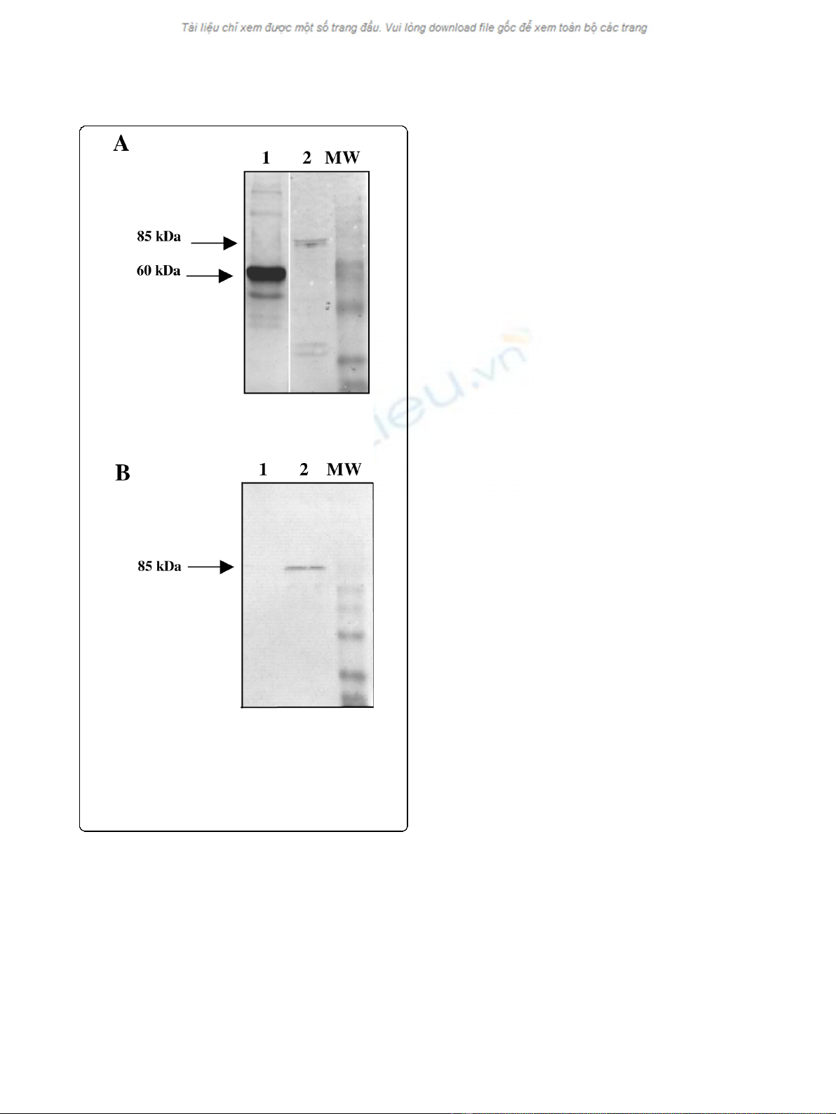
Production of HPV58 pseudovirions
In order to generate HPV58 PsV, 293FT cells were
transfected simultaneously with the pIRES-HPV58 L1/
L2 plasmid encoding the structural proteins of HPV58
and the pGL3 plasmid encoding luciferase. Three days
post-transfection, the nuclear fraction of 293FT cells
was analysed by Western blotting. HPV58 L1 and L2
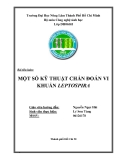
proteins were efficiently expressed (Fig. 1A). Then the
ability of PsV58-31L2 to transduce the HPV31 L2 ΔNLS
gene was investigated by pseudo-infection of COS-7
cells. Western Blot analysis of L2 protein expression
indicated that L2 was detected two days after transduc-
tion (Fig. 1B). In order to rule out the possibility that L2
detected in COS-7 cells was due to the presence of the
HPV58 L2 contained in the pseudovirion structure,
COS-7 cells were transduced with similar PsV packaged
with the GFP gene. The presence of L2 was not evi-
denced in the latter condition (Fig. 1B).
After purification, samples of HPV58 PsV stock were
titered by measuring their end-point luciferase gene
transduction capacities on Cos-7 cells, and compared
with HPV31 PsV obtained in the same cellular system
and experimental conditions. Using endpoint titers with
a cut-off based on the background luminescence of
mock transduced Cos-7 cells, HPV 58 L1/L2 PsV were
Combelas et al.Journal of Translational Medicine 2010, 8:28
http://www.translational-medicine.com/content/8/1/28
Page 4 of 9
shown to be 20 times more efficient than HPV31 L1/L2
(data not shown). In view of this result, HPV58 L1/L2
PsV were selected to develop pseudovirion-based
immunization.
Anti-HPV16-L2 immune response in mice immunized with
heterologous VLPs and pseudovirions
Anti-HPV16 L2 antibodies were not detected in non-
immunized mice (group 1). Anti-L2 antibodies were not
detected in mice immunized with HPV31 L1 VLPs
(group 4), but were detected in all mice immunized
with the LIL2 VLPs (group 5), with a GMT of 1,100.
Anti-L2 antibodies were detected at similar levels in
mice immunized with control PsV (groups 6 and 7),
with GMTs of 855 and 1,212 (p = 0.459). By comparison
with these control pseudovirions, the anti-L2 GMT
(2,600) was higher in mice immunized with PsV58-31L2
(p = 0.001 and p = 0.101, respectively).
Induction of cross-neutralizing antibodies
Homologous HPV31 neutralizing antibodies were
detected in mice immunized with HPV31 L1 or HPV31
L1L2 VLPs and HPV31 HEV PsV (groups 4, 5 and 6),
with GMTs of 2,800 ± 2360, 3,400 ± 460 and 5,198 ±
900, respectively (GMT ± SEM). Low titers of HPV58
neutralizing antibodies were only observed in mice
receiving HPV31 L1L2 VLPs (group 5) and HPV31 PsV
containing the HEV ORF2 irrelevant gene (group 6). No
neutralizing antibodies against HPV16 and HPV18 were
detected in any of the mice from groups 4 to 6 receiving
HPV31 VLP vaccine preparations (Fig. 2).
High levels of homologous neutralizing antibodies
were detected in mice immunized with HPV58 PsV
(groups 7 and 8), with GMTs of 4,650 ± 980 and 5,382
± 2240, respectively. Low levels of neutralizing antibo-
dies to HPV31 (GMT = 50 ± 315) were detected in
mice immunized with PsV58-GFP, and a dramatic
increase in anti-HPV31 neutralizing antibodies (with a
GMT of 733 ± 190) was observed in mice immunized
with PsV58-31L2. Moreover, neutralizing antibodies
against HPV16 and HPV18 were only detected in mice
immunized with the PsV58-31L2, with GMTs of 60 and
400, respectively (Table 1).
Discussion
Since no differences in antibody titers or in protection
were observed in animal studies [40] when immuniza-
tion with L1 and L1/L2 VLPs were compared, it was
generally believed that there was insufficient reason to
introduce L2 protein into the composition of VLPs. In
addition, L2 protein assembled in L1 VLPs is weakly
immunogenic due to the immunodominance of L1 [23].
However, our findings suggested that even in the
absence of adjuvant cross-neutralizing antibodies could
be obtained by incorporating L2 in the composition of
the VLPs (group 5) or pseudovirions encoding irrelevant
genes (groups 6-7) compared to L1 VLPs (group 4),
despite the low anti-L2 immune response (GMT 855 to
1212). Anti-L2 antibody titers are generally several
orders of magnitude lower than the anti-L1 titers
obtained with VLP vaccines. However, even low anti-L2
antibody levels have been shown to be sufficient for pro-
tection [22,26], this being in part explained by the slow
uptake kinetics into cells reported for HPVs [41]. In
Figure 1 Western blot.A/Analysis by Western blotting of the
HPV58 pseudovirion capsid proteins. L1 was detected using the
CamVir-1 monoclonal anti body (lane 1). L2 was detected using
polyclonal anti-HPV16 L2 rabbit antiserum (lane 2). B/Detection of
L2 protein by Western blotting using polyclonal anti-HPV16 L2
rabbit antiserum.Cos-7 cells were transduced with HPV58
pseudovirions encoding GFP (lane 1) or with HPV58 pseudovirion
encoding HPV31 L2 (lane 2).
Combelas et al.Journal of Translational Medicine 2010, 8:28
http://www.translational-medicine.com/content/8/1/28
Page 5 of 9


%20--%3e%3cdefs%3e%3cstyle%3e%20.st0%20{%20fill:%20%23fff;%20}%20.st1%20{%20fill:%20%237800fa;%20}%20%3c/style%3e%3c/defs%3e%3cpath%20class='st1'%20d='M117.78,12.18H43.11c2.9,3.47,4.65,7.94,4.65,12.82,0,5.6-2.3,10.66-6.01,14.29h76.02l7.22-13.56-7.22-13.56Z'/%3e%3cg%3e%3cpath%20class='st0'%20d='M53.58,26.17h-.59v-1.46h.59v-4.96h2.83c1.78,0,2.67.94,2.67,2.82v5.76c0,1.87-.89,2.81-2.67,2.81h-2.83v-4.96ZM55.36,21.37v3.34h1.1v1.46h-1.1v3.34h1.01c.61,0,.91-.37.91-1.1v-5.93c0-.74-.3-1.1-.91-1.1h-1.01Z'/%3e%3cpath%20class='st0'%20d='M65.99,31.14h-1.8l-.31-2.07h-2.19l-.31,2.07h-1.64l1.82-11.39h2.62l1.82,11.39ZM65.28,18.04c-.25.46-.51.77-.75.94-.21.15-.47.22-.79.22-.26,0-.57-.07-.92-.22l-.38-.15c-.14-.05-.26-.07-.37-.07-.3,0-.53.18-.71.54l-.91-.68c.25-.46.51-.77.75-.94.21-.14.48-.21.79-.21.26,0,.57.07.92.21l.38.15c.14.05.26.07.37.07.3,0,.53-.18.71-.54l.91.68ZM61.91,27.52h1.73l-.87-5.76-.87,5.76Z'/%3e%3cpath%20class='st0'%20d='M74.53,26.89v1.52c0,1.91-.89,2.86-2.67,2.86s-2.67-.95-2.67-2.86v-5.93c0-1.91.89-2.86,2.67-2.86s2.67.95,2.67,2.86v1.11h-1.69v-1.22c0-.75-.31-1.12-.93-1.12s-.93.37-.93,1.12v6.15c0,.74.31,1.11.93,1.11s.93-.37.93-1.11v-1.63h1.69Z'/%3e%3cpath%20class='st0'%20d='M81.4,31.14h-1.8l-.31-2.07h-2.19l-.31,2.07h-1.64l1.82-11.39h2.62l1.82,11.39ZM75.9,19.2l1.52-1.91h1.71l1.51,1.91h-1.61l-.76-.95-.75.95h-1.61ZM77.32,27.52h1.73l-.87-5.76-.87,5.76ZM83.1,15.99l-1.76,1.91h-1.26l1.17-1.91h1.86Z'/%3e%3cpath%20class='st0'%20d='M84.86,19.75c1.78,0,2.67.94,2.67,2.82v1.48c0,1.87-.89,2.81-2.67,2.81h-.85v4.28h-1.79v-11.39h2.64ZM84.01,21.37v3.86h.85c.58,0,.87-.36.87-1.08v-1.71c0-.71-.29-1.07-.87-1.07h-.85Z'/%3e%3cpath%20class='st0'%20d='M93.51,19.75c1.78,0,2.67.94,2.67,2.82v1.48c0,1.87-.89,2.81-2.67,2.81h-.85v4.28h-1.79v-11.39h2.64ZM92.66,21.37v3.86h.85c.58,0,.87-.36.87-1.08v-1.71c0-.71-.29-1.07-.87-1.07h-.85Z'/%3e%3cpath%20class='st0'%20d='M98.8,31.14h-1.79v-11.39h1.79v4.88h2.03v-4.88h1.83v11.39h-1.83v-4.88h-2.03v4.88Z'/%3e%3cpath%20class='st0'%20d='M105.36,24.55h2.46v1.62h-2.46v3.34h3.09v1.63h-4.88v-11.39h4.88v1.63h-3.09v3.18ZM108.17,17.29l-1.76,1.91h-1.26l1.17-1.91h1.86Z'/%3e%3cpath%20class='st0'%20d='M112.2,19.75c1.78,0,2.67.94,2.67,2.82v1.48c0,1.87-.89,2.81-2.67,2.81h-.85v4.28h-1.79v-11.39h2.64ZM111.35,21.37v3.86h.85c.58,0,.87-.36.87-1.08v-1.71c0-.71-.29-1.07-.87-1.07h-.85Z'/%3e%3c/g%3e%3ccircle%20class='st1'%20cx='25'%20cy='25'%20r='20'/%3e%3cpath%20class='st0'%20d='M32.78,19.27c2.92,0,4.43,2.55,5.28,5.33l.71,2.17c.14.38-.33.75-.71.75h-5.61c.19-.33.24-.71.09-1.08l-.75-2.45c-.43-1.32-.99-2.64-1.79-3.77.75-.57,1.65-.94,2.78-.94h0ZM25,18.38c3.25,0,4.9,2.78,5.89,5.89l.76,2.45c.14.42-.33.8-.8.8h-11.69c-.42,0-.94-.38-.8-.8l.75-2.45c.99-3.11,2.64-5.89,5.89-5.89h0ZM25,11.35c1.74,0,3.11,1.37,3.11,3.11s-1.37,3.11-3.11,3.11-3.11-1.41-3.11-3.11,1.41-3.11,3.11-3.11h0ZM17.27,19.27c1.08,0,1.98.38,2.73.94-.8,1.13-1.37,2.45-1.74,3.77l-.8,2.45c-.14.38-.05.75.09,1.08h-5.56c-.42,0-.9-.38-.75-.75l.71-2.17c.9-2.78,2.41-5.33,5.33-5.33h0ZM17.27,12.91c1.51,0,2.78,1.27,2.78,2.83s-1.27,2.83-2.78,2.83-2.83-1.27-2.83-2.83,1.27-2.83,2.83-2.83h0ZM32.78,12.91c1.56,0,2.78,1.27,2.78,2.83s-1.23,2.83-2.78,2.83-2.83-1.27-2.83-2.83,1.27-2.83,2.83-2.83h0ZM27.07,28.56v.09c0,.57-.24,1.08-.61,1.46h0v.05c-.38.33-.9.57-1.46.57s-1.08-.24-1.46-.61h0c-.38-.38-.61-.9-.61-1.46v-.09h1.41v.09c0,.19.05.38.19.47v.05c.09.09.28.19.47.19s.38-.09.47-.19v-.05c.14-.09.24-.28.24-.47t-.05-.09h1.41ZM30.99,28.56v.09c0,1.65-.66,3.16-1.74,4.24-1.08,1.08-2.59,1.79-4.24,1.79s-3.16-.71-4.24-1.79l-.05-.05c-1.04-1.08-1.7-2.55-1.7-4.2v-.09h1.41v.09c0,1.27.47,2.4,1.27,3.25h.05c.85.85,1.98,1.37,3.25,1.37s2.4-.52,3.25-1.37c.85-.8,1.37-1.98,1.37-3.25v-.09h1.37ZM34.99,28.56v.09c0,2.78-1.13,5.28-2.92,7.07-1.79,1.79-4.29,2.92-7.07,2.92s-5.23-1.13-7.07-2.92c-1.79-1.79-2.92-4.29-2.92-7.07v-.09h1.41v.09c0,2.4.94,4.53,2.5,6.08,1.56,1.56,3.72,2.5,6.08,2.5s4.52-.94,6.08-2.5c1.56-1.56,2.5-3.68,2.5-6.08v-.09h1.41Z'/%3e%3c/svg%3e)