
RESEARC H Open Access
Development of targeted therapy for bladder
cancer mediated by a double promoter plasmid
expressing diphtheria toxin under the control of
H19 and IGF2-P4 regulatory sequences
Doron Amit
*
, Abraham Hochberg
Abstract
Background: The human IGF2-P4 and H19 promoters are highly active in a variety of human cancers (including
bladder cancer), while existing at a nearly undetectable level in the surrounding normal tissue.
Single promoter vectors expressing diphtheria toxin A-fragment (DTA) under the control regulation of IGF2-P4 or
H19 regulatory sequences (IGF2-P4-DTA and H19-DTA) were previously successfully used in cell lines, animal mod-
els and recently in human patients with superficial cell carcinoma of the bladder (treated with H19-DTA). However
this targeted medicine approach could be limited, as not all cancer patients express high levels of H19. Hence, a
double promoter DTA-expressing vector was created, carrying on a single construct two separate genes expressing
the diphtheria toxin A-fragment (DTA), from two different regulatory sequences, selected from the cancer-specific
promoters H19 and IGF2-P4.
Methods: H19 and IGF2-P4 gene expression was tested in samples of Transitional Cell Carcinoma (TCC) of the
bladder by in-situ hybridization (ISH) and by quantitative Real-Time PCR (qRT-PCR). The therapeutic potential of the
double promoter toxin vector H19-DTA-IGF2-P4-DTA was tested in TCC cell lines and in heterotopic and orthotopic
animal models of bladder cancer.
Results: Nearly 100% of TCC patients highly expressed IGF2-P4 and H19, as determined by ISH and by qRT-PCR.
The double promoter vector exhibited superior tumor growth inhibition activity compared to the single promoter
expression vectors, in cell lines and in heterotopic and orthotopic bladder tumors.
Conclusions: Our findings show that bladder tumors may be successfully treated by intravesical instillation of the
double promoter vector H19-DTA-P4-DTA.
Overall, the double promoter vector exhibited enhanced anti-cancer activity relative to single promoter expression
vectors carrying either gene alone.
Introduction
Bladder cancer is the fourth most commonly diagnosed
malignancy in men and the ninth most commonly diag-
nosed malignancy in women, (NCI annual report 2009).
Urinary bladder neoplasm can be grouped into three
different categories: Superficial, invasive and metastatic.
At presentation, 75% of the tumors are superficial, 20%
are invasive and up to 5% have de novo metastasis. The
wall of the bladder is lined with cells called transitional
cells. More than 90% of urothelial cancers in the bladder
are transitional cell carcinomas (TCC). Other important
histologic types include squamous cell carcinoma and
adenocarcinoma [1].
At presentation, tumors are usually limited to the
bladder mucosa (Ta) or submucosa (T1). These tumors
can be removed by transurethral resection (TUR), but
tend to recur in 50-70% of the patients. Measures to
decrease this high recurrence rate include intravesical
chemotherapy and immunotherapy (BCG - Bacillus
* Correspondence: dyamit@gmail.com
The Hebrew University of Jerusalem, Biological Chemistry, Jerusalem 91904,
Israel
Amit and Hochberg Journal of Translational Medicine 2010, 8:134
http://www.translational-medicine.com/content/8/1/134
© 2010 Amit and Hochberg; licensee BioMed Central Ltd. This is an Open Access article distributed under the terms of the Creative
Commons Attribution License (http://creativecommons.org/licenses/by/2.0), which permits unrestricted use, distribution, and
reproduction in any medium, provided the original work is properly cited.

Calmet-Guerin). These treatments decrease the recur-
rence rate, but are associated with side effects and
frequent failures [1].
The target population of this study is patients with
superficial bladder cancer refractory to conventional
therapies. Conventional therapies have focused on mass
cell killing without specific targeting and often cause
damaging and severe side effects to normal tissues. The
development of targeted therapeutic strategies based on
human cancer gene therapy is an attractive approach.
Based on early studies of our group and others, the
transcriptional regulatory sequences of the H19 and IGF2
genes emerged as candidates for cancer targeted therapy.
H19 and IGF2 (the human P3 and P4 promoters) are
onco-fetal genes and are oncogenes [2-4], expressed in
the fetus and in a broad spectrum of tumors, but rarely
in normal adult tissues [5-7]. H19 is a paternally-
imprinted, oncofetal gene that encodes a RNA (with no
protein product) acting as a “riboregulator”[8], which is
expressed at substantial levels in embryonic tissues, in
different human tumor types, and marginally or not
expressed in the corresponding tissues of the adult [6,9].
The 67-aa IGF2 is a member of the insulin like growth
factor family that is involved in cell proliferation and dif-
ferentiation [10]. The human IGF2 gene contains 9 exons
(E1-9) and 8 introns [10,11], and is transcribed from 4
different promoters (P1-P4) producing 4 different tran-
scripts [11-13]. All four transcripts share a common cod-
ing region and a common 3.9 kb 3-UTR, but variable 5-
UTRs [11]. IGF2 is an imprinted gene that is almost
exclusively expressed from the paternal allele [14-16].
The P3 and P4 promoters are the major IGF2 promoters
during embryogenesis and tumor development, while P1
is exclusively active in adult liver tissue and P2 activity is
rarely detected in adult human tissue [10]. Increased
expression of IGF2 as a result of the loss of its imprinting
is frequently seen in a variety of human tumors [16-18].
In addition, abnormal signal transduction and/or promo-
ter activation was reported as a major mechanism for the
IGF2 overexpression in a variety of tumors including
bladder carcinoma, hepatocellular carcinoma, breast can-
cer, ovarian cancer and prostate cancer [19-22]. The
human H19 gene lies within 200 kb downstream of the
paternally expressed IGF2 gene at 11p.15.5. These two
genes are frequently coordinately regulated, both in
terms of their common expression pattern and are reci-
procal imprinting. Enhancers located downstream of H19
stimulate transcription of both genes [23].
We have shown that IGF2 or H19 are significantly
expressed in 50-84% of human bladder carcinomas,
respectively [7,24]. Our group has previously reported
the construction of single promoter vectors expressing
diphtheria toxin A-chain gene, under the control of
IGF2-P4 or H19 regulatory sequences (IGF2-P4-DTA
and H19-DTA). We showed that these constructs were
able to selectively kill tumor cell lines and inhibit tumor
growth in vitro and in vivo in accordance to the tran-
scriptional activity of the above-mentioned regulatory
sequences [7,25]. Moreover, our group used this thera-
peutic approach (using H19-DTA) in a successful treat-
ment of a patient suffering from bladder cancer for a
period of over 6 years [25], a phase I/IIa clinical trial
using this therapeutic approach has been successfully
completed [26] and the FDA has approved the initiation
of following phase IIb clinical trial. However, there are
TCC cells that do not express H19 and as a result, there
are patients that could not match this treatment.
Thus for the first time, in the present study, a double
promoter DTA-expressing vector was created, carrying
on a single construct two separate genes expressing the
diphtheria toxin A-fragment (DTA), from two different
regulatory sequences, H19 and IGF2-P4 (’H19-DTA-P4-
DTA’vector). This novel approach, create a new family
of plasmids regulated by two regulatory sequences,
which in their natural genome position are both proxi-
mately located and are reciprocally imprinted. This is a
new biology concept, which mimics the unique biology
reciprocity relations phenomenon of IGF2 and H19.
This vector was then used to transfect and to eradicate
tumor cells in culture or to inhibit tumor growth (in vivo),
in heterotopic and orthotopic bladder tumor models.
The activity of the double promoter vector was tested
and compared to the activity of the single promoter
vectors.
The results showed enhanced activity of the double
promoter vector, H19-DTA-P4-DTA, relative to the sin-
gle promoter expression vectors carrying either DTA
sequence alone.
Materials and methods
Cell culture
The human bladder carcinoma cell line T24P was
obtained from the American Type Culture Collection
(ATCC; Rockville, MD). The human bladder carcinoma
cell line HT-1376 was kindly provided by Prof W.
Schulz, Heinrich-Heine University of Dusseldorf, Ger-
many. Cells were grown to confluency in a humidified
incubator with 5% CO2 in polystyrene culture flasks and
were maintained in DMEM-F12(1:1)mediumcontain-
ing 10% fetal calf serum.
RNA Isolation, cDNA Synthesis and PCR
RNA was extracted from cell lines or frozen tissue blocks,
using the RNA STAT-60TM Total RNA/mRNA isolation
reagent, according to the manufacture’s instructions. The
RNA was treated by RNAse-free DNAse I to eliminate
any contaminating DNA. Total cDNA was synthesized
from 2 μgtotalRNAin20μl reaction volume with 10
Amit and Hochberg Journal of Translational Medicine 2010, 8:134
http://www.translational-medicine.com/content/8/1/134
Page 2 of 18

ng/μl of the oligo-(dT)15 primer and 10 units/μl M-MLV
Reverse Transcriptase according to the manufacturer
instructions. 2 μl of cDNA samples were taken for the
amplification of the different transcripts using the differ-
ent primers. The amplification conditions were 95°C for
2 min, followed by 30 cycles of 94°C for 30 sec, 59°C for
45 sec and 72°C for 60 sec, and finally 72°C for 5 min.
The PCR reactions were carried out in 25 μlvolumesin
thepresenceof6ng/μl of each of the forward and the
reverse primers using 0.05 units/μl of Taq polymerase
according to the kit instructions (Takara). The forward
(5’-CCGGCCTTCCTGAACA) and reverse (5’-TTCCGA
TGGTGTCTTTGATGT) primers designed for the
detection of H19 RNA are spanning exons 2-3 and from
exon 5 respectively, in order to validate that the PCR pro-
ductisoftheH19RNAtranscriptandnotfromthe
endogenous H19 gene. The primers designed for the
detection of IGF2-P4 RNA were designed to bind at exon
6(5’-TCCTCCTCCTCCTGCCCCAGCG), for the P4
transcript in the forward direction and the reverse primer
(5’- CAGCAATGCAGCACGAGGCGAAGCC) was
designed to bind the 3’end of exon 7 and the 5’end of
exon 8 without the introns in between. The integrity of
the cDNA was assayed by PCR analysis of the ubiquitous,
cell cycle independent, histone variant, H3.3 [7]. The
PCR products were separated by electrophoresis on 2%
gel agarose, and detected by ethidium bromide dye.
Quantitative Real time PCR (qRT-PCR)
Human TCC samples were obtained from patients
undergoing TUR or radical cystectomy at Hadassah
Hospital (Hadassah Hebrew University Medical Center,
Jerusalem, Israel), following permission of the local IRB.
Samples were analyzed using Mx3000p qRT-PCR detec-
tion system and its appropriate software Mx3000p qRT-
PCR Software version 3.20 (Stratagene, La. Jolla, CA).
Samples contained 10 μl of absolute blue qRT-PCR master
mix (ABgene, Epsom, UK), 2 μl of samples, 500 nM of pri-
mers and 100 nM of TaqMan MGB probes (Applied Bio-
systems, Foster City, CA, USA) [27]. Amplification was
done by an initial step of enzyme activation at 95°C, fol-
lowed by 40 cycles of 95°C for 15 sec and 60°C for 1 min.
The amount of FAM fluorescence released from each tube
was measured as a function of the PCR cycle number. To
estimate the sensitivity of the real-time PCR procedure,
three separate plasmid DNA controls were used with 10
fold serial dilutions of known quantities. For H19 analysis,
starting from 0.2 ng (9 × 10
7
copies) up to 0.2 × 10
-7
ng (≤
9 copies of plasmid DNA) were used. For IGF2-P4 analy-
sis, starting from 0.2 ng (3 × 10
7
copies) up to 0.2 × 10
-7
ng
(≤3 copies of plasmid DNA) were used. Simultaneous
amplifications of standard dilution series were then per-
formed. The number of target copies was determined
using the standard curve created in the same run. The
qRT-PCR assays were accepted when a positive signal was
detected in all positive control dilutions and no signal was
detected in the negative sample controls. The threshold
for high expression level was set as >10,000 DNA copies
number (per 0.2 μg c-DNA). These experiments were per-
formed in triplicates.
DIG-labeled Probe Synthesis
A PCR strategy was used to generate template DNA for
synthesis of labeled RNA probes.
Forward primers for the human H19 and IGF2-P4
genes were designed. Each primer contain Sp6 promoter
sequence in its 5’-end. Accordingly, a reverse primer
was also designed with T7 promoter sequence incorpo-
rated in its 5’-end. The PCR products obtained for the
H19 and IGF2-P4 transcript were purified from the gel
by the DNA and Gel Band Purification Kit (Amersham),
and used as templates for the PCR-based incorporation
of T7 and Sp6 RNA polymerase promoter. The PCR
conditions used to generate the T7/Sp6 templates were
the same as described earlier for the synthesis of H19
and IGF2 specific transcripts. The PCR products (con-
taining T7 and Sp6 promoters) were purified from the
gel, sequenced and found to be identical to the relevant
published sequences in the gene bank. 100 to 200 ng
from the purified products were used as templates for
the T7 and Sp6 polymerase (2 units/μl), according to
the manufacturer instructions in the presence of
2units/μl RNase inhibitor. T7 and Sp6 promoters were
respectively used to drive the synthesis of the antisense
and the control sense Digoxigenin-labeled UTP probes.
The resulting probes were treated by 2 units of RNase
free DNase I, pelleted and resuspended in appropriate
volume of DEPC-treated double distilled water. The
sizes of the synthesized probeswereanalyzedbyrun-
ning on 4% denaturing agarose minigel, and their label-
ing efficiency was determined by dot blot analysis.
In situ hybridization (ISH)
The non radioactive ISH washing and treatments were as
described in [7]. Each section was rehydrated by 30 μlof
the hybridization solution containing about 30 ng of DIG
labeled RNA probe at 52°C. The ISH was performed on
successive slides of TCC tissue for H19 and IGF2-P4
transcripts. The intensity of hybridization signal was indi-
cated as (0) for no staining, (+1) for weak, (+2) for mod-
erate and (+3) for strong signals. The distribution of the
hybridization signal was referred to as up to one third of
the cells, + (1), one to two thirds, ++ (2), and more than
two thirds, +++ (3). Therefore the total scoring (intensity
+ quantity) for each sample varied from 0 (no expression)
to 6 (very high expression). Low expression was set as
total scoring of 0 < X < 3 and high expression was set as
total scoring of 3 ≤X≤6.
Amit and Hochberg Journal of Translational Medicine 2010, 8:134
http://www.translational-medicine.com/content/8/1/134
Page 3 of 18
Plasmid construction
The H19-Luc plasmid which contains the luciferase
gene under the control of the human H19 promoter
region from nucleotide -818 to + 14 was prepared as
described [28]. The H19-Luc plasmid was digested with
XbaI and NcoI, and the insert of the luciferase gene
(luc) was replaced by the Diphtheria toxin A chain
(DTA) coding region to yield the H19-DTA construct.
The DTA gene was prepared from the pIBI30-DT-A
plasmid (kindly donated by Dr. Ian Maxwell, University
of Colorado, USA). The human IGF2-P4 promoter from
the Hup4 vector (described in [11]) (a kind gift from
Prof. P.E. Holthuizen, University of Utrecht, The Neth-
erlands) were constructed by GENEART into the pGL3
basic vector (Luc-1) (Promega,Madison,MI),which
lacks any eukaryotic promoter and enhancer sequences
and carries the Kanamycine resistance gene (insert 812
bp), using BstEII and Hind III restriction sites, resulting
in the expression vector P4-Luc. The DTA containing
vector P4-DTA was designed by replacing the luciferase
gene in P4-Luc with the DTA gene between the XbaI
and NcoI restriction sites. Each of the cloned promoters
and the DTA gene were sequenced and compared to
the published sequences of the gene bank. We con-
structed double promoter expression plasmids, carrying
on a single construct two separate genes expressing the
diphtheria toxin, from two different regulatory
sequences, as follows: H19 + IGF2-P4 promoters (here-
inafter “H19-DTA-P4-DTA"; depicted in Figure 1).
A double promoter control constructs was created,
using the same strategy, expressing the luciferase repor-
ter gene (’H19-Luc-P4-Luc’). The double promoter
expression plasmids were cloned by GENEART™,
(Germany)
Transfection
Cationic polymer (jetPEI) transient transfection
The in vitro jetPEI™transfection reagent compact the
DNA into positively charged particles capable of inter-
acting with anionic proteoglycans at the cell surface and
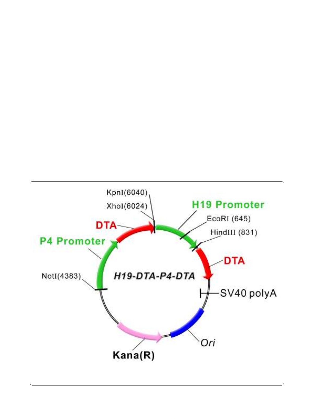
Figure 1 A schematic illustration depicting the construction of the double promoter H19-DTA-P4-DTA expression vector: The coding
sequence of each DTA is under the transcriptional control of both H19 and IGF2-P4 promoter sequences, respectively, Kana (R) - kanamycine
resistance gene.
Amit and Hochberg Journal of Translational Medicine 2010, 8:134
http://www.translational-medicine.com/content/8/1/134
Page 4 of 18

entering cells by endocytosis. The transfection proce-
dure was done as recommended by the manufacturer
(Polyplus-transfection, France). A total of 0.1 × 10
6
cells/well were grown overnight in a twelve-well Nunc
multidish (75 mm). For each well, 2 μgDNAand4μl
of the jetPEI (N/P = 5) were diluted separately with
50 μl of 150 mM NaCl each, and vortex-mixed gently.
The jetPEI solution was added at once to the DNA solu-
tion, the mixture was vortex-mixed for 10 seconds and
the mixture was incubated for 15 minuets at room tem-
perature. The 100 μl jetPEI/DNA mixture was then
applied drop-wise onto the serum containing medium of
each well. The transfection experiment was stopped
after 48 hours.
Luciferase activity
The cells were harvested and the luciferase activity was
determined using the luciferase Assay System kit (Pro-
mega). The light output was measured using a Lumac
Biocounter apparatus. The total protein content of the
lysates was determined by the Bio-Rad protein assay
reagent and the results were normalized to the total
protein and expressed as Light units/μgprotein.
LucSV40 (Luc-4) was used as a positive control for the
efficiency of transfection as it contains the SV40 promo-
ter and enhancer, while Luc-1 that lacks any regulatory
sequences was used as a negative control to determine
the basal nonspecific luciferase expression, which was
found to be negligible in all of the cell lines. All experi-
ments were done in triplicates and the results expressed
as mean and standard error.
In vitro targeted therapy
The cells were cotransfected with 2 μgoftheLucSV40
control vector and with the indicated amounts of the
DTA expressing vector (H19-DTA, P4-DTA or the
DTA double promoter expressing vector H19-DTA-P4-
DTA). The same cells were additionally transfected with
2μg LucSV40 alone in the same experiment. The H19-
DTA, P4-DTA and H19-DTA-P4-DTA cytotoxic activity
was determined by calculating the % of decrease in the
cotransfected LucSV40 activity compared to that of
LucSV40 transfected alone in the same cell type. The
total protein content of the lysates was determined by
the Bio-Rad protein assay reagent and the results were
normalized to the total protein and expressed as Light
units/μg protein. Therefore the reduction in luciferase
activity, reflect the inhibition of protein synthesis activity
by the DTA.
In vivo targeted therapy animal models
All surgical procedures and the care given to the ani-
mals were approved by the local committee for animal
welfare. Animals were kept in the Hebrew University’s
animal facility with water and food ad librum (all
experimental research on animals follow internationally
recognized guidelines). The histopathological examina-
tions of the different tumors were performed in consul-
tation with a trained pathologist.
Heterotopic nude mice model
Confluent T24P and HT-1376 human bladder carci-
noma cells were trypsinized to a single cell suspension
and resuspended in PBS. 2 × 10
6
T24P cells or HT-1376
cells (in 150 μl volume) were subcutaneously injected
into the back of female CD1 nude mice, 6-8 weeks old.
10 days after cell inoculation the developing tumors
were measured in two dimensions and randomized to
different treatments. Animals were separated to different
groupsofthesamesize(n=6).Theabilitytoinhibit
tumor growth by the single promoter DTA expression
vectors (P4-DTA, H19-DTA) and by the double promo-
ter DTA expression vector (H19-DTA-P4-DTA) was
tested. Intratumoral injections of 25 μgofeitherDTA
expressing constructs (treatment groups) or Luc expres-
sing constructs (control groups) were given 10, 12 and
14 days after cells inoculation. In vivo Jet-PEI a 22 kDa
linear form of polyethylenimine (PEI) was used as a
transfection enhancer reagent. PEI/DNA complexes with
a ratio of PEI nitrogen to DNA phosphate of 6 were
prepared in a solution of 5% w/v glucose according to
the manufacture’s instructions. Tumor dimensions were
measured, and the tumor volume was calculated accord-
ing to the formula width
2
× length × 0.5. The animals
were sacrificed 3 days after the last treatment, the
tumors were excised and their ex-vivo weight and
volume were measured. Samples of the tumors were
fixedin4%bufferedformaldehydeandprocessedfor
histological examination for evidence of necrosis and
persistent tumor. Computerized measurements of tumor
surface area and of the necrotic surface area were made
using the Image Pro Plus software (Media cybernetics,
Silver Springs, USA).
Orthotopic bladder cancer model
Female CD1 nude mice, 6-8 weeks old were used to
develop orthotopic superficial bladder tumors. Mice
were anesthetized with intra-peritoneal injection of keta-
mine (85 mg/kg) and xylazine (3 mg/kg). The bladder
was catheterized with a 24 gauge catheter, than drained
and its mucosa was mildly disrupted with 0.1 ml HCl
0.1N for 15-sec. (The bladder is rather resistant to
implantation of cells, and therefore it is necessary to
create abrasions in the bladder mucosa of the anesthe-
tized rodent either by acid, in order to increase “tumor
take”[29]). The acid was immediately neutralized with
0.1 ml NaOH 0.1N, and the bladder was washed three
times with 0.1 ml PBS. Subsequently, a 0.1 ml suspen-
sion of PBS containing 10 × 10
6
T24P human bladder
carcinoma cells was instilled into the bladder. The ure-
thra was ligated with 6/0 silk suture to assure that cells
Amit and Hochberg Journal of Translational Medicine 2010, 8:134
http://www.translational-medicine.com/content/8/1/134
Page 5 of 18


%20--%3e%3cdefs%3e%3cstyle%3e%20.st0%20{%20fill:%20%23fff;%20}%20.st1%20{%20fill:%20%237800fa;%20}%20%3c/style%3e%3c/defs%3e%3cpath%20class='st1'%20d='M117.78,12.18H43.11c2.9,3.47,4.65,7.94,4.65,12.82,0,5.6-2.3,10.66-6.01,14.29h76.02l7.22-13.56-7.22-13.56Z'/%3e%3cg%3e%3cpath%20class='st0'%20d='M53.58,26.17h-.59v-1.46h.59v-4.96h2.83c1.78,0,2.67.94,2.67,2.82v5.76c0,1.87-.89,2.81-2.67,2.81h-2.83v-4.96ZM55.36,21.37v3.34h1.1v1.46h-1.1v3.34h1.01c.61,0,.91-.37.91-1.1v-5.93c0-.74-.3-1.1-.91-1.1h-1.01Z'/%3e%3cpath%20class='st0'%20d='M65.99,31.14h-1.8l-.31-2.07h-2.19l-.31,2.07h-1.64l1.82-11.39h2.62l1.82,11.39ZM65.28,18.04c-.25.46-.51.77-.75.94-.21.15-.47.22-.79.22-.26,0-.57-.07-.92-.22l-.38-.15c-.14-.05-.26-.07-.37-.07-.3,0-.53.18-.71.54l-.91-.68c.25-.46.51-.77.75-.94.21-.14.48-.21.79-.21.26,0,.57.07.92.21l.38.15c.14.05.26.07.37.07.3,0,.53-.18.71-.54l.91.68ZM61.91,27.52h1.73l-.87-5.76-.87,5.76Z'/%3e%3cpath%20class='st0'%20d='M74.53,26.89v1.52c0,1.91-.89,2.86-2.67,2.86s-2.67-.95-2.67-2.86v-5.93c0-1.91.89-2.86,2.67-2.86s2.67.95,2.67,2.86v1.11h-1.69v-1.22c0-.75-.31-1.12-.93-1.12s-.93.37-.93,1.12v6.15c0,.74.31,1.11.93,1.11s.93-.37.93-1.11v-1.63h1.69Z'/%3e%3cpath%20class='st0'%20d='M81.4,31.14h-1.8l-.31-2.07h-2.19l-.31,2.07h-1.64l1.82-11.39h2.62l1.82,11.39ZM75.9,19.2l1.52-1.91h1.71l1.51,1.91h-1.61l-.76-.95-.75.95h-1.61ZM77.32,27.52h1.73l-.87-5.76-.87,5.76ZM83.1,15.99l-1.76,1.91h-1.26l1.17-1.91h1.86Z'/%3e%3cpath%20class='st0'%20d='M84.86,19.75c1.78,0,2.67.94,2.67,2.82v1.48c0,1.87-.89,2.81-2.67,2.81h-.85v4.28h-1.79v-11.39h2.64ZM84.01,21.37v3.86h.85c.58,0,.87-.36.87-1.08v-1.71c0-.71-.29-1.07-.87-1.07h-.85Z'/%3e%3cpath%20class='st0'%20d='M93.51,19.75c1.78,0,2.67.94,2.67,2.82v1.48c0,1.87-.89,2.81-2.67,2.81h-.85v4.28h-1.79v-11.39h2.64ZM92.66,21.37v3.86h.85c.58,0,.87-.36.87-1.08v-1.71c0-.71-.29-1.07-.87-1.07h-.85Z'/%3e%3cpath%20class='st0'%20d='M98.8,31.14h-1.79v-11.39h1.79v4.88h2.03v-4.88h1.83v11.39h-1.83v-4.88h-2.03v4.88Z'/%3e%3cpath%20class='st0'%20d='M105.36,24.55h2.46v1.62h-2.46v3.34h3.09v1.63h-4.88v-11.39h4.88v1.63h-3.09v3.18ZM108.17,17.29l-1.76,1.91h-1.26l1.17-1.91h1.86Z'/%3e%3cpath%20class='st0'%20d='M112.2,19.75c1.78,0,2.67.94,2.67,2.82v1.48c0,1.87-.89,2.81-2.67,2.81h-.85v4.28h-1.79v-11.39h2.64ZM111.35,21.37v3.86h.85c.58,0,.87-.36.87-1.08v-1.71c0-.71-.29-1.07-.87-1.07h-.85Z'/%3e%3c/g%3e%3ccircle%20class='st1'%20cx='25'%20cy='25'%20r='20'/%3e%3cpath%20class='st0'%20d='M32.78,19.27c2.92,0,4.43,2.55,5.28,5.33l.71,2.17c.14.38-.33.75-.71.75h-5.61c.19-.33.24-.71.09-1.08l-.75-2.45c-.43-1.32-.99-2.64-1.79-3.77.75-.57,1.65-.94,2.78-.94h0ZM25,18.38c3.25,0,4.9,2.78,5.89,5.89l.76,2.45c.14.42-.33.8-.8.8h-11.69c-.42,0-.94-.38-.8-.8l.75-2.45c.99-3.11,2.64-5.89,5.89-5.89h0ZM25,11.35c1.74,0,3.11,1.37,3.11,3.11s-1.37,3.11-3.11,3.11-3.11-1.41-3.11-3.11,1.41-3.11,3.11-3.11h0ZM17.27,19.27c1.08,0,1.98.38,2.73.94-.8,1.13-1.37,2.45-1.74,3.77l-.8,2.45c-.14.38-.05.75.09,1.08h-5.56c-.42,0-.9-.38-.75-.75l.71-2.17c.9-2.78,2.41-5.33,5.33-5.33h0ZM17.27,12.91c1.51,0,2.78,1.27,2.78,2.83s-1.27,2.83-2.78,2.83-2.83-1.27-2.83-2.83,1.27-2.83,2.83-2.83h0ZM32.78,12.91c1.56,0,2.78,1.27,2.78,2.83s-1.23,2.83-2.78,2.83-2.83-1.27-2.83-2.83,1.27-2.83,2.83-2.83h0ZM27.07,28.56v.09c0,.57-.24,1.08-.61,1.46h0v.05c-.38.33-.9.57-1.46.57s-1.08-.24-1.46-.61h0c-.38-.38-.61-.9-.61-1.46v-.09h1.41v.09c0,.19.05.38.19.47v.05c.09.09.28.19.47.19s.38-.09.47-.19v-.05c.14-.09.24-.28.24-.47t-.05-.09h1.41ZM30.99,28.56v.09c0,1.65-.66,3.16-1.74,4.24-1.08,1.08-2.59,1.79-4.24,1.79s-3.16-.71-4.24-1.79l-.05-.05c-1.04-1.08-1.7-2.55-1.7-4.2v-.09h1.41v.09c0,1.27.47,2.4,1.27,3.25h.05c.85.85,1.98,1.37,3.25,1.37s2.4-.52,3.25-1.37c.85-.8,1.37-1.98,1.37-3.25v-.09h1.37ZM34.99,28.56v.09c0,2.78-1.13,5.28-2.92,7.07-1.79,1.79-4.29,2.92-7.07,2.92s-5.23-1.13-7.07-2.92c-1.79-1.79-2.92-4.29-2.92-7.07v-.09h1.41v.09c0,2.4.94,4.53,2.5,6.08,1.56,1.56,3.72,2.5,6.08,2.5s4.52-.94,6.08-2.5c1.56-1.56,2.5-3.68,2.5-6.08v-.09h1.41Z'/%3e%3c/svg%3e)