Changes in rat liver mitochondria with aging
Lon protease-like activity and
N
e
-carboxymethyllysine accumulation in the matrix
Hilaire Bakala
1
, Evelyne Delaval
1
, Maud Hamelin
1
, Jeanne Bismuth
1
, Caroline Borot-Laloi
1
,
Bruno Corman
2
and Bertrand Friguet
1
1
Laboratoire de Biologie et Biochimie Cellulaire du Vieillissement, Universite
´Paris7-Denis Diderot, Paris, France;
2
Service de Biologie Cellulaire, Commissariat a
`l’Energie Atomique/Saclay, Gif-sur-Yvette, France
Aging is accompanied by a gradual deterioration of cell
functions. Mitochondrial dysfunction and accumulation of
protein damage have been proposed to contribute to this
process. The present study was carried out to examine the
effects of aging in mitochondrial matrix isolated from rat
liver. The activity of Lon protease, an enzyme implicated in
the degradation of abnormal matrix proteins, was meas-
ured and the accumulation of oxidation and glycoxidation
(N
e
-carboxymethyllysine, CML) products was monitored
using immunochemical assays. The function of isolated
mitochondria was assessed by measuring respiratory chain
activity. Mitochondria from aged (27 months) rats exhi-
bited the same rate of oxygen consumption as those from
adult (10 months) rats without any change in coupling
efficiency. At the same time, the ATP-stimulated Lon
protease activity, measured as fluorescent peptides released,
markedly decreased from 10-month-old rats (1.15 ±
0.15 FUÆlgprotein
)1
Æh
)1
) to 27-month-old-rats (0.59 ±
0.08 FUÆlgprotein
)1
Æh
)1
). In parallel with this decrease in
activity, oxidized proteins accumulated in the matrix upon
aging while the CML-modified protein content assessed
by ELISA significantly increased by 52% from 10 months
(11.71 ± 0.61 pmol CMLÆlgprotein
)1
) to 27 months
(17.81 ± 1.83 pmol CMLÆlgprotein
)1
). These results
indicate that the accumulation of deleterious oxidized and
carboxymethylated proteins in the matrix concomitant
with loss of the Lon protease activity may affect the ability
of aging mitochondria to respond to additional stress.
Keywords: aging; mitochondria; matrix; Lon protease;
carboxymethyllysine.
A striking characteristic of normal aging in long-lived
animals is the gradual decline in their physiological func-
tions. This decline is associated with an increase in reactive
oxygen species (ROS) production [1] and an accumulation
of macromolecules damaged by post-translational nonenzy-
matic modifications which alter the structure and function
of tissue and cellular proteins [2–5]. Under oxidative stress,
carbohydrates, lipids and proteins are the major targets of
reactive oxygen species. Proteins can be damaged either
directly or indirectly through the reactive carbonyl com-
pounds derived from the oxidation of carbohydrates and
lipids [6,7]. These carbonyl compounds react with protein
amino-groups to give glycoxidation products such as
N
e
-carboxymethyllysine (CML) [8]. This glycoxidation
process modifies cell proteins and the cumulative effects
lead to the tissue alterations and cell dysfunction typical of
aging and diabetes [9,10]. Recent data also indicate that
glycoxidative processes affect mitochondrial membrane
phospholipids [11]. Mitochondria are in fact a major
intracellular source of ROS during oxidative phosphoryla-
tion and the increased production of ROS is implicated in
the aging process [12–16]. Mitochondria are also the major
targets of these ROS, which may damage mitochondrial
proteins themselves, leading to dysfunction of these
organelles [17,18]. Oxidative damage mainly concerns
the activities of electron transport complexes of the inner
mitochondrial membrane which are specifically modified
during aging [19–21].
No attempt has yet been made to correlate the alterations
in mitochondrial function with biochemical changes in the
liver mitochondrial matrix of aging rats. Nevertheless, an
age-related decrease in the expression of several genes
involved in mitochondrial bioenergetics and mitochondrial
biogenesis occurs in aging mice; the largest age-associated
alteration affects the matrix enzyme Lon protease [22]. This
ATP-stimulated protease is homologous to bacterial Lon
protease [23] and has been found in several mammals
including human tissues and cell lines [24–26]. It is
responsible for the degradation of abnormal proteins and
certain short-lived specific proteins [27,28].
In this study we have investigated the age-associated
biochemical changes in mitochondrial matrix proteins
from the rat liver. For this purpose we related the activity
of Lon protease with the level of damaged proteins in the
matrix, focusing on the accumulation of oxidized and
CML-proteins as a marker of glycoxidative stress.
Correspondence to H. Bakala, Laboratoire de Biologie et Biochimie
Cellulaire du Vieillissement, Universite
´Paris7-Denis Diderot,
T23-33 1
er
e
´tage CC 7128, 2 Place Jussieu, 75252 Paris, France.
Fax: + 33 1 44 27 82 34; Tel.: + 33 1 44 27 82 35;
E-mail: bakala@paris7.jussieu.fr
Abbreviations: ROS, reactive oxygen species; AGE, advanced glycat-
ion end products; CML, carboxymethyllysine; ABTS, 2,2¢-azinobis
(3-ethylbenzo-6-thiazolinesulfonic acid); RCR, respiratory
control ratio.
(Received 22 January 2003, accepted 27 March 2003)
Eur. J. Biochem. 270, 2295–2302 (2003) FEBS 2003 doi:10.1046/j.1432-1033.2003.03598.x

Our results indicate that mitochondrial matrix proteins
undergo oxidative and glycoxidative modifications. These
damaged proteins accumulate with aging, in parallel with
a large decrease in Lon protease activity.
Materials and methods
Animals
Experiments were performed on male Wistar rats (WAG/
Rij) born and raised in the animal care facilities of the
Commissariat a
`l’Energie Atomique (CEA Gif-sur-Yvette,
France). This strain remains lean even when fed ad libitum
and does not suffer from age-associated nephropathy [29].
The animals were fed a commercial diet (DO4; UAR,
Villemoisson sur Orge, France) composed of 17% protein,
0.71% phosphorus, 0.78% calcium, 0.62% potassium,
0.27% sodium and 0.22% magnesium, with a total of
12.1 kJÆg
)1
. Water was provided ad libitum. Cohorts were
composed of adult and senescent animals (10 and 27 months
old, respectively). All studies were conducted in accordance
with the animal care policy of the National and European
regulations.
Chemicals
Na(CN)BH
3
, glyoxylic acid, BSA (Fraction V) and HRP–
conjugated anti-mouse IgG were purchased from Sigma.
Anti-AGE mAb (clone no. 6D12) was from Trans Genic
Inc. (Japan) and Oxyblot protein oxidation detection kit
from Intergen.
Chemical modification of BSA
(
N
e
-carboxymethyllysine-BSA)
Carboxymethylated bovine serum albumin (CML-BSA)
was prepared according to Murata et al. [30]. Briefly,
100 mg BSA was incubated at 37 Cfor24hwith0.15
M
glyoxylic acid and 0.45
M
Na(CN)BH
3
in 1 mL of 0.2
M
sodium phosphate buffer (pH 7.4), and then extensively
dialyzed against phosphate-buffered saline (NaCl/P
i
).
The CML content of the modified BSA was measured by
amino acid analysis following hydrolysis of the modified
protein in 6
M
HCl, 0.2% phenol (Laboratoire de Micro-
sequenc¸age des Prote
´ines, Institut Pasteur, Paris, France).
There were 43.2 CML residues and 16.1 Lys residues in
BSA-modified protein, while native BSA contained 57.9 Lys
in a total of 692 residues. As the expected value in the
primary sequence is 59 Lys, the error in the CML–adduct
rate can be estimated to be as low as 2%. The CML content
was expressed as 0.644 nmol CML per lg BSA, and this
solution was used as the standard in a competitive ELISA.
Isolation of mitochondria
A 10% tissue homogenate was prepared in an ice-cold
medium containing 220 m
M
mannitol, 70 m
M
sucrose,
2m
M
Hepes, 0.1 m
M
EDTA and 0.5% (w/v) BSA,
pH 7.4. Nuclei and unbroken cells were pelleted by
centrifugation for 10 min at 800 gand 0 C. The super-
natant was centrifuged at 8000 gfor 10 min at 0 C. The
mitochondrial pellet was washed three times with the
homogenization medium and used for polarographic
measurements.
For determination of matrix protease activity, mitochon-
dria were suspended in 50 m
M
Tris/HCl buffer, pH 7.9,
then disrupted by sonication (four times for 10 s). The
resulting suspension was centrifuged at 15 000 gfor 10 min
and then at 100 000 gfor 45 min. The supernatant (con-
taining matrix protein) was stored at )80 C for further
determinations of protease activity and the level of
carboxymethylated protein. Protein was assayed by the
Bradford method.
To estimate the contamination of mitochondrial prepar-
ation with lysosomes, we used acid phosphatase activity as a
marker.
Measurements of mitochondrial respiration
Oxygen consumption was measured polarographically with
a Clark electrode in the sample, as described by Aprille and
Asimakis [31] in a thermostatically controlled closed 2 mL
chamber (30 C). The rate of oxygen consumption was
measured in the presence of 310 nmol ADP and 10 m
M
succinate or 5 m
M
glutamate and 5 m
M
malate (state 3) and
when all the ADP has been consumed (state 4 or resting
state). Oxygen-consumption rates are expressed as ng atoms
of oxygen consumed per minute and per mg protein. The
rate of oxygen consumption in state 3 and in state 4,
respiratory control ratio (RCR) of oxygen consumptions in
states 3 and 4 and the ADP/O ratio were calculated. Oxygen
consumption in the presence of 40 l
M
of dinitrophenol
(uncoupled state) was also checked.
Enzymatic activities
ATP-stimulated Lon protease activity was determined using
casein-fluoresceine isothiocyanate (0.5 mgÆmL
)1
) as sub-
strate. Casein was incubated with mitochondrial matrix
extract (70 lgprotein)in70lL buffer (final concentration:
50 m
M
Tris/HCl, pH 7.9, 10 m
M
MgCl
2
, with or without
8m
M
ATP) for 1 h at 37 C. The reaction was terminated
by adding 30 lL of 40% trichloroacetic acid and 50 lLof
3% BSA. After centrifugation at 15 000 gfor 30 min,
100 lLof2
M
sodium borate was then added to 80 lLof
supernatant. Fluorescence was measured with excitation/
emission wavelengths of 495/515 nm. Activity is expressed
as fluorescence units per hour of incubation and per lg
protein (FUÆlgprotein
)1
Æh
)1
) corrected from the fluores-
cence of casein alone, which represents around 25% of the
measured fluorescence.
Acid phosphatase activity was determined at each step
of the isolation procedure using the acid phosphatase
assay (Sigma kit), which measures the accumulation of
p-nitrophenol from p-nitrophenyl phosphate disodium, at
410 nm. The reaction was stopped after 30 min incubation
at room temperature.
Determination of CML content in mitochondrial matrix
proteins by competitive ELISA
Each well of a 96-well microtiter plate (Nunc-immuno Plate,
Nunc, Denmark) was coated with 100 lLCML-BSA
(6.4 nmol CMLÆmL
)1
)in50m
M
sodium carbonate buffer,
2296 H. Bakala et al.(Eur. J. Biochem. 270)FEBS 2003

pH 9.6 by incubation overnight at 4 C. The wells were
washed three times with NaCl/P
i
containing 0.05% (v/v)
Tween 20 (buffer A) and free binding sites were blocked by
incubation for 1 h at room temperature with 100 lLNaCl/
P
i
containing 6% (w/v) skimmed milk. The wells were then
washed with buffer A, 50 lL of competing antigen (test
samples at 0.100 mgÆmL
)1
or serial dilutions of standard
CML-BSA from 0.64 m
M
to 128 m
M
) was added, followed
by 50 lL monoclonal antibody clone 6D12 (diluted
1 : 1000 in NaCl/P
i
). The plate was incubated for 2 h at
room temperature, washed and then incubated with 50 lL
horseradish peroxidase-conjugated anti-mouse IgG (50 lL
per well, second antibody diluted 1 : 1000) for 2 h at room
temperature. The wells were washed, 100 lLofsubstrate
solution [40 m
M
2,2¢-azinobis(3-ethylbenzo-6-thiazolinesul-
fonic acid (ABTS) and 200 lL of 30% hydrogen peroxide in
20 mL acetate-phosphate buffer] were added per well and
incubated for 30 min at 37 C. The absorbance (A)was
measured at 405 nm on a micro-ELISA plate reader
(Spectra Rainbow, SLT Labinstruments, Austria). Results
are expressed as the ratio B/B
0
, calculated as [experimental
Aminus background A(no antibody)]/[total A(no
competitor) minus background A], vs. CML added as pmol
CMLÆlgprotein
)1
.
Western blot analysis
Western blot of CML proteins. Matrix protein samples
(10 lg protein per lane) were electrophoresed on 10% (w/v)
SDS/PAGE for 90 min at 100 V. One of two identical gels
was stained with Coomassie blue to analyzed the pattern of
matrix proteins. The proteins from the second gel were then
transferred electrophoretically to a nitrocellulose membrane
(Bio-Rad) for 1 h at 100 V. The membrane was saturated
with 5% (w/v) skimmed milk in NaCl/P
i
/0.1% Tween 20
overnight at 4 C, followed by four washes (10 min each)
with NaCl/P
i
containing 0.2% (v/v) Tween 20 (wash buffer).
The membrane was then incubated for 2 h at room
temperature with anti-CML mAb clone 6D12 (diluted
1 : 1000 in NaCl/P
i
/0.1% Tween 20), washed four times
with wash buffer, incubated for 1 h with anti-mouse IgG
coupled to horseradish peroxidase (1 : 2500 dilution) and
given a final wash. The proteins were revealed with an
ECL reagent (Amersham-Pharmacia Biotech).
Western blot of carbonylated proteins. Carbonylated
proteins were analyzed using the oxyblot kit according to
the manufacturer’s instructions (Oxyblot Detection, Inter-
gen). Briefly, samples (10 lg protein per lane) were treated
with 10 m
M
2,4-dinitrophenolhydrazine in 2
M
HCl, incu-
bated at room temperature and neutralized. The derivatized
proteins were separated by SDS/PAGE, transferred to a
nitrocellulose membrane and treated as previous Western
blotting (see below). The primary antibody used was against
2,4-dinitrophenol, and detection was performed using the
ECL reagent.
Western blot of Lon protease. A polyclonal antibody
against Lon protease was raised in rabbit against a synthetic
peptide corresponding to amino acids 208–221 of rat Lon.
This antibody mainly recognized one protein band with an
estimated molecular mass of 100 kDa corresponding to the
molecularmassofLon.
For Western blot analysis, we previously verified that
signals were linear in the range from 10–60 lgoftotal
protein. We used routinely 20 lg of mitochondrial matrix.
Proteins were transferred onto nitrocellulose membrane
(Bio-Rad), which was then incubated with Lon antibody
(1 : 1000 dilution) for 1 h at room temperature and antigen
were detected by chemiluminescence with Amersham’s
ECL reagent.
Electron microscopy
Electron micrographs were taken of purified rat liver
mitochondria. The mitochondrial pellet was washed with
NaCl/P
i
, fixed by immersion in 2% paraformaldehyde/
0.2% glutaraldehyde in NaCl/P
i
for 2 h at room tempera-
ture, contrasted with 1% uranyl acetate. The resulting
material was dehydrated and embedded in LRWhite.
Ultrathin 60 nm sections were cut with a Leica ultramicro-
tome, postfixed with 2% osmium tetroxide and examined in
a Philips 400 electron microscope.
Statistical analysis
Results are presented as means ± SEM and differences
between groups were assessed by means of Student’s
unpaired t-test. Significance was set at P< 0.05.
Results
Age-related changes in the structure and respiratory
function of isolated mitochondria
As shown in Table 1, there was no difference in mito-
chondrial oxygen consumptions between 10-month- and
27-month-old-rats, whatever the substrate used. The
Table 1. Biochemical respiratory parameters in rat liver mitochondria from 10- and 27-month-old rats. Data represent mean values ± SEM
(n, number of animals). Oxygen consumption rates were measured polarographically in the presence of 10 m
M
succinate or 5 m
M
glutamate plus
5m
M
malate. State 3 respiration was determined after adding 310 nmoles ADP.
Glutamate (n¼4) Succinate (n¼7)
10 months 27 months 10 months 27 months
State 4 (ng atom OÆmin
)1
Æmg
)1
) 30.3 ± 0.3 26.5 ± 3.5 46.0 ± 3.5 47.1 ± 7.6
State 3 (ng atom OÆmin
)1
Æmg
)1
) 120.8 ± 7.4 117.8 ± 9.4 189.7 ± 15.8 166.9 ± 14.1
RCR 3.98 ± 0.26 4.52 ± 0.32 4.15 ± 0.20 3.79 ± 0.36
P/O 2.85 ± 0.13 2.94 ± 0.13 2.05 ± 0.08 1.91 ± 0.06
FEBS 2003 Protein alteration in mitochondrial matrix with aging (Eur. J. Biochem. 270) 2297

ADP/O ratios obtained with succinate or glutamate indicate
no change in coupling efficiency. The yield of mitochondrial
preparation as well as the respiratory control ratio (state 3/
state 4) did not change significantly during aging. Oxygen
consumption rates were the same in the presence of 2,4-
dinitrophenol or in the presence of ADP (state 3) at all ages
considered.
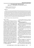
Electron micrography showed few alterations of crests
and of mitochondrial membranes leading to some disrupted
organelles (Fig. 1). Mitochondria preparations were free of
contamination and both sets exhibited similar purity.
ATP-stimulated protease activity in aging rats
Results obtained by use of acid phosphatase determination
in rat preparations indicated a very low lysosomal contami-
nation (4% of mitochondrial matrix vs. total liver homo-
genate).
The activity of ATP-stimulated protease was determined
in the presence or absence of ATP (Fig. 2A). Protease
activity in the absence of ATP was lower in aging rats than
in adults. Activity decreased by 51% between 10-month-
old and 27-month-old rats (1.15 ± 0.15 and 0.59 ±
0.08 FUÆlgprotein
)1
Æh
)1
, respectively). Whatever the age,
addition of ATP stimulated the degradation of the substrate
casein about 2.5-fold but activity still decreases in an age-
dependent manner.
No significant difference could be detected in the level of
Lon protein from the mitochondrial matrix of 10-month-
and 27-month-old rats, as shown by Western blot analysis
(Fig. 2B,C).
Fig. 1. Assessment of the purity of mitochondrial preparations. Electron
micrograph of isolated liver mitochondria from (A) 10-month-old and
(B) 27-month-old rats. All the recognizable organelles are mito-
chondria (arrow) showing different degrees of matrix density (star) and
dilatation of the cristae (arrowhead). Disrupted mitochondria are
visible (double arrow). Magnification, 30 000.
Fig. 2. Lon protease quantification and activity in liver mitochondrial
matrix from 10- and 27-month-old rats. Activitywasdeterminedinthe
absence of ATP or in the presence of 8 m
M
ATP (A, white and grey
columns respectively). Matrix proteins (20 lg) were subjected to
Western blotting using a polyclonal antibody against Lon protease (B)
and quantified by densitometric scanning (C), results being expressed
in arbitrary units. Values are the mean ± SEM for five independent
determinations. The P-value for enzymatic activity was significant
(**P< 0.01 for 10-month-old rats).
2298 H. Bakala et al.(Eur. J. Biochem. 270)FEBS 2003

CML adduct content in mitochondrial matrix proteins
We used the monoclonal antibody clone 6D12 in competi-
tive ELISA to evaluate the CML-protein content (Fig. 3).
The CML content increased significantly by 52% from
11.71 ± 0.61 pmol CMLÆlgprotein
)1
(n¼8) in 10-
month-old rats to 17.81 ± 1.83 pmol CMLÆlgprotein
)1
(n¼9) in 27-month-old rats (P¼0.007). These data
indicate an age-associated accumulation of CML adducts
in mitochondrial matrix.
Western blotting of modified proteins
We identified the major proteins in the mitochondrial
matrix by performing SDS/PAGE separation and staining
the gel with Coomassie blue or Western blotting. Analysis
of the SDS/PAGE revealed a broad spread of proteins
with apparent molecular mass ranging from 10–170 kDa
(Fig. 4A). The samples from the two different ages exhibited
a comparable pattern of bands, although two bands of 60
and 150 kDa strongly visible at 10 months (lane 1) were
absent at 27 months, and an important band of 70 kDa
(lane 2) emerged in this latter sample. Western blotting with
mAb 6D12 was used to detect matrix proteins undergoing
CML modification with aging. Proteins from all molecular
masses were immunolabelled in samples of both ages
(Fig. 4B). The 10-month-old matrix proteins (lane 1)
contained 14 bands intensely stained over an apparent
range of 10–170 kDa, with the most prominent band at
60 kDa (lane 1). In the 27-month-old preparations (lane 2)
only eight main bands are stained, with two intense signals
of 70 and 50 kDa, while the bands at 60 and 150 kDa
vanished.
With oxyblot (Fig. 4C), antibodies stained carbonylated
proteins mainly in band ranges of 30–60 and 70–120 kDa at
10 months (lane 1). These protein bands became strongly
stained at 27 months, particularly those with apparent
molecular masses of 30, 55, 75 and 105 kDa (lane 2).
These results strongly support the hypothesis that CML-
proteins and oxidized proteins do occur selectively in the
liver mitochondrial matrix and that their recruitment varies
with aging.
Discussion
We used isolated rat liver mitochondria to analyse the
matrix defects that occur with aging. Mitochondria, similar
to the cytosol, contain a proteolytic system that controls the
metabolic stability of mitochondrial proteins and ensures
the elimination of damaged proteins [28]. This continuous
Fig. 3. Determination of CML-protein content in liver mitochondrial
matrix with aging. The CML content in the matrix proteins from
10- and 27-month-old rats was measured by competitive ELISA using
mAb 6D12. Results are expressed as pmol CMLÆlgprotein
)1
(n, number of animals). ***P¼0.007 vs. 10-month-old rats.
Fig. 4. Western blot analyses of liver mitochondrial matrix proteins from 10-month- and 27-month-old rats. Samples (10 lg) were subjected to SDS/
PAGE under reducing conditions. The gel was stained with Coomassie blue (A) or subjected to Western blotting using either 6D12 monoclonal
antibody to detect CML-modified proteins (B) or oxyblot kit to reveal oxidized proteins (C). Samples from 10-month-old (lane 1) and from
27-month-old rats (lane 2). Standard molecular masses (lane 3). The arrow and arrowhead show the disappearance and appearance of bands,
respectively.
FEBS 2003 Protein alteration in mitochondrial matrix with aging (Eur. J. Biochem. 270) 2299