CAS E REP O R T Open Access
Granulomatous pyoderma preceding chronic
recurrent multifocal osteomyelitis triggered by
vaccinations in a two-year-old boy: a case report
Neslihan Karaca
1
, Guzide Aksu
1*
, Can Ozturk
2
, Nesrin Gulez
1
, Necil Kutukculer
1
Abstract
Introduction: Chronic recurrent multifocal osteomyelitis is a rare, systemic, aseptic, inflammatory disorder that
involves different sites. Pathogenesis of chronic recurrent multifocal osteomyelitis is currently unknown.
Case presentation: A two-year-old Caucasian boy, diagnosed with chronic recurrent multifocal osteomyelitis with
granulomatous pyoderma following routine vaccinations is presented for the first time in the literature.
Conclusion: We conclude that antigen exposures might have provoked this inflammatory condition for our case.
Skin and/or bone lesions following vaccinations should raise suspicion of an inflammatory response such as
chronic recurrent multifocal osteomyelitis only after thorough evaluation for chronic infection, autoimmune,
immunodeficiency or vasculitic diseases.
Introduction
Chronic recurrent multifocal osteomyelitis (CRMO) is a
rare, systemic, noninfectious, inflammatory disorder that
is characterised by recurrent, nonsuppurative, multiple
osteolytic bone lesions. It accounts for 2% to 5% of all
osteomyelitis cases [1,2].
Itmainlyaffectsmetaphysesofthelongboneswith
repetitive exacerbations and spontaneous remissions and
is frequently associated with a cutaneous inflammatory
condition such as pustulosis palmoplantaris, Sweet syn-
drome, psoriasis and pyoderma gangrenosum [3,4].
We hereby present a case of chronic recurrent multi-
focal osteomyelitis initially presenting as granulomatous
pyoderma following routine vaccinations.
Case presentation
A two-year-old Caucasian boy, the first child of non-
consanguineous healthy parents, presented with history
of recurrent skin lesions. These lesions occurred after
BCG (Bacillus Calmette-Guerin) and DTP (diphteria,
tetanus and pertussis) vaccinations at the age of two
months and after each hepatitis B vaccination thereafter.
Skin lesions were initially papular, then vesicular with
purulent exudate, progressing to multiple ulcers and
draining sinuses spreading from the injection site.
On admission, skin examination revealed violaceous,
tender; 5-7 mm sized superficial ulcerations with drain-
ing sinuses on right forearm, left deltoid area and right
cheek (Figure 1). There were no constitutional symp-
toms or other abnormality in physical examination.
Laboratory results were as follows; white blood cell
count 9220/mL with 56% polymorph nuclear cells, 40%
lymphocytes, 4% monocytes on peripheral smear, hemo-
globin 11.4 g/dL, platelets 426.000/mL, erythrocyte sedi-
mentation rate (ESR) was 24 mm/h and the C-reactive
protein was 0.43 mg/dL. X-rays of humerus, radius and
ulna were normal. Possibilities of combined immunode-
ficiency, hyper IgM syndrome types I/III, chronic granu-
lomatous disease, IL-12/interferon-gamma pathway
defects were excluded: Immunoglobulin G-M-A serum
concentrations, lymphocyte subsets, expression of CD40
on B cells, CD40 ligand on active T cells, complement
levels (C3, C4), adenosine deaminase level, phagoburst
test, expression of CD119 (interferon-gamma receptor)
and IL-12 receptor b-I on lymphocytes were within nor-
mal ranges. Functional and genetic studies related with
IL-12 b1 and IFN-greceptors were normal. Histopathol-
ogyofskinbiopsyspecimenshowed‘noncaseating
* Correspondence: guzide.aksu@ege.edu.tr
1
Ege University School of Medicine, Department of Pediatric Immunology,
Izmir, Turkey
Full list of author information is available at the end of the article
Karaca et al.Journal of Medical Case Reports 2010, 4:325
http://www.jmedicalcasereports.com/content/4/1/325 JOURNAL OF MEDICAL
CASE REPORTS
© 2010 Karaca et al; licensee BioMed Central Ltd. This is an Open Access article distributed under the terms of the Creative Commons
Attribution License (http://creativecommons.org/licenses/by/2.0), which permits unrestricted use, distribution, and reproduction in
any medium, provided the original work is properly cited.

granuloma’. Regarding the initial appearance of the
lesions after BCG vaccination and histopathology, scro-
fuloderma was considered. However, acid-fast bacillus
smears and initial cultures for nonspecific bacteria and
mycobacteria were negative. The patient was empirically
treated with isoniazid 5 mg/kg, rifampicin 10 mg/kg and
pyrazinamide 25 mg/kg and skin lesions improved
gradually.
Five months after initiation of anti-mycobacterial
treatment, an elevation in ESR to 74 mm per hour was
obtained. On physical examination, he did not have new
skin lesions. Nonspecific and mycobacterial cultures of
blood and urine, peripheral and bone marrow aspiration
smears, Mantoux skin test, serological investigations for
Brucella and Salmonella, abdominal ultrasonography
were normal. Rheumatic and auto-inflammatory diseases
including sarcoidosis and vasculitis were searched out;
HLA-B27 antigen, anti-nuclear antibody, antineutrophi-
lic cytoplasmic antibody and rheumatoid factor were
negative. Genetic analyses for ‘Familial Mediterranean
Fever’,‘Tumor Necrosis Factor Receptor-Associated
Periodic Syndrome’and IL-1 receptor defects were per-
formed with negative results except IL-1 receptor
antagonist intron 2 variable tandem repeat polymorph-
ism (IL-1RN-1/1). Ciprofloxacin was added to anti-
mycobacterial treatment and ESR decreased to normal
(18 mm per hour). After a period of two months with
no complaints, he developed new purulent skin lesions
on the left forearm and left medial malleolus. Increased
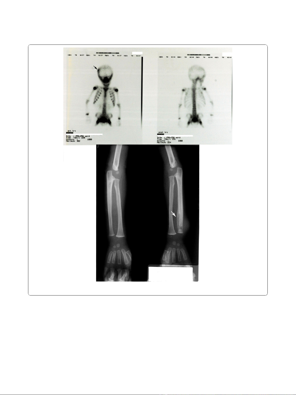
activity was seen on right frontoparietal bone with bone
Tc 99m MDP scintigraphy; X-ray of the distal metaphy-
seal region of the radius revealed osteolytic lesions (Fig-
ure 2). Bone biopsy was planned but parental consent
was not given for the procedure. The patient was treated
with intravenous teicoplanin for three weeks. Cultures of
abscess material, taken before antibiotic treatment, for
bacteria (including acid-fast bacilli) and fungi yielded
negative results. During the following three months, he
was given anti-mycobacterial treatment including isonia-
zid and rifampicin for nine months and pyrazinamid for
five months. After this period, the patient was read-
mitted with pain and swelling on both ankles. The
ankles were swollen and warm. X-ray of the left distal
tibia showed ‘osteolytic lesions surrounded by reactive
hyperostosis’consistent with chronic osteomyelitis. He
was diagnosed as CRMO based on four clinical exacer-
bations and repeatedly negative cultures. Prednisolone
(0.8 mg/kg/day) treatment was started and all other
medications were stopped. X-ray of tubular bones
showed disappearance of all osteolytic lesions two
months after the initiation of corticosteroid therapy. He
was complaint-free during the following 18 months.
According to the initial presentation with multiform
skin lesions affecting left deltoid area and right cheek
just after BCG vaccination at the age of two months and
the biopsy findings of the skin lesions, our patient raised
the suspicion of scrofuloderma and empirical anti-
mycobacterial treatment was given [5]. Mycobacterial
disease was not supported by skin tests, lesional smears
or repeated cultures before treatment. Possible inherited
defects in the defence against mycobacterial infections
such as IL-12 band IFN-greceptor deficiencies were
ruled out with functional and genetic investigations. On
admission, X-rays of left humerus, radius and ulna were
normal.
During follow-up, he had new skin lesions as well as
recurrent, sterile, multifocal, osteolytic bone lesions with
reactive hyperostosis interpreted as chronic osteomyeli-
tis. As broad-spectrum antibiotics and anti-mycobacter-
ial treatment were not effective, he was treated by
corticosteroids and complete remission was then
obtained. The prompt response to steroid rather than
Figure 1 Violeceous, tender, superfically ulcerated plaques on right deltoid area, right cheek and right forearm.
Karaca et al.Journal of Medical Case Reports 2010, 4:325
http://www.jmedicalcasereports.com/content/4/1/325
Page 2 of 5
antibiotic treatment raised the possibility of an inflam-
matory condition rather than an immunodeficiency. Col-
lectively, we diagnosed the patient as having CRMO at
29 months of age with a history of disease course of
more then three months and failure to cultivate a
micro-organism.
Discussion
The etiology of CRMO remains unknown. Rheumatic
disease, bacterial subacute osteomyelitis and malignancy
are the main differential diagnoses. These were excluded
in our case. The histopathology of bone lesions is vari-
able. Chronic lesions demonstrate a predominance of
lymphocytes with the occasional presence of plasma
cells. Non-caseating granulomatous foci occasionally
coexist [6,7]. The diagnosis of CRMO remains a chal-
lenge. Schultz et al. [8] suggested that the disease can
be diagnosed in the presence of a prolonged course
more than three months, evidence of bone inflamma-
tion, negative bone cultures by an open bone biopsy and
Figure 2 Bone Tc 99m MDP scintigraphy (a) showing increased activity on right frontoparietal bone: (b) plain radiograph of the
patient showing osteolytic lesions in distal metaphyseal region of the radius.
Karaca et al.Journal of Medical Case Reports 2010, 4:325
http://www.jmedicalcasereports.com/content/4/1/325
Page 3 of 5

the presence of multiple bone lesions. The impossibility
to perform a bone biopsy, due to lack of parental con-
sent, represents a relevant limitation to the correct
interpretation of the clinical and pathological findings
observed. Recurrent, multifocal, sterile osteomyelitis in
X-rays and bone scintigraphy findings with negative cul-
tures supported the diagnosis of CRMO for our case.
CRMO primarily affects bone but is often accompa-
nied by chronic inflammatory neutrophilic dermatoses
such as palmoplantar pustulosis, psoriasis, severe acne,
Sweet syndrome, pyoderma gangrenosum or superficial
granulomatous pyoderma [4,9]. In our case, it was pre-
ceded with granulomatous pyoderma. Little is known
about the simultaneous presence of chronic musculoske-
letal inflammation and skin disorders. CRMO has been
considered to be the pediatric variant of SAPHO (syno-
vitis, acne, pustulosis, hyperostosis and osteitis) syn-
drome [7]. In a report presenting ten cases with SAPHO
syndrome during childhood, the ages at onset ranged
from 2.9 to 13.5 years [7]. The age that the first lesions
appeared in our patient was two months, which is extre-
mely young for disease onset. The increased prevalence
of HLA-B27, sacroiliitis, inflammatory bowel disease and
psoriasis in patients with SAPHO syndrome has led it to
be classified as a spondyloarthropathy [10]. Our case
was HLA-B27 negative and lacked these rheumatologic
manifestations.
In most patients, cultures from bone lesions are ster-
ile. Propionibacterium acnes has been found in the
affected area, in a few cases. There is no response to
antibiotics. Some authors suppose P. acnes as a trigger
in the pathogenesis of the disease [11]. However, these
bacteria might also be contaminants during biopsy. As
all clinical symptoms preceded various vaccinations in
our patient, it can be speculated that antigen exposures
might have triggered this inflammatory condition.
Although most reported cases of CRMO are sporadic,
there is evidence for a genetic component to its etiology.
There is an autosomal recessive syndromic form of
CRMO (Majeed syndrome) which is caused by muta-
tions in LPIN2 [12,13]. In addition, mice with a muta-
tion on chromosome 18 develop a syndrome resembling
human CRMO, suggesting a possible genetic predisposi-
tion [14]. There is also evidence to suggest that the
bony inflammation in CRMO is a result of an aberrant
immune response directed against bone [15]. In our
case, the bony symptoms have improved after treatment
of antiinflammatory drugs.
CRMO is generally treated with nonsteroidal anti-
inflammatory drugs, corticosteroids, analgesics and anti-
biotic therapy is not recommended [2]. In our patient,
skin lesions and multifocal osteomyelitis responded well
to oral prednisolone treatment.
Conclusion
Clinicians should be aware of CRMO, because it typi-
cally occurs during childhood and should be included in
the differential diagnosis of patients with signs and
symptoms of recurrent osteomyelitis and granulomatous
pyoderma to avoid prolonged antibiotic treatment.
Granulomatous pyoderma with CRMO triggered by
vaccinations was not previously reported. This novel
association may serve to enlighten the currently
unknown pathogenesis of CRMO.
Consent
Written informed consent was obtained from the par-
ents of the patient for publication of this case report
and accompanying images. A copy of the written con-
sent is available for review by the Editor-in-Chief of this
journal.
Acknowledgements
We thank Dr J L Casanova and his co-workers in Laboratory of Human
Genetics of Infectious Diseases Necker-Enfants Malades Medical School for
their help in the study of functional and genetic studies related with IL-12
receptor b1 and IFN-greceptor and Medical Genetic Department of Ege
University for IL1 receptor mutation analyses.
Author details
1
Ege University School of Medicine, Department of Pediatric Immunology,
Izmir, Turkey.
2
SB Tepecik Egitim Hastanesi, Department of Pediatrics, Izmir,
Turkey.
Authors’contributions
All authors have analysed and interpreted the patient data regarding the
auto-inflammatory disease. All authors have read and approved the final
manuscript.
Competing interests
The authors declare that they have no competing interests.
Received: 8 December 2009 Accepted: 18 October 2010
Published: 18 October 2010
References
1. Giedion A, Holthusen W, Masel LF, Vischer D: Subacute and chronic
symmetrical osteomyelitis. Ann Radiol 1972, 15:329-342.
2. Huber AM, Lam PY, Duffy CM, Yeung RS, Ditchfield M, Laxer D, Cole WG,
Graham K, Allen RC, Laxer RM: Chronic recurrent multifocal osteomyelitis:
Clinical outcomes after more than five years of follow-up.J Pediatr 2002,
141:198-203.
3. Haliasos E, Soder B, Rubenstein DS, Henderson W, Morrell DS: Pediatric
Sweet Syndrome and immunodeficiency successfully treated with
intravenous immunoglobulin. Pediatr Dermatol 2005, 22:530-535.
4. Omidi CJ, Siegfried EC: Chronic recurrent multifocal osteomyelitis
preceding pyoderma gangrenosum and occult ulcerative colitis in a
pediatric patient. Pediatr Dermatol 1998, 15:435-438.
5. Farina MC, Gegundez MI, Pigue E, Esteban J, Martvn L, Requena L, Barat Q,
Guerrero MF: Cutaneous tuberulosis: a clinical, histopathologic and
bacteriologic study. J Am Acad Dermatol 1995, 33:433-440.
6. Björsten B, Boquist L: Histopathological aspects of chronic recurrent
multifocal osteomyelitis. J Bone Joint Surg Br 1980, 62(3):376-380.
7. Beretta-Piccoli BC, Sauvain MJ, Gal I, Schibler A, Saurenman T, Kressebuch H,
Bianchetti MG: Synovitis, acne, pustulosis, hyperostosis, osteitis (SAPHO)
syndrome in childhood: a report of ten cases and review of the
literature. Eur J Pediatr 2000, 159:594-601.
Karaca et al.Journal of Medical Case Reports 2010, 4:325
http://www.jmedicalcasereports.com/content/4/1/325
Page 4 of 5

8. Schultz C, Holterhus PM, Seidel A, Jonas S, Barthel M, Kruse K, Bucsky P:
Chronic recurrent multifocal osteomyelitis in children. Pediatr Infect Dis
1999, 18:1008-1013.
9. Majeed HA, Kalaawi M, Mohanty D, Teebi AS, Tunjekar MF, Al-Gharbawy F,
Majeed SA, Al-Gazzar AH: Congenital dyserythtropoietic anemia and
chronic recurrent multifocal osteomyelitis in three related children and
the association with Sweet syndrome in two siblings. J Pediatr 1998,
115:730-734.
10. Van Dornuum S, Barraciough D, McColl G, Wicks I: SAPHO: rare or just not
recognized? Semin Arthritis Rheum 2000, 30:70-77.
11. Kotilainen P, Merilahti-Palo R, Lehtonen OP, Manner I, Helander I,
Mottonen T, Rintala E: Propionibacterium acnes isolated from sternal
osteitis in a patient with SAPHO syndrome. J Rheumatol 1996,
23:1302-1304.
12. Majeed HA, El-Shanti H, Al-Rimawi H, Al-Masri N: On mice and men: an
autosomal recessive syndrome of chronic recurrent multifocal
osteomyelitis and congenital dyserythropoietic anemia. J Pediatr 2000,
137:441-442.
13. Ferguson PJ, Chen S, Tayeh MK, Ochoa L, Leal SM, Pelet A, Munnich A,
Lyonnet S, Majeed HA, El-Shanti H, et al:Homozygous mutations in lpin2
are responsible for the syndrome of chronic recurrent multifocal
osteomyelitis and congenital dyserythropoietic anemia (Majeed
syndrome). J Med Genet 2005, 42:551-557.
14. Byrd L, Grossmann M, Potter M, Shen-Ong G: Chronic recurrent multifocal
osteomyelitis: A new recessive mutation on chromosome 18 of the
mouse. Genomics 1991, 11:794-798.
15. Bousvaros A, Marcon M, Treem W, Waters P, Issenman R, Couper R,
Burnell R, Rosenberg A, Rabinovich E, Kirschner BS: Chronic recurrent
multifocal osteomyelitis associated with chronic inflammatory bowel
disease in children. Dig Dis Sci 1999, 44:2500-2507.
doi:10.1186/1752-1947-4-325
Cite this article as: Karaca et al.: Granulomatous pyoderma preceding
chronic recurrent multifocal osteomyelitis triggered by vaccinations in a
two-year-old boy: a case report. Journal of Medical Case Reports 2010
4:325.
Submit your next manuscript to BioMed Central
and take full advantage of:
• Convenient online submission
• Thorough peer review
• No space constraints or color figure charges
• Immediate publication on acceptance
• Inclusion in PubMed, CAS, Scopus and Google Scholar
• Research which is freely available for redistribution
Submit your manuscript at
www.biomedcentral.com/submit
Karaca et al.Journal of Medical Case Reports 2010, 4:325
http://www.jmedicalcasereports.com/content/4/1/325
Page 5 of 5


![Vaccine và ứng dụng: Bài tiểu luận [chuẩn SEO]](https://cdn.tailieu.vn/images/document/thumbnail/2016/20160519/3008140018/135x160/652005293.jpg)